

Etiologie retence horních stálých špičáků. Přehledový článek.
Etiology of impacted permanent maxillary canines. A review.
MDDr. Dana Kloudová, MUDr. Martin Horáček
Ortodontické oddělení Stomatologické kliniky dětí a dospělých 2. LF UK a FN v Motole, Praha
Department of Orthodontics, Clinic of Stomatology, 2nd Medical Faculty of Charles University and University Hospital Motol, Prague
Souhrn
Cílem článku bylo přinést ucelený přehled dosavadních poznatků o retenci horních špičáků, přičemž pozornost byla zaměřena na palatinální retenci. S využitím vědeckých databází bylo po řádném prostudování nakonec využito 94 nejdůležitějších studií a monografií.
U vestibulární retence se literatura víceméně shoduje, za hlavní příčinu se považuje nedostatek místa v zubním oblouku. U palatinální retence situace tak jednoznačná nebyla. V etiologii palatinální retence dominují dva hlavní názorové proudy: genetická teorie a teorie vedení. Zatímco u genetické teorie přetrvává názor, že příčina palatinální retence horního stálého špičáku je ryze genetická, teorie vedení zastává názor, že retence špičáku je způsobena odchylkami ve tvaru horního postranního řezáku – ageneze, mikrodoncie a čípkovitý tvar korunky. Postranní řezák proto nemůže plnit funkci vodící lišty, podél které špičák prořezává do dutiny ústní. Dalšími možnými příčinami retence horního stálého špičáku mohou být lokální obstrukce, patologie a traumata. Jejich podíl na retenci je však pouze minoritní.
(Ortodoncie 2023, 32, č. 4, s. 219–234
Abstract
The presented article presents a comprehensive survey of current knowledge on impacted maxillary canines with focus on palatal impaction. We worked with the relevant databases and eventually selected the 94 most important studies and monographies.
In case of vestibular impaction the literature more or less agrees on that the main cause is the lack of space in the dental arch. However, in case of palatal impaction the situation is not so clear. In etiology of palatal impaction there prevail two opinions: genetic theory and guidance theory. Genetic theory sees genetics as the only cause, while guidance theory says impacted canine is the result of deviations in the shape of upper lateral incisor – agenesis, microdontia, peg shaped crown. Thus, lateral incisor cannot take on the role of guiding crest along which canine erupts into the oral cavity. Other potential causes include local obstruction, pathologies, traumas. However, they appear in only a minimum of cases.
(Ortodoncie 2023, 32, No. 4, p. 219–234)

Klíčová slova: horní stálé špičáky, retence, etiologie
Key words: permanent maxillary canines, impaction, etiology
Cíl
Cílem předloženého článku (literární rešerše) je přinést dosavadní nejdůležitější poznatky o etiologii retence stálých horních špičáků. Důraz byl kladen na palatinální retenci, vestibulární retenci je v souladu se světovou literaturou věnována pozornost pouze marginální. K naplnění uvedeného cíle byly nastudovány základní i nejnovější studie, které se snaží vysvětlit, proč k retenci špičáků dochází. Největší prostor je věnován dvěma hlavním názorovým proudům, teorii vedení a genetické teorii, okrajově traumatům, lokálním obstrukcím a lokáním patologiím jakožto minoritním faktorům majícím vliv na neprořezání stálého horního špičáku do dutiny ústní. Všechny studie byly vyhledány s použitím klíčových slov (horní stálé špičáky, retence, etiologie) ve vědeckých databázích (Pubmed, ScienceDirect, Scopus, Web of Science, Cochrane, Google Scholar, Medline a Embase). Celkově bylo vyhledáno dvě sta dvacet dva článků. S ohledem na validitu informací byly vybrány zejména prospektivní randomizované klinické studie, retrospektivní studie, systematická review a metaanalýzy, okrajově soubory kazuistik. Po důsledném prostudování plných textů dvěma recenzenty bylo nakonec vybráno 97 nejrelevantnějších studií přinášejících vysokou přidanou hodnotu do oblasti studie zabývající se etiologií retence špičáku.
Úvod
Horní stálý špičák je jeden z nejdůležitějších zubů lidské dentice. Jedná se o nejdelší zub (rozměry korunky spolu s kořenovou částí), který je pevně zaklíněný v alveolární kosti, což z něj dělá jeden z nejstabilnějších zubů [1]. Kromě mastikační funkce, a to zejména trhání potravy, je rovněž velmi důležitý z hlediska estetiky, která dnes patří mezi hlavní důvody návštěvy pacienta v ortodontické praxi [2]. Literatura také uvádí, že kromě výše uvedených funkcí se špičák podílí na vedení mandibuly do centrické pozice [3]. Špičáky se navíc podílejí i na vzhledu tváře jedinců, poněvadž tvoří podporu tvářovým svalům [1]. Jejich nepřítomnost v zubním oblouku se negativně odráží na dynamice temporomandibulárního kloubu a okolních zubů a vede k vyšší resorpci jejich kořenů [4–6]. Uvádí se také, že u jedinců bez stálých špičáků se pětkrát častěji vyskytuje malokluze III. třídy dle Angle [7].
Obecně existují dva důvody neprořezání stálého špičáku: ageneze a retence. Retencí se myslí anomálie, kdy zub neprořezal z různých důvodů do dutiny ústní, a to po období jeho fyziologického prořezávání, nebo jeho poloha je tak anomální, že prořezání do dutiny ústní je velice nepravděpodobné [8]. Ageneze špičáku ve srovnání s jinými zuby (zejména třetími moláry, postranními řezáky a druhými premoláry) je velmi vzácná, uvádí se rozmezí 0,07–0,13 % [9, 10]. Na druhé straně retence špičáků je relativně častým důvodem návštěvy ortodontické praxe: horní špičáky jsou po třetích molárech nejčastější retinované zuby [11–15]. V období dětství a dospívání jsou to dokonce nejčastější retinované zuby [16].
Prevalence retence horních špičáků se uvádí nejčastěji v rozmezí 1,7–4,7 % [17]. Pro USA byla naměřena prevalence Dachim a Howelem ještě pod dolní hranicí uvedeného rozmezí, pouze 0,92 % [11]. Starší švédské studie uvádějí prevalenci kolem 1,8 % [18, 19], zatímco novější literatura uvádí nižší prevalenci: 1,2 % [17]. Tento pokles v prevalenci retinovaných špičáků v průběhu asi třiceti let se vysvětluje pozitivním vlivem interceptivní léčby – na 54716 pacientech se ukázalo, že po zavedení řádné interceptivní léčby klesá prevalence této anomálie [17]. Horní hranici v prevalenci, tj. 4,7 %, naměřili v Chorvatsku [20]. Nicméně existují i další studie, které přinesly vyšší hodnoty: tak např. v Řecku až 8,4 % [21]. Prevalence v Turecku je blízká evropské prevalenci: 1,7 % [22]. Pro další asijské země platí srovnatelné hodnoty [23, 24].
Literatura až na malé výjimky uvádí, že retence špičáku se více vyskytuje u žen. Brin [25] sice uvádí poměr téměř vyrovnaný (M:F – 1:1,1), u ostatních studií jsou rozdíly mezi pohlavími výraznější: 1:2 pro íránskou populaci [26], 1:3 pro italskou [27], pro švédskou 65:35 % [17]. Retence horních špičáků může být vestibulární nebo palatinální, přičemž palatinální retence je mnohem častější: Peck [28] uvádí poměr 85:15, Jacoby [29] 6,6:1 a další studie 3:1 [31]. I v těchto poměrech ovšem existují rasové odlišnosti – u asijské populace je tomu opačně, výrazně vyšší výskyt je u vestibulární retence [32].
Literatura uvádí dva hlavní důvody vestibulární retence horních špičáků: nedostatečný prostor v zubním oblouku, stěsnání [29, 33] a nepříznivý vertikální vývojový směr [34]. Normální erupční dráha horních špičáků je mírně bukálně k linii zubního oblouku, redukovaný prostor, který zbude pro špičák po erupci řezáku a prvního premoláru snadno vede k vestibulární retenci [35]. Dalším z možných důvodů vzniku vestibulární retence je změna velikosti korunek zubů v populaci, jež je genderově podmíněna [35]. Bylo zjištěno, že nedostatek prostoru v zubním oblouku je způsoben větší velikostí řezáků a prvního moláru v meziodistálním rozměru, a to zejména u žen (Autoři se v článku nevyjadřují ke zmenšování šířky postranních řezáků, druhých premolárů a k častějšímu výskytu agenezí). Tento fakt by měl pomoci vysvětlit vyšší výskyt vestibulární retence horního špičáku u žen. Velikost premoláru nebývá změněna. Stejná situace nastává i u oboustranné vestibulární retence, což ještě zhoršuje stěsnání. Na druhé straně u mužů s vestibulární retencí horního špičáku byly naměřeny velikosti zubů velice podobné rozměrům zubů v kontrolní skupině (nicméně nevýhodou tohoto výzkumu je malý rozsah souboru, což může být do jisté míry matoucí). V některých případech dochází ovšem k vestibulární retenci špičáku i při nadbytku místa v zubním oblouku. Příčinou je u těchto retencí nejčastěji geneticky podmíněné nesprávné umístění zárodku dočasného špičáku. Z toho důvodu se zub vyvíjí na nesprávném místě nebo v nesprávném sklonu, což negativně ovlivňuje vývoj jeho stálého nástupce [36]. Jak bylo uvedeno výše, v evropské oblasti je mnohem častější palatinální retence špičáku, zbývající část článku se bude tedy výhradně zabývat pouze palatinální retencí těchto zubů.
Etiologie – rozdělení
Jedno z prvních rozdělení etiologie retence horních špičáků bylo na primární a sekundární [37]. Mezi primární příčiny byly zařazeny resorpce dočasných zubů, trauma zárodku dočasného zubu, narušení sekvence prořezávání zubů, nedostatek místa v zubním oblouku, rotace zubního zárodku, šikmou erupční dráhu špičáku, předčasný uzávěr kořene a erupce špičáku do oblasti rozštěpu. Sekundární příčiny retence špičáku byly abnormální tah svalu (studie neuvádí přesně které), horečnaté a endokrinní onemocnění a nedostatek vitamínu D. Někteří autoři ještě doplňují další celkové vlivy jako různá onemocnění a syndromy, nejčastěji hypopituitarismus, hypotyreóza, dysostosis cleidocranialis, Downův syndrom, achondroplazie a osteoporóza [38, 39]. Becker a Chaushu (2015) příčiny retence horního špičáku rozdělili do čtyř skupin: místní obstrukce v tvrdých zubních tkáních, místní patologie, odchylky v normálním vývoji řezáků, dědičné a genetické vlivy. Většina studií zabývající se etiologií retence horních špičáků však rozděluje zjednodušeně pouze na teorii vedení a genetickou teorii [17, 32, 41–43].
Nedostatek místa
Nedostatek místa v zubním oblouku je považováno za příčinu retence pouze u vestibulárně uložených retinovaných horních špičáků [29]. Ve studii Jacobyho [29] pouze 17 % vestibulárně retinovaných špičáků nebylo doprovázeno stěsnáním. V české populaci bylo zjištěno, že 20,8 % pacientů mělo nedostatek místa v zubním oblouku [30]. U palatinálně retinovaných špičáků se v 85 % případů dokonce vyskytoval nadbytek místa, a to zejména z důvodu nadměrného růstu v bázi maxily, ageneze nebo čípkovitých postranních řezáků, stimulace růstu postranních řezáků a prvních premolárů. V těchto případech se špičák „ponoří“ do kosti s následkem palatinální retence. Pro palatinálně retinované špičáky proto některé studie navrhují příčinu nedostatku místa v zubním oblouku nahradit termínem nadměrný prostor v horní čelisti [28]. Klinicky dostatek místa v zubním oblouku má vysvětlovat skutečnost, že u velké části pacientů s palatinální retencí horního špičáku se jedná o tzv. neextrakční případy [44].
Trauma
Přímou příčinou retence špičáku může být i trauma, i když tato příčina není příliš častá. Bylo popsáno, že trauma v oblasti horních řezáků může v některých případech vést k resorpci těchto zubů, což negativně ovlivní dráhu špičáku, s následkem palatinální retence zubu [45]. Kromě resorpce kořenů řezáků na ipsilaterální straně dochází ke změně erupční dráhy špičáku z důvodu výrazného sklonu postranního řezáku jakožto důsledku distálního sklonu apexu [46]. S retencí horního středního řezáku jakožto důsledku úrazu dochází nejčastěji k vestibulární retenci špičáku, mnohem méně často k palatinální retenci a někdy také k transpozici špičák – laterální řezák. U vestibulární retence špičáku v těchto případech někdy dochází také k tzv. pseudotranspozici,
U pacientů, kteří mají v anamnéze trauma zubů ve frontální oblasti, je proto nutné věnovat zvýšenou pozornost v rámci pravidelných prohlídek s cílem zachytit včas retenci špičáku. Nejsou ojedinělé ani případy, kdy po traumatu došlo k posunu zárodků špičáků do oblasti kořenů středních řezáků, což vedlo nejen k retenci špičáků, ale také k pokročilé resorpci kořene středního řezáku a jeho následné eliminaci [47]. Literatura také uvádí, že traumata nemusí negativně ovlivnit pouze tvrdé zubní tkáně, ale mohou způsobit rovněž i laceraci měkkých tkání rtů a tváří. Síly způsobené jejich poškozením se mohou přenést na horní čelist a následně vést k posunu neprořezaného zubu nebo k dilaceraci jeho kořene [40].
Lokální obstrukce
Jednou z možných mechanických příčin retence horních špičáků může být nedostatečná resorpce kořene dočasného špičáku, který tím tvoří překážku pro fyziologickou erupci jeho nástupce (obr. 1) [48]. Oponenti této teorie ovšem tvrdili, že kromě absence kontrolní skupiny je neresorbovaný kořen dočasného špičáku pouze důsledkem velké vzdálenosti perikoronárního vaku stálého špičáku, který se za normálních okolností podílí na resorpci kořene dočasného špičáku [40]. Není tedy příčinou retence stálého špičáku. Další lokální obstrukcí bývají nadpočetné zuby nebo odontomy, jejich četnost je ovšem relativně nízká (obr.. 2) [49]. Tyto patologické útvary v blízkosti zubu mohou vést k jeho posunu a následně k odchýlení erupční dráhy. Překážkou v erupci špičáku výjimečně bývá i distorotace prvního premoláru, čímž se palatinální kořen tohoto zubu dostává přímo do erupční dráhy špičáku a brání tím jeho prořezání [40].
Lokální patologie
Málo častou příčinou retence špičáku jsou zánětlivé procesy a jeho důsledky v oblasti kořene dočasného zubu [40]. Vhodným postupem u takového chronického periapikálního granulomu je extrakce dočasného špičáku. Tento neléčený stav může vest ke vzniku radikulární cysty. Další možností progrese patologie jsou cystické změny ve folikulárním vaku stálého špičáku. Narůstající hydrostatický tlak ve folikulární cystě nejen, že může způsobit retenci špičáku, nýbrž tento tlak může převýšit sílu erupce a stálý špičák může být dokonce zatlačen zpět ve své erupční dráze [40].
Teorie vedení
První hlavní teorií etiologie retence horních stálých špičáků je tzv. teorie vedení, která přímo vychází z vývoje erupce špičáku, respektive jeho odchylek [25, 33, 50, 51]. Horní stálý špičák má nejkomplikovanější erupční dráhu ze všech zubů horní čelisti [37]. To popsal jako jeden z prvních Broadbent a s malými úpravami platí dodnes [52]. Ve věku 2,5 let je zárodek prvního horního premoláru nad prvním dočasným molárem, nad ním pak leží zárodek stálého špičáku [38]. Ačkoliv kalcifikace tvrdých zubních tkání korunky špičáku začíná přibližně stejně jako u řezáků a prvního stálého moláru, trvá špičáku asi dvakrát delší dobu, než dojde kompletně k erupci zubu do dutiny ústní. Na rozdíl od ostatních zubů nemá špičák konstantní místo vývoje, ve svém vývoji zaujímá hned několik pozic [2]. Pro správný proces erupce stálého špičáku hraje velmi důležitou roli horní postranní řezák: jak bylo uvedeno výše, ve věku kolem tří let je korunka uložena vysoko v maxile. Je skloněna mírně meziálně a lingválně. S nástupem erupční aktivity se pak posouvá směrem rovně dolů, dokud nenarazí na apex horního postranního řezáku, který mu tvoří jakousi vodící lištu, podél které zub prořezává do dutiny ústní [36]. Tyto poznatky jsou plně v souladu s erupcí zubů horního frontálního úseku [52]. Horní stálé střední řezáky mají po erupci relativně velkou vzdálenost mezi korunkami s typickým diastematem, které je způsobeno místem vývoje erupce postranních řezáků, jež je na distální straně kořenů stálých řezáků. Kořeny středních řezáků jsou korunkami postranních řezáků tlačeny k sobě, což se v koronární oblasti projeví mezerou. Poté, co špičáky začnou prořezávat podél kořene postranních řezáků, způsobí napřímení dosud meziálně skloněných apexů postranních řezáků, zatímco korunky se přibližují k sobě. Tímto procesem dochází k uzávěru mezer a k estetickému postavení korunek zubů. Končí tím přechodné období zvané „ugly duckling“. Celková erupční dráha špičáku je ze všech zubů nejdelší, byla změřena, resp. zprůměrována na 22 mm [53]. Je proto pro něj velmi snadné se na této komplikované cestě odchýlit od erupční dráhy a zůstat retinován. Právě od tohoto názoru je odvozena teorie vedení, podle které palatinální retence špičáku je způsobena chybou v průběhu erupce špičáku. Tyto chyby jsou dle teorie vedení způsobeny odchylkami v tvaru postranního řezáku, který neplní dostatečně pro špičák funkci vodící lišty.
Již v 60. letech 20. století si někteří autoři všimli, že palatinální retence horního špičáku se velmi často vyskytuje s agenezí stálého postranního řezáku [54, 55]. Došli k závěru, že při chybějícím zubu ztratil stálý špičák vodící lištu, podél které by mohl prořezat do dutiny ústní. Nicméně pozměněný tvar postranního řezáku nebyl zpočátku považován za možnou příčinu retence, právě naopak, i výrazně čípkovitý postranní řezák byl považován za zcela dostatečnou vodící lištu pro stálý špičák [55]. Pravděpodobnost, že retence horního stálého špičáku má spojitost s pozměněným tvarem horního postranního řezáku, se nezkoumala až do 80 let [51], kdy se zjistila vysoká prevalence ve změně morfologie postranních řezáků u pacientů s palatinálně retinovanými špičáky (obr. č. 3). 25 % pacientů s retinovaným horním špičákem mělo sice normální tvar, ale menší meziodistální šířku, 17,2 % laterálních řezáků bylo čípkovitých a 5,5 % řezáků se nezaložilo. Pozměněný tvar korunky tedy hraje důležitější roli než ageneze. Anomálie tvaru anatomické korunky horního postranního řezáku se u pacientů s palatinální retencí horní špičáku vyskytuje až 6x častěji než u kontrolní skupiny [25]. 93 % z kontrolní skupiny mělo zcela normální tvar korunky [33].
U pacientů s palatinální retencí horního špičáku je také typický opožděný vývoj dentice [56]. Kořeny postranních řezáků (zejména u menších a čípkovitých) se u pacientů s palatinální retencí horního špičáku vyvíjejí pomalu, jsou užší, kratší a objemově menší [57, 58]. Pro prořezávající špičák netvoří dostatečnou vodící lištu. Špičák se nemůže opřít o prozatím nevyvinutý kořen a zanoří se v alveolární kosti. Ženy mají signifikantně více opožděný dentální věk než muži. Tento poznatek je plně v souladu s tvrzením, že palatinální retence je častější u žen. U vestibulární retence špičáků nebyl opožděný dentální věk pozorován, což je v souladu s názorem, že vestibulární retence má jinou etiologii než palatinální retence.
Zajímavé je také porovnat velikosti a tvary postranního řezáku jakožto vodící lišty pro špičák mezi pacienty s palatinálně retinovaným špičákem a kontrolní skupinou, v rámci skupiny pacientů s retinovaným špičákem na postižené a nepostižené straně, srovnání výsledků mezi jednostrannou a oboustrannou palatinální retencí a srovnání mezi pohlavími [58]. Ve skupině pacientů s jednostrannou retencí vyšly rozměry meziodistální šířky korunky horního středního a postranního řezáku velice podobně na postižené i nepostižené straně. U kontrolní skupiny měli muži větší velikost zubů než ženy, rovněž i u pacientů s retencí špičáku měly ženy menší rozměry korunek než muži. Ve skupině s retinovaným špičákem měli muži tendenci k menším rozměrům korunek řezáků než muži v kontrolní skupině. Na druhé straně muži s oboustrannou retencí měli menší rozměry zubů než muži s jednostrannou retencí. Velikost korunek zubů je tedy výrazně ovlivněna tím, zdali je retence špičáku jednostranná, nebo oboustranná [59].
Tvar postranního řezáku nebývá nikdy změněn u labiopalatinální retence [60]. Jedná se o specifický typ retence horního špičáku, kdy špičák leží labiálně k postrannímu řezáku a sklon korunky je palatinálně k hornímu střednímu řezáku. Výskyt tohoto typu retence není častý, často ale bývá spojen s úplnou nebo částečnou transpozicí špičáku a postranního řezáku. Na labiopalatinální retenci nás může upozornit již klinické vyšetření: ipsilaterální horní střední řezák má spíše distální sklon, zatímco tvarově nezměněný postranní řezák je skloněn spíše meziálně. Proč k tomuto typu retence dochází, když má postranní řezák zcela anatomický tvar a splňuje tedy všechny požadavky na ideální vodící lištu pro eruptující špičák, není dosud potvrzeno [60].
Koncept teorie vedení ovšem nevylučuje zcela vliv genetiky, jak by se mohlo na první pohled zdát [25, 36, 51, 61, 62]. Naopak se připouští [25, 36, 51, 61, 62], že anomálie tvaru postranního řezáku (na druhé straně stálý špičák je geneticky velmi stabilní zub), ageneze, mikrodoncie a čípkovitý tvar korunky a změny kořene, mají zcela jistě souvislost s vlivem genetiky. Směr a samotný proces erupce stálého špičáku je však hlavně utvářen okolními vlivy, zejména pokud vzniká nesoulad mezi procesem erupce a opožděným vývojem kořene postranního řezáku [40].
Genetická teorie
Druhou hlavní teorií etiologie retence horních špičáků je genetická teorie. Literatura uvádí pět hlavních kategorií, které potvrzují genetický původ retence horních špičáků: výskyt dalších zubních anomálií u pacientů s palatinálně retinovaným špičákem, bilaterální výskyt palatinálně retinovaných špičáků, genderové a populační rozdíly v prevalenci a výskyt v rodině [28].
Na možný vztah mezi palatinálně retinovanými špičáky a výskytem dalších zubních anomálií poukázaly již studie ze 60. let [54, 63]. Autoři si všimli, že palatinálně retinované horní špičáky se často vyskytují zároveň se zuby s odlišnou pozicí, změnou v počtu zubů (mnohem častější ageneze než nadpočetné zuby, dominují třetí moláry, postranní řezáky a druhé premoláry) a změnou ve velikosti zubů, dominuje mikrodoncie a čípkovitý postranní řezák (obr. 4). Pro českou populaci vztah retence horních špičáků a dalších zubních anomálií zkoumali Racek a Sottner [64–66]. Až u 32 % pacientů z celkem 92 pacientů s retinovaným horním špičákem byly přítomny ageneze zubů nebo změny ve velikosti, což je čtyřikrát více než u kontrolní skupiny. Velmi podobných výsledků bylo dosaženo i u studií, kde bylo k měření použito CBCT [42, 43, 68]. Ageneze třetích molárů, ačkoliv se jedná o vývojový trend, je mnohem častější u pacientů s retinovaným špičákem než u kontrolní skupiny (54 % vs. 5 %) [69]. Je zajímavé, že mnohem častější byla ageneze třetích molárů v maxile [70]. Zatímco oboustranná retence má signifikantní vztah spíše s agenezí třetích molárů, jednostranná retence horních špičáků má spíše signifikantní vztah s agenezí horního postranního řezáku [71]. Další dentální anomálií spojenou s palatinální retencí horních špičáků je abnormální pozice zubního zárodku dolního druhého premoláru [68]. Během erupce je toto distální umístění sice korigováno, nicméně na druhé straně je často spojeno s agenezí těchto zubů [72]. K těmto anomáliím dochází na základě genetického mechanismu, který má stejný podíl na přidružené palatinální retenci horních špičáků. Kromě výše uvedených anomálií jsou s palatinální retencí horních špičáků, i když v menší míře, spojeny ještě infraokluze dočasných molárů a hypoplazie skloviny [28, 73–76]. Výše uvedené studie dle Pecka potvrzují, že výskyt palatinálně retinovaného horního špičáku není jen náhodným jevem, nýbrž se jedná o polygenně podmíněný fenomén [28].
Palatinální retence horních špičáků je také často spojená s familiárním výskytem [77, 78]. Obecně existují tři názory na dědičnost této anomálie. Podle první je palatinální retence horních špičáků způsobena polygenní dědičností, což bylo potvrzeno kvantitativní analýzou tří generací skupin pacientů [63–66]. Novější studie s využitím DNA zjistily, že na palatinální retenci horních špičáků se podílejí geny MSX1 a PAX9, což jsou geny, které také způsobují agenezi třetích molárů. Jejich ageneze je právě často spojována s retencí špičáků. Tyto poznatky by měly potvrdit platnost polymorfní dědičnosti palatinální retence horních špičáků [79]. Druhá (starší) teorie upřednostňuje spíše autosomální dědičnost [52]. Autoři však údajně nerozlišovali mezi palatinální a vestibulární retencí [28]. Třetí názor spíše upřednostňuje kombinaci genetického vlivu a následné mechanické reakce [66].
Populační rozdíly v prevalenci palatinálně retinovaných špičáků jsou zřejmé. Nejnižší jsou u negroidní rasy: u Američanů afrického původu se palatinální retence horních špičáků vyskytovala kolem dolní hranice, tj. 1,2 % [80]. Studie srovnávající prevalenci mezi europoidní a mongoloidní rasou (Číňany) potvrdila dvakrát větší výskyt anomálie u europoidní rasy [81]. Nižší výskyt palatinální retence u mongoloidní rasy (resp. Číňany a obyvateli Hong Kongu) literatura vysvětluje vyšším výskytem vestibulární retence: pouhých 28 % všech retencí bylo palatinálně [82]. Tento rozdíl ve srovnání s europoidní rasou je dán genetickou predispozicí užšího předního zubního oblouku a skeletální šířky u čínské populace [32]. U evropské populace byla prevalence v rozmezí 1,7–4,7 %, jak bylo uvedeno na začátku článku. Rovněž byly již uvedeny i poměry výskytu mezi pohlavími: u všech ras dominovaly výrazně ženy [16, 24–26]. Vyšší podíl žen byl také zjištěn i u jiných anomálií: hypodoncie (M:F – 1,3:1,6) [83, 84] a transpozice stálý horní špičák – první horní premolár (M:F – 1:3,8) [85].
Asi nejslabším tvrzením, které vysvětluje genetický původ palatinální retence horních stálých špičáků, je oboustranný výskyt anomálie. Ten se u retinovaných špičáků vyskytuje v rozmezí 19–45 % [51, 64, 66, 86, 87]. Někteří autoři ovšem považují horní hranici za dostatečně vysokou pro potvrzení genetického původu [28, 42]. Navíc ostatní dentální anomálie jasně genetického původu se oboustranně vyskytují v podobné míře: ageneze horních postranních řezáků v rozmezí 29–46 % [83, 88–90], transpozice horní špičák-horní první premolár v rozmezí 23–43 % [82]. Navíc dosavadní data o oboustranné retenci špičáků mohou být často podhodnocena z důvodu neschopnosti rozeznat palatinální retenci od jiných typů retence [28]. Pro podporu genetické teorie s ohledem na oboustranný výskyt mluví také skutečnost, že jednostranně retinované horní špičáky mají polohu umístění apexu velice podobnou umístění apexů oboustranně retinovaných špičáků, která je výrazně odlišná od polohy apexů fyziologicky prořezaných horních špičáků [42].
Odpůrci genetické teorie právě nejčastěji poukazují na nízký oboustranný výskyt patologie [40]. Tvrdí, že pravá a levá strana jsou si geneticky identické, proto není možné, aby se patologie vyskytla jen na jedné straně. Existuje sice možnost různého stupně exprese poškozeného genu na pravé nebo levé straně, ale projevit se v určité míře musí (jako např. u dysostosis cleidocranialis, kde může být výskyt nadpočetných zubů na obou stranách odlišný). U geneticky daného tvaru horního postranního řezáku se jedná o tyto tři varianty exprese genu: ageneze, mikrodoncie a čípkovitý tvar korunky. Jejich vzájemné oboustranné kombinace se skutečně vyskytují, o oboustranné retenci špičáku, byť s různým stupněm retence, to ovšem neplatí [40]. Toto tvrzení potvrzují i výše uvedená data.
Genetickou teorii vyvrací i studie na dvojčatech. U jednovaječných (tj. identických) dvojčat lze očekávat mnohem vyšší výskyt retinovaných špičáků než u dvouvaječných dvojčat. Výsledky výzkumu ovšem ukázaly, že retence špičáků u dvojvaječných dvojčat je zhruba stejná jako u jednovaječných [91]. Proti genetické teorii vystupuje i skutečnost, že palatinální retence horních špičáků je sedmkrát častěji spojena s čípkovitým postranním řezákem než s jeho agenezí [36]. Podle genetického mechanismu by ovšem retence měla být spojena s vyšším stupněm exprese defektního genu, tj. agenezí, což neodpovídá realitě. Mnohem vyšší asociace retence a změna tvaru postranního řezáku (jakožto slabšího stupně exprese genu) vyvrací platnost genetické teorie [40].
Diskuse
Ačkoliv moderní výzkum zabývající se etiologií retence horních stálých špičáků probíhá již desítky let, shody v dosavadní literatuře bylo dosaženo jen částečně. U vestibulární retence špičáku se literatura víceméně shoduje, hlavní příčinou je především nedostatek místa v zubním oblouku. Jednota v názorech se vyskytuje i u traumat, lokálních obstrukcí a patologií. Tyto příčiny mohou vést k retenci špičáku, ale pouze v minimu případů. U palatinální retence stojí proti sobě dvě teorie: teorie vedení a genetická teorie. U obou teorií přitom existují studie, které je potvrzují více a další méně. Např. u ageneze postranního řezáku mnohé studie potvrzují jejich vliv na retenci špičáku [92–94], existují ale i početné studie snižující její vliv [19, 28, 95]. Rovněž u vlivu změny tvaru korunky postranního řezáku se publikované studie někdy významně odlišují v procentech anomálních postranních řezáků, mající souvislost s retinovaným špičákem [25, 26, 32, 51]. Genetické teorie považují zase teorii vedení za nevědeckou, neboť jediný pravý důvod retence špičáku spočívá ryze v genetice [28]. To ostatně připouští i některé nové studie obhajující teorii vedení. Zastánci teorie vedení připouští, že existuje situace, kdy je retence špičáku způsobena ryze geneticky – při nesprávném umístění zubního zárodku v zubním oblouku [40]. Často nacházíme oboustranný výskyt. U genetické teorie mnohé studie oboustranný výskyt uvádějí na velmi nízkých hodnotách [96, 97].
Nicméně všechny studie se shodují, že retenci špičáku je nutno vždy léčit, neboť může vést ke komplikacím, z těch nejčastějších se uvádějí resorpce kořenů postranních zubů, ztráta vitality nebo ankylóza špičáku, vracející se záněty, bolesti a zkrácení perimetru zubního oblouku [98].
Závěr
Cílem článku bylo přinést relevantní a srozumitelný přehled poznatků zabývajících se etiologií retence stálých horních špičáků. S ohledem na mnohem vyšší výskyt u europoidní rasy byla pozornost zaměřena zejména na palatinální retenci. Bylo ukázáno, že téměř po sedmdesáti letech trvajícího novodobého výzkumu nebylo dosaženo shody, zdali je retence špičáku způsobena geneticky, nebo spíše absencí adekvátní vodící lišty způsobené agenezí či malformací horního postranního řezáku. Tato problematika zůstává tedy stále otevřená a je výzvou pro další výzkum, neboť její vyřešení by mělo pozitivní dopad na klinickou praxi.
Autoři nemají komerční, vlastnické nebo finanční zájmy na produktech nebo společnostech popsaných v tomto textu
Aim
The aim of the article (literature research) is to present the most important current knowledge on etiology of impacted permanent maxillary canines. The focus is on palatal impaction, vestibular impaction is mentioned only marginally. To achieve the aim, we researched basic as well as the most recent studies on the cause of canine impaction. The attention is paid to two main opinions – guidance theory and genetic theory; we also deal with traumas, local obstructions and local pathologies as minor factors affecting non-eruption of permanent maxillary canine into the oral cavity. All studies were searched with the help of key words (permanent maxillary canines, impaction, etiology) in scholarly databases (Pubmed, ScienceDirect, Scopus, Web of Science, Cochrane, Google Scholar, Medline and Embase). We found 222 articles. With regard to validity of information we selected mainly prospective randomized clinical studies, retrospective studies, systematic reviews and metaanalyses, and some case studies. After detailed study of the texts by two reviewers we finally selected 97 most relevant works with high added value in the area of impacted canine etiology.
Introduction
Permanent maxillary canine is one of the most important tooth in the human dentition. It is the longest tooth (crown plus root), firmly wedged in the alveolar bone, i.e., one of the most stable tooth [1]. Apart from the function in mastication, tearing of food in particular, it is also very important from the esthetic viewpoint which is today one of the major reasons for patients seeking orthodontic treatment [2]. In the literature we read that canine also contributes to the mandible guiding into central position [3]. Canines also contribute to appearance of individuals as they support facial muscles [1]. Their absence in the dental arch is negatively reflected in the dynamics of temporomandibular joint and adjacent teeth, and leads to more profound absorption of their roots [4–6]. It is reported that in individuals with missing permanent canines there is higher occurence of maloccslusion of Angle Class III [7].
Basically, there are two reasons for unerupted permanent canine: agenesis and impaction. By impaction we mean the anomaly, when a tooth did not erupt into the oral cavity during the period of its physiological eruption, or its position is so anomalous that eruption into the oral cavity is virtually impossible [8]. Canine agenesis is very rare, between 0.07–0.13%, compared with other teeth (namely third molars, lateral incisors and second premolars) [9, 10]. On the other hand, impacted canines are a relatively frequent reason to see an orthodontist. Third molars are the most often impacted teeth [11–15]. During childhood and puberty canines are even the most frequently impacted teeth [16].
Impacted upper canines prevalence is approx. 1.7–4.7% [17]. For the USA Dachi and Howel stated the prevalence below the lower value, i.e. only 0.92% [11]. Older Swedish studies give prevalence about 1.8% [18, 19], while more recent literature gives lower values: 1.2% [17]. The decrease in the prevalence of impacted canines during approx.30 years is explained by the positive impact of interceptive treatment – the study working with 54716 patients showed that after correct interceptive treatment the anomaly prevalence decreases [17]. The upper limit of prevalence, i.e. 4.7%, was recorded in Croatia [2]). Nevertheless, there are other studies giving higher values: e.g. in Greece as many as 8.4% [21]. The prevalence in Turkey is similar to that in Europe: 1.7% [22]. Other Asian countries give similar values [23, 24].
With some exceptions, the literature confirms higher incidence of impacted canines in females. Brin [25] gives almost identical numbers (M:F – 1:1.1), in other works the differences are more significant: 1:2 – Iran population [26], 1:3 – Italian population [27], 65:35% in Swedish population [17]. Impaction of maxillary canines can be vestibular or palatal, where palatal impaction is more frequent: Peck [28] gives the ratio 85:15, Jacoby [29] 6.6:1, and other studies 3:1 [31]. However, there are differences among individual ethnic groups– in Asian population the situation is contrary – vestibular impaction is significantly more frequent [32].
The literature gives two major reasons of vestibular impaction of maxillary canines: insufficient space within the dental arch, crowding [29, 33], and unfavourable vertical development [34]. Normal path of eruption of maxillary canines runs a bit buccally to the dental arch line, the reduced space for canine after the eruption of incisor and first premolar leads easily to vestibular impaction [35]. Another reason of vestibular impaction is the change in the size of crowns in the population which is gender specific [35]. It was found that the lack of space in the dental arch is caused by bigger incisors and first molar in mesiodistal dimension, especially in women. The fact can help to explain higher incidence of vestibularly impacted upper canine in women. The size of premolar is not usually affected. The same situation occurs in bilateral vestibular impaction, which is further worsened by crowding. On the other hand, in males with vestibularly impacted maxillary canine there were found teeth dimensions very similar to those in the control group (disadvantage of the research is in its small sample which may be confusing). In some cases, we can found vestibular impaction of canines even when there is excess space in the dental arch. This is usually due to genetically determined incorrect place of germ of deciduous canine. Therefore, the tooth develops in incorrect place or in incorrect inclination which has negative impact on its permanent successor [36]. As given above, in Europe palatal impaction of canine is more frequent, therefore, from this point onwards our article deals only with palatally impacted canines.
Etiology – division
One of the first categorization of etiology of impacted maxillary canine was to primary and secondary causes [37]. Primary causes included resorption of deciduous teeth, trauma of deciduous tooth germ, oblique eruption path of canine, premature closing of the root, and canine eruption into the cleft area. Secondary causes included abnormal muscle traction (the studies do not spedify which muscles), feverish and endocrine illnesses and syndroms, most frequently hypopituitarism, hypothyreosis, dysostosis cleidocranialis, Down syndrom, achondroplasia, osteoporosis [38, 39]. Becker and Chaushu (2015) divided the causes into four groups: local obstruction in hard dental tissues, local pathologies, deviations in normal development of incisors, hereditary and genetic factors. However, most studies dealing with etiology of impacted upeer canines divide the causes simply to theory of guidance and genetic theory [17, 32, 41–43].
Lack of space
Lack of space in the dental arch is considered as the cause of impaction only in cases of vestibular impacted maxillary canines [29]. In the study by Jacoby [29] only 17% of vestibularly impacted canines were not accompanied with crowding. It was found that in the Czech population there is even excess of space in 85% of cases, especially due to excessive growth in the maxilla base, lateral incisors agenesis or peg shape of lateral incisors, stimulation of growth of lateral incisors and first premolars. In such cases canine „dives“ into the bone which results in palatal impaction. Therefore, some studies suggest instead of lack of space in the dental arch to use the term excessive space in the maxilla [28]. Clinically sufficient space in the dental arch explains the fact that in many patients with palatally impacted upper canine the therapy does not involve extraction [44].
Trauma
Trauma can be the direct cause of canine impaction, however, not too often. Trauma in the maxillary incisors may sometimes result in the resorption of these teeth, which has negative impact on canine eruption path leading to palatally impacted tooth [45]. Apart from the resorption of incisors roots on ipsilateral side, the eruption path of canine is altered due to significant inclination of lateral incisor which is the result of distal inclination of the apex [46]. Trauma leads most often to vestibular impaction of canine, less often to palatal impaction, and sometimes to transposition canine – lateral incisor. In vestibularly impacted canine there sometimes occurs the so-called pseudotransposition.
In patients with the history of trauma of the front segment of dentition we must pay extra attention in order to diagnose impacted canine as early as possible. There are even cases when trauma led to the movement of canine germs into the area of central incisors roots which resulted not only in impacted canines, but also in advanced resorption of central incisor root and its following elimination [47]. In the literature we also read that trauma may have negative impact not only on hard dental tissues but can lead also to laceration of soft tissue of lips and cheeks. Forces can transpose into the maxilla and lead to the shift of unerupted tooth or to dilaceration of its root [40].
Local obstruction
One of the possible mechanical causes of impacted maxillary canines can be insufficient resorption of the root of deciduous canine which then obstructs the physiological eruption of its successor (Fig. 1) [48]. However, the opponents to this theory stated that apart from the absent control group, the unresorbed root of deciduous canine is only due to the great distance of pericoronary sac of permanent canine which normally contributes to the resorption of the root of deciduous canine [40]. This is not the cause of impaction of permanent canine. Another local obstruction can be supernumerary teeth or odontoma, their frequency is relatively low (Fig. 2) [49]. These pathologies near the tooth may lead to its movement and deviation from the eruption path. Very rarely eruption of canine may be obstructed by distorotation of first premolar – its palatal root gets directly into the canine eruption path and prevents its eruption [40].
Local pathologies
Less frequent cause of impacted canine is inflammation and its consequences for the deciduous tooth root area [40]. In such chronic periapical granuloma we recommend extraction of deciduous canine. When the condition is untreated a radicular cyst may develop. Other pathologies include cystic changes in follicular sac of permanent canine. Increasing hydrostatic pressure in follicular cyst can be the cause of impacted canine, but the pressure may also exceed the eruption force and permanent canine can be pressed back in its eruption path [40].
Guidance theory
The first major theory of impacted maxillary permanent canines is the so-called theory of guidance, directly based on the development of canine eruption, or its deviation [25, 33, 50, 51]. Upper Permanent maxillary canine has the most complicated eruption path of all teeth in the maxilla [37]. One of the first authors to desctibe the canine eruption path was Broadbent, and the description with minor modifications is still valid [52]. At the age of 2.5 yrs the germ of first maxillary premolar is placed above deciduous molar, and above it the germ of permanent canine is located [38]. Though calcification of hard dental tissues of canine crown starts approximately at the same time as in incisors and first permanent molar, in case of canine it takes twice as long before the tooth erupts into the oral cavity. Unlike other teeth, canine has no stable place of development and takes on several positions in the course of its development [2]. Correct eruption of permanent canine is significantly affected by maxillary lateral incisor: as stated above, around the age of 3 yrs the crown is located high in the maxilla. It is slightly inclined mesially and lingually. With the beginning of eruption, it shifts straight down until it reaches the apex of maxillary lateral incisor that is a kind of a guiding crest along which the tooth erupts into the oral cavity [36]. This fully agrees with the eruption of maxillary front segment [52]. Permanent maxillary central incisors had relatively great distance between the crowns after the eruption with typical diastema caused by the place of the lateral incisors eruption that is on the distal side of the roots of permanent incisors. Roots of central incisors are pushed together by the crowns of lateral incisors which is represented in the coronary area as a diastema. After canines start to erupt along the root of lateral incisors, the straightening of mesially inclined apexes of lateral incisors occurs and the crowns get closer together. The process leads to diastema closure and esthetic position of the crowns. The interim period, the so called „ugly duckling stage“ is finished. The eruption path of canine is the longest of all teeth, it is about 22 mm on average [53]. Therefore, canine can easily deviate from its eruption path and become impacted. This is the basis of the theory of guidance according to which palatal impaction of canines is caused by the error during canine eruption. Such errors are the results of deviations in the shape of lateral incisor that does not meet its role of a guiding crest.
As soon as in the 60 s of the last century some authors noticed that palatal impaction of maxillary canine is quite frequently accompanied with missing permanent lateral incisor [54, 55]. They concluded that due to the missing incisor canine lost its guiding crest along which it could erupt into the oral cavity. Nevertheless, an altered shape of lateral incisor was not considered a potential cause of impaction at the beginning, rather on the contrary – even peg shaped lateral incisor was considered a sufficient guiding crest for permanent canine [55]. The possible relationship between impacted permanent maxillary incisor and altered shape of maxillary lateral incisor was not paid attention until the 80 s of the 20th century [51] when high prevalence of altered morphology of lateral incisors was found in patients with palatally impacted canines (Fig. 3). 25% of patients with impacted maxillary canine had a normal shape, but shorter mesiodistal width; 17.2% of lateral incisors were peg shaped, and 5.5% of incisors were not established. Therefore, the altered shape of the crown is more important than agenesis. Anomaly in the shape of the anatomical crown of maxillary lateral incisor in patients with palatal impaction of maxillary canine is 6 x more frequent than in the control group [25]. 93% patients of the control group had perfectly normal shape of the crown [33].
For patients with palatally impacted canine delayed development of the dentition is typical [56]. Roots of lateral incisors (especially small and peg shaped) develop slowly, they are narrower, shorter and had smaller volume [57, 58]. They do not form adequate guiding crest for erupting canine. Canine has no support in the underdeveloped root and it sinks in the alveolar bone. In women dental age is significantly more delayed than in men. This corresponds to the fact that palatal impaction is more frequent in women. In vestibular impaction of canines the delayed dental age was not found which corresponds to the idea that vestibular impaction has different etiology than palatal impaction.
It is interesting to compare the size and shapes of lateral incisor as the guiding crest of canine between patients with palatally impacted canine and the control group, within the group of patients with impacted canine on the affected and unaffected side, to compare the results between unilateral and bilateral palatal impaction, and to compare the situation between sexes [58]. In the group of patients with unilateral impaction mesiodistal width of the crown of upper central and lateral incisor is very similar on affected and unaffected sides. In the control group men had bigger teeth than women, in patients with impacted canines the crowns of women were smaller than those of men. In the group with impacted canine men showed tendency to smaller incisor crowns than men in the control group. On the other hand, men with bilateral impaction had smaller teeth than men with unilateral impaction. The size of crown is thus significantly affected by the fact whether the impaction is unilateral or bilateral [59].
In case of labiopalatal impaction the shape of lateral incisor is never altered [60]. It is a specific kind of maxillary canine impaction when the canine is placed labially to lateral incisor and crown inclines palatally to maxillary central incisor. This type of impaction is rather rare, it is often accompanied with overall or partial transposition of canine and lateral incisor. Labiopalatal impaction may be noticed during clinical examination: ipsilateral maxillary central incisor inclines rather distally while non altered lateral incisor inclines rather mesially. Why this kind of impaction occurs when the lateral incisor has perfect anatomical shape and meets all criteria of an ideal guidance for erupting canine is still not fully explained [60].
However, the theory of guidance does not exclude the impact of genetics [25, 36, 51, 61, 62]. On the contrary, it is admitted [25, 36, 51, 61, 62] that anomalies in the shape of lateral incisor (on the other hand, permanent canine is genetically very stable tooth), agenesis, microdontia and peg shape of the crown as well as changes of the root are clearly associated with genetics. Direction and process of permanent canine eruption is affected by the environment, especially in case where there is not harmony between the eruption process and the delayed development of lateral incisor root [40].
Genetic theory
The second major theory of etiology of impacted maxillary canines is genetic theory. The literature gives five main categories confirming genetic origin of maxillary canines impaction: other dental anomalies in patients with palatally impacted canine, bilateral palatally impacted canines, differences in the prevalence due to gender and ethnic group, and impaction of canines running in the family [28].
Potential relationship between palatally impacted canines and incidence of other dental anomalies was confirmed as soon as in the 60 s of the last century [54, 63]. The authors noticed that palatally impacted maxillary canines are often accompanied by teeth with incorrect location, changes in the number of teeth (more often missing than supernumerary teeth, dominated by third molars, lateral incisors and second premolars), and by the change in the size of teeth (especially microdontia and peg shaped lateral incisor) (Fig. 4). The relationship between impacted maxillary canines and other dental anomalies in the Czech population was studied by Racek and Sottner [64–66]. In as many as in 32% out of 92 patients with impacted maxillary canine there were found missing teeth or compromised size of teeth, which is four times more than in the control group. Very similar results were brought by the studies using CBCT [42, 43, 68]. Agenesis of third molars, though it is the trend in the development, is more frequent in patients with impacted canine than in the control group (54% vs. 5%) [69]. It is interesting that missing third molars were more frequent in the maxilla [70]. While bilateral impaction is significantly related rather to missing third molars, unilateral impaction of maxillary canines is significantly related to missing maxillary lateral incisor [71]. Another dental anomaly associated with palatal impaction of maxillary canines is abnormal position of dental germ of lower second premolar [68]. In the course of eruption, the distal position is corrected, however, it is usually associated with agenesis of these teeth on the opposite side [72]. These anomalies are based on the genetic mechanism that also contributes to the associated palatal impaction of maxillary canines. To a lesser degree, palatal impacted maxillary canines are associated also with infraocclusion of deciduous molars and enamel hypoplasia [28, 73–76]. According to Peck, the above mentioned studies confirm that the incidence of palatal impacted maxillary canine is not just a random phenomenon, but it is determined polygenically [28].
Palatal impaction of maxillary canines often runs in families [77, 78]. There exist three opinions on the heredity of the anomaly. According to the first, palatal impaction of maxillary canines is due to polygenic heredity, which was confirmed with qualitative analysis of three generations of patients [63–66]. More recent studies using DNA found that palatal impaction of maxillary canines is caused by genes MSX1 and PAX9, i.e. genes causing agenesis of third molars as well. The agenesis is often associated with impacted canines. The findings should confirm the validity of polymorphic heredity of palatal impaction of maxillary canines [79]. The second (older) theory prefers autosomal heredity [52]. However, the authors allegedly did not distinguish between palatal and vestibuar impaction [28]. The third opinion prefers the combination of genetics and subsequent mechanical response [66].
It is clear that there are differences in the prevalence of palatal impacted canines among individual ethnic groups. The lowest prevalence is in the Negroid ethnic group: in Afroamericans palatal impaction of maxillary canines was around the lower limit, i.e. 1.2% [80]. The study comparing prevalence between Caucassians and Mongoloids (Chinese) confirmed twice as many anomalies in Caucassians [81]. The lower prevalence in Mongoloids (Chinese and Hong-Kong inhabitants respectively) is explained by the higher number of vestibular impaction: only 28% out of all impactions were palatal [82]. The difference is due to genetic predisposition to narrower front dental arch and skeletal width in Chinese population [32]. In Caucassians the prevalence was within the interval 1.7–4.7% as already stated at the beginning of this article. We have already cited the differences between sexes: in all ethnic groups women dominated significantly [16, 24–26]. The higher proportion of women was found for other anomalies, too: hypodontia (M:F – 1.3:1.6) [83, 84], transposition permanent maxillary canine – first upper premolar (M:F – 1:3.8) [85].
The weakest statement explaining the genetic origin of palatal impaction of permanent maxillary canines is the bilateral occurence of the anomaly. In impacted canines the bilateral anomaly is found in 19–45% of patients [51, 64, 66, 86, 87]. However, some authors consider the high limit as sufficient to prove the genetic origin [28, 42]. Moreover, other dental anomalies of clearly genetic origin occur bilaterally in similar proportions: mising lateral incisors in 29-46% [83,88-90], transposition maxillary canine-upper first premolar in 23-43% [82]. The current data on bilateral impaction of canines are often undervalued due to inability to distinguish between palatal impaction and other types of impaction [28]. The genetic theory is also supported by the fact that the position of the apex of unilateral impacted maxillary canines is very similar to that of apexes of bilaterally impacted canines. The position of apexes differs significantly from that of physiologically erupted upper canines [42].
The opponents of the genetic theory point out the low number of cases of bilateral impaction [40]. They say the left and right sides are genetically identical, and therefore it is impossible that the pathology occurs at only one side. There is possibly different degree of expression of the damaged gene on the right or left side, however it must be expressed to some extent (as in e.g. dysostosis cleidocranialis where the occurence of supernumerary teeth may be different in both sides). In genetically determined shape of maxillary lateral incisor there are three varieties of the gene expression: agenesis, microdontia, and peg shaped crown. Their bilateral combinations do occur, however, it cannot be said about bilateral canine impaction, though with the different degree of impaction [40]. This is confirmed also by the above cited data.
Genetic theory is disproved by the study of twins. In identical twins we can expect much higher incidence of impacted canines than in fraternal twins. However, the results of the research showed approximately the same incidence of impacted canines in fraternal an identical twins [91]. Genetic theory is doubted by the fact that palatal impaction of maxillary canines is 7 x more often associated with peg shaped lateral incisor than with its agenesis [36]. Impaction should be associated with a higher degree of defective gene expression though, i.e. with agenesis, which was not confirmed. Much more frequent association of impaction and altered shape of lateral incisor (as a weaker degree of the gene expression) undermines the validity of genetic theory [40].
Discussion
Though modern research in etiology of impacted maxillary permanent canines has been going on for the decades, the literature is not unanimous. In case of vestibular impaction the literature more or less agrees on that the main cause is the lack of space in the dental arch. The same agreement is on traumas, local obstructions and pathologies. These may lead to canine impaction, however only in the minimum of cases. In palatal impaction there are two theories: theory of guidance and genetic theory. There are studies proving or disproving one or the other. e.g. in case of lateral incisor agenesis a number of studies confirm its impact on canine impaction [92–94]; however, there are also studies suggesting the opposite [19, 28, 95]. In case of the impact of lateral incisor crown shape there are also significant differences in per cents of anomalous lateral incisors and their relationship to impacted canine [25, 26, 32, 51]. On the other hand, genetic theories consider the theory of guidance as not scientific because the true cause of impacted canine is exclusively genetic [28]. This is also ackonowledged by some recent studies advocating the theory of guidance. Such authors admit that there is a situation when impacted canine is caused exclusively by genetics – when the germ is located incorrectly in the dental arch [40]. Often we found this bilaterally. In genetic theory a number of studies cite very low proportion of bilateral occurence [96, 97].
Nevertheless, all the studies agree on that impacted canine should be always treated as it may lead to complications, such as resorption of roots of lateral teeth, loss of vitality or canine ankylosis, recurrent inflammations, pain and decreased perimeter of the dental arch [98].
Conclusion
The aim of the article was to give relevant and comprehensible survey of knowledge on the etiology of impacted maxillary permanent canines. With regard to the fact that the impaction is more frequent in Caucassians, the attention was paid specifically to palatal impaction. It was shown that after almost seventy years of modern research there has not been achieved agreement on whether impacted canines are caused by genetics or rather by the lack of adequate guiding crest due to agenesis or malformation of maxillary lateral incisor. The question is yet to be answered and it is a challenge for future research. The solution would have positive impact in clinical practice.
Authors have no commercial, proprietary or financial interests in products or companies mentioned in the article.
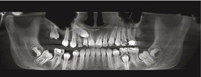
Obr. 1 / Fig. 1
Snímek z CBCT 12leté dívky s perzistujícím dočasným špičákem a palatinálně retinovaným horním špičákem se zvětšeným dentálním folikulem. Některé starší práce hlásaly, že perzistující dočasný špičák je přímou příčinou retence špičáku. Podle novějších názorů to neplatí / Picture from the CBCT of a 12-year-old girl with overretained (persisting) deciduous canine and palatal impacted maxillary canine with enlarged dental follicle. Some older studies claim that an overretained (persisting) deciduous canine is direct cause of canine impaction. However, according to the newest view, deciduous canine is not a cause of maxillary canine impaction
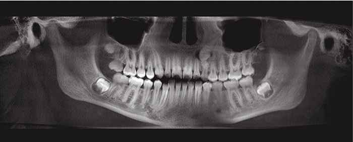
Obr. 2 / Fig. 2
Snímek z CBCT 13leté dívky s osmi malými odontomy. Dentální folikul stálého špičáku je zvětšený / Picture from the CBCT of 13-year-old girl with eight small odontomas. Dental follicle of permanent canine is enlarged
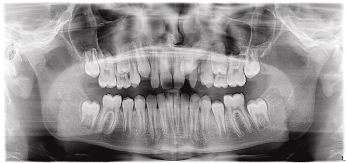
Obr. 3 / Fig.3
Ortopantomogram 11letého hocha s levým čípkovým laterálním řezákem a retinovaným levým stálým špičákem / a panoramic X ray of 11-year-old boy with left peg-shaped lateral incisor and impaction of left permanent canine
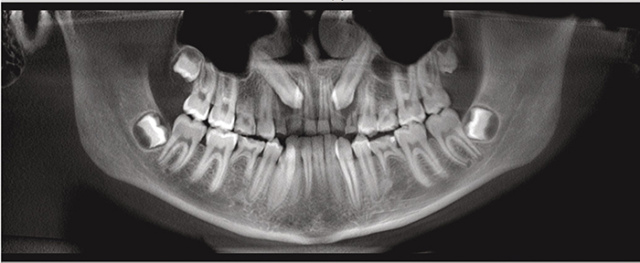
Obr. 4 / Fig. 4
Snímek z CBCT 12leté dívky s oboustrannou retencí horních špičáků doprovázenou dalšími dentálními anomáliemi – bilaterální ageneze horních a dolních druhých premolárů / A picture from CBCT of 12-year-old female with bilateral maxillary canine impaction associated with other dental anomalies – bilateral agenesis of maxillary and mandibular second premolars.

Literatura / References
1. Fuller JL, Denehy GE, Schulein TM. The permanent canines. In: Concise dental anatomy and morphology. Iowa: University of Iowa College of Dentistry; 2001.p. 58–68.
2. Dewel BF. The upper cuspids: its development and impaction. Angle Orthodont. 1949;19:79–90.
3. D’Amico A. The canine teeth – normal functional relation of the natural teeth of man. JS kalif Dent Assoc. 1958;26:194–208.
4. Okeson JP. Management of temporomandibular disorders and occlusion. St. Louis: Mosby Year Book; 1993. p. 121.
5. Sasakura H, Yoshida T, Murayana S, Hanada K, Nakajima T. Root resorption of upper permanent incisor caused by impacted canine. An analysis of 23 cases. Int. J. Oral. Surg. 1984;13:299–306.doi: 10.1016/S0300-9785(84)80037-X.
6. Ericson S, Kurol J. Incisor resorption caused by maxillary cuspids: a radiographic study. Angle Orthodont. 1987;57:332–346.
7. Scaife RR Jr, Holt JE. Natural occurrence of cuspid guidance. J. Prosth. Dent. 1969;22:225–229.doi: 10.1016/0022-3913(69)90249-2.
8. Kamínek et. al. Ortodoncie. Praha: Galén; 2020. p. 11.
9. Lum YM, Lim ST. Four cases of congenitally missing permanent cuspids. Sing. Dent. 1976;2:49–51.
10. Kambalimath HV, Jain S, Patil RU, Asokan A, Kambalimath D. Permanent maxillary canine agenesis: A rare case report. Int. J. Clin. Pediatr. Dent. 2015;8:242–6.doi: 10.5005/jp-journals-10005-1322.
11. Dachi SF, Howell FV. A survey of 3,874 routine full mouth radiographs. Oral Surg. Oral Pathol. 1961;14:1165–9.doi: 10.1016/0030-4220(61)90204-3.
12. Chu FC, Li TK, Lui VK, Newsome PR, Chow RL, Cheung KL. Prevalence of impacted teeth and associated pathologies – a radiographic study of the Hong Kong Chinese population. Hong Kong Med. J. 2003;9:158–63
13. Pedro FL, Bandéca MC, Volpato LE, Marques AT, Borba AM, Musis CR, Borges AH. Prevalence of impacted teeth in a Brazilian subpopulation. J. Contemp. Dent. Pract. 2014;15:209–13.doi: 10.5005/jp-journals-10024-1516.
14. Shah RM. Boyd MA, Vakil TF. Studies of permanent tooth anomalies in 7886 Canadian individuals. J. Can. Dent. Assoc. 1978;44:262–4.
15. Rayne J. The unerupted maxillary canine. Dent. Prac. Dent. Rec. 1969;19:194–204.
16. Tiwana PS, Kushner GM. Management of impacted teeth in children. Oral Maxillofac. Surg. Clin. North Am. 2005;17:365–73.doi: 10.1016/j.coms.2005.09.001.
17. Lövgren ML, Dahl O, Uribe P, Ransjö M, Westerlund A. Prevalence of impacted maxillary canines – an epidemiological study in a region with systematically implemented interceptive treatment. Eur. J. Orthodont. 2019;41(5):454–9.doi: 10.1093/ejo/cjz056.
18. Thilander B, Jacobsson SO. Local factors in impaction of maxillary canines. Acta Odontol. Scand.1968;26(2):145–68.doi: 10.3109/00016356809004587.
19. Ericson S, Kurol J. Radiographic assessment of maxillary canine eruption in children with clinical signs of eruption disturbances. Eur. J. Orthodont. 1986;8 (3):133–40.doi: 10.1093/ejo/8.3.133.
20. Prskalo K, Zjaca K, Skarić-Jurić T, Anić-Milosević S, Lauc T. The prevalence of lateral incisor hypodontia and canine impaction in Croatian population. Coll. Antropol. 32 (4):1105–9.
21. Fardi A, Kondylidou-Sidira A, Bachour Z, Parisis N, Tsirlis A. Incidence of Impacted and Supernumerary Teeth – A radiographic Study in a North Greek Population. Med. Oral Patol.Oral Cir. Bucal. 2011;16(1): e56–61.doi: 10.4317/medoral.16.e56.
22. Aktan AM, Kara S, Akgünlü, F, Malkoç S. The incidence of canine transmigration and tooth impaction in a Turkish subpopulation. Eur. J. Orthodont. 2010;32(5):575-81.doi: 10.1093/ejo/cjp151.
23. Kifayatullah J, Bangash T, Ayub A, Khan D. Prevalence and pattern of impacted canines in Najran, South Western Saudi Arabian population. Pakistan Oral Dent. J. 2015;35:67–71.
24. Alif, SM, Haque S, Nimmi N, Ashraf A, Khan SH, Khan MH. Panoramic Radiological Study to Identify Locally Displaced Maxillary Canines in Bangladeshi Population. Imaging Sci Dent. 2011;41:155–9.doi: 10.5624/isd.2011.41.4.155.
25. Brin I, Becker A, Shalhav M. Position of the maxillary permanent canine in relation to anomalous or missing lateral incisors: a population study. Eur. J. Orthodont. 1986;8(1):12–6.doi: 10.1093/ejo/8.1.12.
26. Rohani K, Eslamian L, Nahvi G. Correlation between maxillary cuspid impaction with available space and anomalies of maxillary lateral incisors. Dent. Res. J. (Isfahan). 2021;18:27.doi: 10.4103/1735-3327.313122.
27. Sacerdoti, R, Baccetti T. Dentoskeletal Features Associated with Unilateral or Bilateral Palatal Displacement of Maxillary Canines. Angle Orthodont. 2004;74 (6):725–32.
28. Peck S, Peck L, Kataja M. The pallataly displaced canine as a dental anomaly of genetic origin. Angle Orthodont. 1994;64:249–56.
29. Jacoby H. The etiology of maxillary canine impactions. Amer. J. Orthodont. 1983;84(2):125–32.doi: 10.1016/0002-9416(83)90176-8.
30. Černochová.P. Analýza lokálních faktorů predisponujících k poruchám prořezávání horního stálého špičáku. Prakt. zub. Lék., 2007;55(6):101–6
31. Fournier, A, Turcotte JZ, Bernard Ch. Orthodontic considerations in the treatment of maxillary impacted canines. Amer. J. Orthodont. 1982;81(3):236–9.doi: 10.1016/0002-9416(82)90056-2.
32. Yan B, Sun Z, Fields H, Wang L, Luo L. Etiologic factors for buccal and palatal maxillary canine impaction: A perspective based on cone-beam computed tomography analyses. Amer. J. Orthodont. dentofacial Orthop. 2013;143(4):527–34.doi: 10.1016/j.ajodo.2012.11.021.
33. Becker A, Zilberman Y, Tsur B. Root length of lateral incisors adjacent to palatally-displaced maxillary cuspids. Angle Othodont. 1984;54(3):218–25.
34. Rayne J. The unerupted maxillary canine. Dent. Pract. Dent. Rec. 1969;19(6):194–204.
35. Chaushu S, Sharabi S, Becker A. Tooth size in dentitions with buccal canine ectopia. Eur. J. Orthodont. 2003;25(5):485–91.doi: 10.1093/ejo/25.5.485.
36. Becker A, Gillis I, Shpack N. The etiology of palatal displacement of maxillary canines. Clin. Orthodont. Res. 1999;2(2):62–6.doi: 10.1111/ocr.1999.2.2.62.
37. Moyers, RE. Handbook of orthodontics. Chicago: Year Book Medical Publishers, Inc. 1963. p. 83–88.
38. Lappin MM. Practical management of the impacted maxillary cuspid. Amer. J. Orthodont. 1951;37(10):769–78.doi: 10.1016/0002-9416(51)90048-6.
39. Thilander B, Rönning O. Introduction to orthodontics. Stockholm: Tandläkarförlaget. 1985.
40. Becker A, Chaushu S. Etiology of maxillary canine impaction: A review. Amer. J. Orthodont. dentofacial Orthop. 2015;148(4):557–67.doi: 10.1016/j.ajodo.2015.06.013.
41. Sajnani AK. Permanent maxillary canines – review of eruption pattern and local etiological factors leading to impaction. J. Investig. Clin. Dent. 2015;6(1):1–7.doi: 10.1111/jicd.12067.
42. Sewoong O, Yong-II K, Seong-Sik K, Soo-Byung Park, Sung-Hun K. Comparison of root apex’s position between unilateral and bilateral palatally impacted canines: A pilot study. Amer. J. Orthodont. dentofacial Orthop. 2023;163(3):311–8.doi: 10.1016/j.ajodo.2021.11.017.
43. Kim SH, Kim YM, Sewoong O, Kim SS, Park SB, Son WS et al. How far is the root apex of a unilateral impacted canine from the root apices‘ arch form. Amer. J. Orthodont. dentofacial. Otrhop. 2017;151(2): 351–6.doi: 10.1016/j.ajodo.2016.06.047.
44. Landberg BJ, Peck S. Adequacy of maxillary dental arch width in patients with palatally displaced canines. Amer. J. Orthodont. dentofacial Orthop. 2000;118(2):220–3.doi: 10.1067/mod.2000.104819.
45. Brin I, Solomon Y, Zilberman Y. Trauma as a possible etiologic factor in maxillary canine impaction. Amer. J. Orthodont. dentofacial Orthop. 1993;104(2):132–7.doi: 10.1016/S0889-5406(05)81002-9.
46. Chaushu S, Zilberman Y, Becker A. Maxillary incisor impaction and its relationship to canine displacement. Amer. J. Orthodont. dentofacial Orthop. 2003;124(2):144–50.doi: 10.1016/S0889-5406(03)00344-5.
47. Toyoglu S, Zolcu U, Toyuglu U. Developmental disturbance of maxillary lateral incisor after trauma. Dent. Traumatol. 2007;23(2):85–6.doi: 10.1111/j.1600-9657.2006.00380.x.
48. Lappin MM. Practical management of the impacted maxillary canine. Amer. J. Orthodont. dentofacial Orthop. 1951;37:769–78.doi: 10.1016/0002-9416(51)90048-6.
49. Becker A. General principles related to the diagnosis and treatment of impacted teeth. In: Orthodontic treatment of impacted teeth. Oxford: Wiley Blackwell; 2012. p. 8.doi: 10.1002/9781118709641.
50. Bishara SE. Impacted maxillary canines: a review. Amer. J. Orthodont. dentofacial. Otrhop 1992;101(2):159–71.doi: 10.1016/0889-5406(92)70008-X.
51. Becker A, Smith P, Behar R. The incidence of anomalous lateral incisors in relation to palatally-displaced cuspids. Angle Orthodont. 1981;51(1):24–9.
52. Broadbent BH. Ontogenetic development of occlusion. Angle Orthodont. 1941;11:223–41.
53. Coulter J. Richardson A. Normal eruption of the maxillary canine quantified in three dimensions. Eur. J. Orthodont. 1997;19(2):171–83.doi: 10.1093/ejo/19.2.171.
54. Bass TB. Observations on the misplaced upper canine tooth. Dent. Pract. 1967;18:25–33.
55. Miller BH. The influence of congenitally missing teeth on the eruption of the upper canine. Dent. Pract. Dent. Rec. 1963;13:497–504.
56. Chaushu S, Sharabi S, Becker A. Dental morphologic characteristics of normal versus delayed developing dentitions with palatally displaced canines. Amer. J. Orthodont. dentofacial. Orthop. 2002;121(4):339–46.doi: 10.1067/mod.2002.121559.
57. Dubovska I. el. al. Lateral incisor morphology in patients with impacted canine: A CT Study, IOSR-JDMS 2015;14:1–5.
58. Liuk WI, Olive J R, Griffin M, Monsour P. Associations between palatally displaced canines and maxillary lateral incisors, Amer. J. Orthodont. dentofacial Orthop. 2013;143:622–32.doi: 10.1016/j.ajodo.2012.11.025.
59. Becker A, Sharabi S, Chaushu S. Maxillary tooth size variation in dentitions with palatal canine displacement. Eur. J. Orthodont. 2002;24(3):313–8.doi: 10.1093/ejo/24.3.313.
60. Chaushu S, Vryonidou M, Becker A, Leibovich A, Dekel E, Dykstein E.et al. The labiopalatal impacted canine: Accurate diagnosis based on the position and size of adjacent teeth: A cone-beam computed tomography study. Amer. J. Orthodont. Dentofacial. Orthop. 2023;163(5):690–9.doi: 10.1016/j.ajodo.2022.05.021.
61. Baccetti T. A controlled study of associated dental anomalies. Angle Orthodont. 1998;68(3):267–74.
62. Zilberman Y, Cohen B, Becker A. Familial trends in palatal canines, anomalous lateral incisors, and related phenomena. Eur. J.Orthodont.1990;12:135–9.doi: 10.1093/ejo/12.2.135.
63. Weise W, Anbuhl B. Beitrag zur Keimverlagerung des Eckzahnes. Dtsch. Zahnärztl. Z 1969;24:803–8.
64. Racek J, Sottner L. Příspěvek k dědičnosti retence špičáků. Čs.Stomat.1977;77:209–13.
65. Sottner L, Racek J. Stanovení dědivosti. Model: retence špičáku. Čas. Lék. Česk. 1978;117:1060–2.
66. Racek J, Sottner L. Naše názory na dědičnost retence špičáku. Sborn. Lék. 1984;86:355–60.
67. Sottner L, Racek J, Marková M, Sládková M. Genetika v ortodoncii. Sborn. Lék. 1987;89:15–9.
68. Baccetti T, Leonardi M, Armi P. A randomized clinical study of two interceptive approaches to palatally displaced canines. Eur. J. Orthodont. 2008;30:381–5.doi: 10.1093/ejo/cjn023.
69. Alhaija ESA, Wazwaz FT. Third molar tooth agenesis and pattern of impaction in patients with palatally displaced canines. Angle Orthodont. 2019;89(1):64–70.doi: 10.2319/031318-203.1.
70. Hattab FN, Rawashdeh MA, Fahmy MS. Impaction status of third molars in Jordanian students. Oral Surg. Oral Med. Oral Pathol. Oral Radiol. Endod. 1995;79(1):24–9.doi: 10.1016/S1079-2104(05)80068-X.
71. Sacerdoti R, Baccetti T. Dentoskeletal features associated with unilateral or bilateral palatal displacement of maxillary canines. Angle Orthodont. 2004;74(6):725–32.
72. Shalish M, Chaushu S. Malposition of unerupted mandibular second premolar in children with palatally displaced canines. Angle Orthodont. 2009;79(4):796–9.doi: 10.2319/081808-435.1.
73. Baccetti T. A controlled study of associated dental anomalies. Angle Orthodont. 1998;68(3):267–74.
74. Vastardis H. The genetics of human tooth agenesis: new discoveries for understanding dental anomalies. Amer. J. Orthodont. dentofacial Orthop 2000;117:650–6.doi: 10.1016/S0889-5406(00)70173-9.
75. Stahl F, Grabowski R. Maxillary canine displacement and genetically determined predisposition to disturbed development of the dentition. J Orofac Orthop. 2003;64:167–77.doi: 10.1007/s00056-003-0221-y.
76. Baccetti T. Leonardi M, Giuntini V. Distally displaced premolars: A dental anomaly associated with palatally displaced canines. Amer. J. Orthodont. dentofacial Orthop. 2010;138(3):318–22.doi: 10.1016/j.ajodo.2008.09.036.
77. Richardson A, McKay C. Delayed eruption of maxillary canine teeth, Part I – etiology and diagnosis. Proc. Br. Paedodont. Soc. 1982;12:15–25.
78. Rayne J. The unerupted maxillary canine. Dent. Pract. Dent. Rec. 1969;19:194–204.
79. Devi MSA, Padmanabhan S. Role of polymorphisms of MSX1 and PAX9 genes in palatal impaction of maxillary canines. J. Orthodont. 2019;46 (1):14–9.doi: 10.1177/1465312518820537.
80. Kramer RW, Williams AC. The incidence of impacted teeth. Oral Surg. Oral Med. Oral Pathol. 1970;29:237–41.doi: 10.1016/0030-4220(70)90091-5.
81. Montelius GA. Impacted teeth, a comparative study of Chinese and Caucasian dentitions. J. Dent. Res. 1932;12:931–8.doi: 10.1177/00220345320120060701.
82. Oliver RG, Mannion JE, Robinson JM. Morphology of the lateral incisor in cases of unilateral impaction of maxillary canine. Brit. J. Orthodont. 1989;19:9–16.doi: 10.1179/bjo.16.1.9.
83. Davis PJ. Hypodontia and hyperdontia of permanent teeth in Hong Kong schoolchildren. Community Dent Oral Epidemiol. 1987;15:218–20.doi: 10.1111/j.1600-0528.1987.tb00524.x.
84. Haavikko K. Hypodontia of permanent teeth. An orthopantomographic study. Suom Hammaslääk Toim. 1971;67:219–25.
85. Peck L, Peck S, Attia Y. Maxillary canine – first premolar transposition, associated dental anomalies and genetic basis. Angle Orthodont. 1993;63:99–109.
86. Power SM, Short MBE. An investigation into the response of palatally displaced canines to the removal of deciduous canines and an assessment of factors contributing to favorable eruption. Brit. J. Orthodont. 1993;20:215–23.doi: 10.1179/bjo.20.3.215.
87. Ericson S, Kurol J. Early treatment of palatally erupting maxillary canines by extraction of the primary canines. Eur. J. Orthodont.1998;10:283–95.doi: 10.1093/ejo/10.4.283.
88. Muller TP, Hill IN, Peterson AL, Blayney JR. A survey of congenitally missing permanent teeth. J. Amer. Dent. Assoc.1970;81:101–7.doi: 10.14219/jada.archive.1970.0151.
89. Meskin LH, Gorlin RJ. Agenesis and peg-shaped permanent maxillary lateral incisors. J. Dent. Res. 1963;42:1476–9.doi: 10.1177/00220345630420062501.
90. Ringqvist M, Thilander B. The frequency of hypodontia in an orthodontic material. Svesk Tandläk Tidskr. 1969;62:535–41.
91. Camilleri S, Lewis CM, McDonald F. Ectopic maxillary canines: segregation analysis and a twin study. J. Dent. Res. 2008;87:580–3.doi: 10.1177/154405910808700606.
92. Al-Nimri KS, Bsoul E. Maxillary palatal canine impaction displacement in subjects with congenitally missing maxillary lateral incisors. Amer. J. Orthodont. dentofacial Orthop 2011;140(1): 81–6.doi: 10.1016/j.ajodo.2009.11.016.
93. Lempesi E, Karamolegkou M, Pandis N, Mavragani M. Maxillary canine impaction in orthodontic patients with and without agenesis: a cross-sectional radiographic study. Angle Orthodont. 2014; 84(1):11–7.doi: 10.2319/022413-155.1.
94. Laganá G, Venza N, Borzabadi-Farahani A, Fabi F, Danesi C, Cozza P. Dental anomalies: prevalence and associations between them in a large sample of non-orthodontic subjects, a cross-sectional study. BMC Oral Health. 2017;17(1):62.doi: 10.1186/s12903-017-0352-y.
95. Khalaf K, Miskelly J, Voge E, Macfarlane TV. Prevalence of hypodontia and associated factors: a systematic review and meta-analysis. J. Orthodont. 2014;41(4):299–316.doi: 10.1179/1465313314Y.0000000116.
96. McKay C. The unerupted maxillary canine, an assessment of the role of surgery in 2500 treated cases. Br. Dent. J. 1978;145(7): 207–10.doi: 10.1038/sj.bdj.4804146.
97. Fleury JE, Deboets D, Assaad-Auclair C, Maffre N, Sultan P. La canine incluse. Rev. Stomatol. Chir. Maxillofac. 1985;86:122–31.
98. Mitsea A, Palikaraki G, Karamesinis K, Vastardis H, Gizani S, Sifakakis I. Evaluation of lateral incisor resorption caused by impacted maxillary canines based on CBCT : A systematic review and meta-analysis. Children (Basel). 2022;9 (7):1006.doi: 10.3390/children9071006.
MDDr. Dana Kloudová
Ortodontické oddělení
Stomatologická klinika dětí a dospělých
2. lékařská fakulta UK a FN Motol
V Úvalu 84, 130 06 Praha 5