


Stabilita incizálního schůdku, hloubky skusu a horní mezimolárové šířky po segmentální osteotomii maxily
Stability of overjet, overbite and upper intermolar width following segmental osteotomy of the maxilla
MDDr. Nina Žuffová1, MUDr. Hana Böhmová1, MUDr. Martin Kotas, Ph.D.2
1 Ortodontické oddělení, Stomatologická klinika LFP UK a FN Plzeň / Department of Orthodontics, Clinic of Stomatology, Medical Faculty of Komensky University and University Hospital Plzeň
2 Klinika ústní, čelistní a obličejové chirurgie FN Brno / Clinic of oral, jaw and facial surgery, University Hospital Brno
DOI10.61110/50101
Souhrn
Cíl: Cílem pozorování bylo zjistit stabilitu incizálního schůdku, hloubky skusu a horní mezimolárové šířky po korekci ortodontické anomálie ortodonticko-chirurgicky metodou segmentální H-osteotomie maxily.
Materiál a metodika: Do retrospektivní studie bylo zařazeno 40 pacientů, kteří podstoupili segmentální osteotomii maxily. U každého pacienta byla zhotovená kompletní ortodontická dokumentace před léčbou (T1), před operací (T2), po skončení ortodontické léčby (T3) a průměrně 25 měsíců po sejmutí fixního aparátu, minimálně 1 rok od ukončení léčby a minimálně 1,5 roku po ortognátní operaci (T4). V každém časovém intervalu byl zhotoven digitální model zubů, na kterém byla měřena hodnota incizálního schůdku, hloubky skusu a horní mezimolárové šířky.
Výsledky: Při dlouhodobé kontrole jevila velikost incizálního schůdku klinicky i statisticky nevýznamný pokles v průměru o 0,10,5 mm. V období dlouhodobé kontroly došlo ke klinicky nevýznamnému poklesu hloubky skusu o 0,1±<0,5 mm, přitom v ani jednom případě nedošlo ke klinicky významnému otevření skusu. Průměrná hodnota horní mezimolárové šířky před začátkem léčení byla 38,93,1 mm, před ortognátní operací se její velikost ortodontickou přípravou zvětšila v průměru o 0,5 ± 1,7 mm. Při ukončení aktivní léčby její hodnota dále vzrostla o 0,9 ± 1,7 mm, tj. o 1,4 mm ± 2,6 mm vůči výchozímu stavu. Při dlouhodobé kontrole byl zjištěn průměrný úbytek horní mezimolárové šířky -0,4 ± 0,7 mm.
Závěr: Za největší riziko potenciální dentální recidivy po ortognátní operaci lze tedy i u segmentální H-tomie maxily považovat transverzální rozměr chrupu. Nestabilita horní mezimolárové šířky po léčbě může vést i ke vzniku zkříženého skusu a tedy kompromitování výsledku. Příznivé změny v oblasti horních řezáků navozené léčbou zůstaly klinicky nezměněny a stabilní.
(Ortodoncie 2024, 33, č. 1, s. 30–43)
Aim: he aim is to assess stability of overjet, overbite, and upper intermolar width after the orthodontic-surgical correction of orthodontic anomaly with segmental H-osteotomy of the maxilla.
Material and method: The retrospective study works with 40 patients who underwent the segmental osteotomy of the maxilla. Each patient had a complete orthodontic documentation before the therapy (T1), prior to the surgery (T2), after a completion of an orthodontic treatment (T3) and approx. 25 months after a removal of fixed appliance, minimum of 1 year after the completed orthodontic treatment and at least 1.5 years after the orthognathic surgery (T4). In every time interval, a digital model of a dentition was made to measure the overjet, the overbite and the upper intermolar width.
Results: During a long-term check-up the value of the overjet showed a clinically and statistically insignificant decrease by 0.1.5 mm. The overbite also showed a clinically insignificant decrease by 0.1 ±0.5 mm; in none of the cases there was a clinically significant bite opening. The average value of the upper intermolar width was 38.9 ±3.1 mm before the beginning of therapy, prior to orthognathic surgery the value increased due to orthodontic treatment by 0.5 ±1.7 mm. After the active therapy completion, the value increased by 0.9 ±1.7 mm, i.e. by 1.4 ± 2.6 mm compared to the initial condition. In a long-term check up, the average loss of uper intermolar width by 0.4 ±0.7 mm was recorded.
Conclusion: Change in a transversal dimension of the dentition is considered the greatest risk of potential dental relapse following segmental H-osteotomy of the maxilla. Instability of the upper intermolar width following the therapy may result in crossbite, and thus compromise the outcomes. Favourable changes in the upper incisors induced by the therapy remained clinically unchanged and stable.
(Ortodoncie 2024, 33, No. 1, p. 30–43)

Klíčová slova: segmentální osteotomie maxily, stabilita
Key words: egmental osteotomy of the maxilla, stability
Úvod
U pacientů, u kterých je ortodontická vada natolik závažná, že ani růstová modifikace, ani kompenzační ortodontická léčba nenabídne přijatelné řešení, je vhodným léčebným postupem kombinovaná ortodonticko-chirurgická léčba s úpravou postavení čelistí nebo dentoalveolárních segmentů [1].
Komplexnost a variabilita dento-skeleto-faciálních anomálií jednotlivých pacientů vedla k vývoji operační techniky Le Fort I, při které se současně mění poloha několika maxilárních dento-oseálních segmentů vůči sobě vzájemně. Mnoho chirurgických modifikací této morfologicky velmi variabilní techniky usnadňuje současné anteroposteriorní, vertikální a horizontální pohyby anteriorní a posteriorní části maxily pod přímou vizuální kontrolou. Maxilu lze segmentalizovat parasagitálně, transverzálně nebo cirkumpalatálně. Touto technikou si lze usnadnit uzávěr mezer, nivelizaci a vyrovnání Speeovy křivky pomocí vertikálních interdentálních osteotomií nebo osteotomiích po neúplně uzavřených poextrakčních mezerách. Rozšiřováním nebo zužováním maxily lze korigovat bukální nonokluzi nebo zkřížený skus. Tyto nekomplikované a často relativně malé (2–5 mm) pohyby maxily obcházejí potřebu ortodonticky kompenzovat skeletální problém bukálním nebo palatinálním sklonem [2]. V současnosti se segmentální osteotomie maxily provádí téměř vždy jako součást ortognátní operace horní čelisti v linii Le Fort I.
Dle odborné literatury je časovou hranicí pro hodnocení výsledné stability léčby 1 rok od ortognátní chirurgie, resp. ukončení kombinované chirurgicko-ortodontické léčby [3, 4, 5, 6]. Změny, které nastanou v prvním roce po ortognátní operaci, přímo souvisí s hojením po chirurgickém zákroku, s pooperačním ortodontickým doléčením a mají souvislost s krátkodobou adaptací kraniofaciálního systému na nové podmínky. Změny, které nastanou po prvním roce po operaci, již souvisí s dlouhodobou adaptací, případně u některých pacientů se zbytkovým růstem [3, 4, 5, 6].
Toto sdělení se zabývá stabilitou tří vybraných dentálních charakteristik – incizálního schůdku, hloubky skusu a horní mezimolárové šířky v období minimálně 1 rok od ukončení léčby a minimálně 1,5 roku po ortognátní operaci metodou segmentální H-osteotomie maxily.
Materiál a metodika
Do retrospektivní studie bylo zařazeno 40 pacientů, kteří podstoupili ortodonticko-chirurgickou léčbu metodou segmentální H - osteotomie maxily ke korekci skeletální anomálie v časovém období od února 2013 do listopadu 2021. Vzhledem k tomu, že segmentální osteotomie maxily je indikována u komplexních vad ve všech třech rovinách [2, 7, 8, 9], obsahoval daný soubor případy monomaxilární i bimaxilární ortognátní operace, které byly indikovány a prováděny v souladu s potřebami korekce dané čelistní anomálie. Do souboru byly zařazené tyto vady: skeletální II. třída, skeletální III. třída, pacienti s nízkou, průměrnou, ale i s vysokou dolní obličejovou třetinou, transverzální komprese horní čelisti, asymetrie maxily, laterogenie.
U všech pacientů proběhla předoperační ortodontická příprava, modifikovaná vzhledem k typu jejich vady. Všichni pacienti byli léčeni vestibulárním fixním aparátem, předoperační fáze léčby končila po dekompenzaci zubních oblouků na čtyřhranném ocelovém oblouku 0.019 x 0.025‘‘ v drážce 0.022‘‘ s přihlédnutím na specifika přípravy na segmentální osteotomii maxily. V průběhu ortodontické přípravy byla zpravidla zachována šířka i tvar horního zubního oblouku, a také zachován průběh horní Speeovy křivky. Před chirurgickým zákrokem byl horní ocelový oblouk rozdělen do segmentů. Těsně po ortognátní operaci byly parciální ocelové oblouky rigidně spojené do jednoho celku.
Design segmentální osteotomie byl u všech pacientů sledovaného souboru uniformní, byla provedena segmentální H-osteotomie v úrovni Le Fort I se separací horní čelisti na frontální segment v rozsahu od laterálního řezáku ke kontralaterálnímu řezáku, 2 distální segmenty a patrový segment (viz obr. č. 1) [10]. V indikovaných případech, tedy pokud byl prostor mezi kostními fragmenty větší než 3 mm, byly během vlastní ortognátní operace využity kostní štěpy [11]. Při operačním zákroku byly u každého pacienta použity operační dlahy. Ani jeden z pacientů nenosil v pooperačním období operační dlahu pro stabilizaci, stabilita byla zabezpečena rigidní osteofixací, dále artikulací zubních oblouků a elastickými mezičelistními tahy.
U všech pacientů ze souboru byla v rámci léčby provedena ortodontická a chirurgická dokumentace (digitální modely a fotografie chrupu, 2D a 3D fotografie obličeje, standardní a CBCT rentgenogramy) před léčbou (T1), před ortognátní operací (T2) a po skončení ortodontické léčby (T3). Všechna rentgenologická a fotografická vyšetření v čase T1-T3 byla zhotovena z diagnostických a léčebných důvodů, nikoliv z důvodů experimentálních. Při dlouhodobé kontrole, tj. minimálně 1,5 roku po operaci a zároveň minimálně 1 rok po sejmutí fixního aparátu (T4), byla zhotovena rentgenologická a fotografická dokumentace stavu po předchozím informovaném souhlasu pacienta. Průměrný interval mezi ukončením aktivní léčby a zhotovením dokumentace v retenční fázi byl 2 roky a 1 měsíc, tj. 25 měsíců.
Provedení studie bylo schváleno Etickou komisí fakultní nemocnice Plzeň a lékařské fakulty Univerzity Komenského v Plzni č. j. 75-23. Tento výzkum nebyl financovaný z grantových projektů, veřejných ani komerčních zdrojů ani ze zdrojů neziskových organizací.
U všech pacientů byl v každém časovém intervalu (T1–T4) zhotoven digitální model chrupu optickým skenerem Trios 3 (3Shape A/S, Denmark). Na těchto digitálních modelech byla následně v programu Ortho System 2019.1 (3Shape A/S, Denmark) realizována měření dentálních lineárních a úhlových parametrů a dentálních indexů. Výsledky těchto měření spolu s výsledky měření skeletálních paramentů na CBCT rentgenogramech byly autorsky publikovány v širší studii [12]. Toto sdělení pojednává následující tři lineární dentální parametry charakterizující sledovaný soubor z hlediska transverzálního, sagitálního a vertikálního: v transverzální rovině byla měřena horní mezimolárová šířka, v sagitální rovině incizální schůdek, vertikálně hloubka skusu.
Chyba lineárních měření na modelech byla zjišťována pro všechny časové fáze společně, a to vždy opakovaným dvojím měřením na 40 náhodně vybraných modelech chrupu, přičemž z každého sledovaného časového období bylo náhodně vybráno 10 modelů (tj. celkem 40 modelů ze 160). Druhé měření bylo realizováno v odstupu jednoho měsíce týmž pozorovatelem a za stejných podmínek jako první měření. Opakovatelnost měření byla kvantifikována koeficientem opakovatelnosti (coefficient of repeatability) podle Blanda a Altmana.
U měření prováděných na digitálních modelech se Bland-Altmanův koeficient opakovatelnosti pohyboval v rozmezí 0,2 mm–0,3 mm u měření na řezácích (incizální schůdek a hloubka skusu). Měření v oblasti molárů jevilo vyšší chybu, koeficient opakovatelnosti jevil chybu měření 0,6 mm. Soustavná chyba prokázána nebyla.
Statistické výpočty byly provedeny v programech MS Excel 2016 (Microsoft, USA) a MedCalc for Windows, v. 20.2 (MedCalc Software, Belgium). Byly zjištěny základní statistické charakteristiky souboru – popisná statistika míry střední polohy a míry rozptýlenosti. Ke zjištění potencionálních rozdílů sledovaných znaků před léčbou a po léčbě bylo užito párového t-testu a Wilcoxonova párového testu. Ke korelační analýze bylo využito lineární korelace a vhodně Pearsonova a Spearmanova korelačního koeficientu.
Výsledky
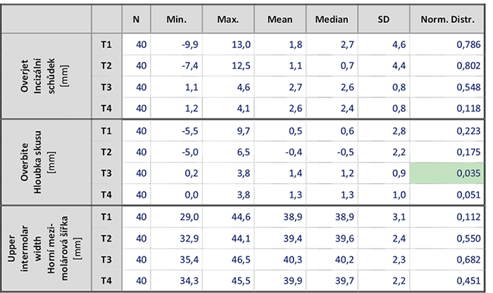
Přehled hodnot dentálních lineárních charakteristik sledovaného souboru v čase před léčbou (T1), v čase před operací (T2), v čase po ukončení aktivní léčby (T3) a při dlouhodobé kontrole (T4) souhrnně podává tab. č. 1. Rozložení hodnot u hloubky skusu po léčbě jako jediné nesplňuje podmínky normálního rozložení, proto je v následných statistikách v případech statistických odhadů týkajících se hloubky skusu po léčbě použito metod neparametrických testů.
V tab. č. 2 jsou uvedeny změny hodnot dentálních lineárních a úhlových charakteristik sledovaného souboru v časových obdobích před léčbou → po přípravě na operaci (T1 → T2), před léčbou → po léčbě (T1 → T3), před léčbou → při dlouhodobé kontrole (T1 → T4), po přípravě na operaci → po léčbě (T2 → T3), po přípravě na operaci → při dlouhodobé kontrole (T2 → T4) a po ukončení léčby → při dlouhodobé kontrole (T3 → T4), jejich statistická významnost a údaj, zda naměřené rozdíly hodnot dosahují hranice chyby měření. S ohledem na požadovaný rozsah práce jsou následné statistické odhady redukovány zejména na časová období před léčbou → po léčbě (T1 → T3) a po ukončení léčby → při dlouhodobé kontrole (T3 → T4).
Průměrná hodnota incizálního schůdku zjištěná na modelech zhotovených před začátkem léčení byla 1,8 ± 4,6 mm s mediánem 2,7 mm, minimem –9,9 mm a maximem 13,0 mm. Ortodontickou přípravou se průměrná velikost incizálního schůdku mírně snížila o 0,7 ± 2,2 mm. Po ukončení aktivní léčby průměrná velikost incizálního schůdku vzrostla o 1,5 ± 4,3 mm vůči hodnotě před operací, tj. o průměrně 0,8 ± 4,6 mm vůči výchozí velikosti schůdku. Při dlouhodobé kontrole jevila velikost incizálního schůdku klinicky i statisticky nevýznamný pokles v průměru o -0,1 ± 0,5 mm (tab. 2, obr. 2).
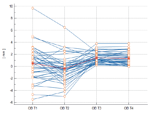
Průměrná hodnota hloubky skusu na 40 digitálních modelech zhotovených před začátkem léčení byla 0,5 ± 2,8 mm s mediánem 0,6 mm. Vzhledem k faktu, že soubor hodnot hloubky skusu naměřený při ukončení léčby nesplňoval podmínku normálního rozdělení, bylo nutno užít i neparametrické metody statistického odhadu. V průběhu ortodontické přípravy se hloubka skusu zmenšila průměrně o 0,9 ± 2,0 mm s mediánem –0,45 mm, tj. do záporných hodnot a otevřeného skusu. Po ukončení aktivní fáze léčení byla naměřena průměrná hodnota hloubky skusu 1,4 ± 0,9 mm s mediánem 1,2 mm, tj. došlo k opětovnému prohloubení a zavření skusu o průměrně 1,8 ± 2,0 mm (resp. 1,8 ± 2,0 mm vůči původnímu stavu). Při dlouhodobé kontrole byla zjištěna průměrná hodnota hloubky skusu 1,3 ± 1,0 mm s mediánem 1,3 mm. V období dlouhodobé kontroly došlo ke klinicky nevýznamnému poklesu o 0,1 ± 0,5 mm s mediánem –0,1 mm; tj. hodnotě pod hranicí chyby měření 0,25 mm. Přitom v ani jednom případě nedošlo ke klinicky významnému otevření skusu, i když v 11 případech ze 40 (27,5 %) se výsledná hloubka skusu při dlouhodobé kontrole nacházela v rozmezí 0–0,5 mm. Stav zůstal z klinického hlediska nezměněn a stabilní (obr. 3 a tab. 3).
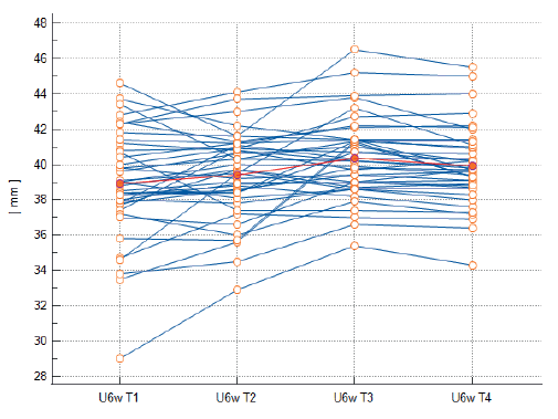
Průměrná hodnota horní mezimolárové šířky před začátkem léčení byla 38,9 ± 3,1 mm s mediánem 38,9 mm. Před ortognátní operací v čase T2 se její velikost ortodontickou přípravou zvětšila v průměru o 0,5 ± 1,7 mm. Při ukončení aktivní léčby její hodnota dále vzrostla o 0,9 ± 1,7 mm, tj. o 1,4 ± 2,6 mm vůči výchozímu stavu. Při dlouhodobé kontrole byl zjištěn průměrný úbytek horní mezimolárové šířky –0,4 ± 0,7 mm (obr. 4 a tab. 2).
Při srovnání změn lineárních parametrů mezi sebou (T1-T3), nebo změn po léčbě (T3-T4), nebo změn navozených léčbou (T1-T3) se změnami, ke kterým došlo po léčbě v období retence (T3-T4), nebyl korelační analýzou zjištěn vliv žádného z daných parametrů na změnu hloubky skusu ani incizální schůdek.
Při srovnání změn navozených léčbou a změn, ke kterým došlo po léčbě v období retence, byla korelační analýzou zjištěna pouze jedna významná korelace mezi změnami jednotlivých hodnot, a to středně silná a signifikantní korelace mezi rozšířením horní mezimolárové vzdálenosti léčbou a zužováním se horní mezimolárové vzdálenosti v období retence (tab. 4).
Diskuse
Hodnota incizálního schůdku před zahájením léčby se pohybovala od –9,9 mm do 13 mm, jednalo se o vady všech sagitálních tříd. Při ukončení aktivní léčby se průměrná hodnota schůdku pohybovala na hodnotě 2,6 mm, a také se velmi významně zmenšil rozptyl hodnot na rozsah 1,1–4,6 mm. Při dlouhodobé kontrole incizální schůdek nevýznamně poklesl průměrně o 0,1 mm a jeho hodnoty zůstaly velmi homogenní a klinicky vyhovující v rozmezí 1,2–4,1 mm. Výsledky dané studie jsou srovnatelné s odbornou literaturou [13, 14, 15, 16], kde se potvrzuje, že segmentální osteotomie horní čelisti je v sagitální rovině velice stabilní. Ze systematické studie Haase Jr. [14] vyplývá, že skeletální změny jsou poměrně malé a recidiva se objevuje v dentálních parametrech. Konkrétně se jedná o změny sagitální polohy horních řezáků, které reagují na změny v dalších rovinách, a tedy dochází k jejich retruzi nebo jejich protruzi. V této studii se nestabilita polohy horních řezáků nepotvrdila, nicméně inklinace horních řezáků ani sagitální skeletální změny polohy horní čelisti nebyly zkoumány. V souladu s pozorováním v této studii je i studie Arpornmaeklonga [15]. Na kefalometrických snímcích porovnával dlouhodobou stabilitu ventrálního posunu horní čelisti, a to jak u skupiny pacientů s čelistí operovanou vcelku, tak u skupiny, kde se čelist dělila na fragmenty. Stabilita předsunu byla ve studii potvrzena, autoři dokonce tvrdí, že byla vyšší u skupiny, kde se maxila segmentovala, a to zejména u větších posunů ventrálně (více než 6 mm) nebo u kombinace předsunutí a extruze horní čelisti. Autoři si tyto výsledky vysvětlují tím, že segmentováním horní čelisti se dosáhne pooperačně lepší interkuspidace a okluzní stability, a tedy i dlouhodobé stability. Zmiňují také další faktory jako dentoalveolární kompenzační mechanismus, použití kostních štěpů a rigidní osteofixace. V souladu s pozorováním v této studii je studie Brandtnera [16], který sledoval skupinu 47 pacientů po segmentální osteotomii maxily. V rámci dlouhodobé kontroly, průměrně 8,8 let po ortognátní operaci, nezaznamenal klinicky významné změny hloubky skusu a incizálního schůdku.
Dalším zkoumaným lineárním parametrem v této studii byla hloubka skusu. Před zahájením terapie se hloubka skusu v celém souboru pohybovala od –5,5 do 9,7 mm. V souboru bylo přítomno 17 pacientů s klinicky nevyhovující hloubkou skusu ≤ 0 mm. V průběhu ortodontické předoperační přípravy se hloubka skusu zmenšovala průměrně o 0,9 mm a docházelo tedy k otevírání skusu. Při ukončení aktivní léčby se hloubka skusu u všech pacientů pohybovala v rozsahu 0,2–3,8 mm, žádný z pacientů (a to bez ohledu na původní typ vertikální vady) neměl na konci léčby otevřený skus, ačkoliv 20 pacientů z celkového souboru bylo možné dle skeletálních morfometrických a morfologických znaků při zahájení léčby charakterizovat diagnózou long face syndrom. V operační fázi byla u 24 pacientů provedena CCW rotace horní čelisti nebo celého maxilo-mandibulárního komplexu, přičemž ani tento postup neměl vliv na klinický úspěch zkoumané metody. V dlouhodobé retenci docházelo k malému klinicky nevýznamnému otevírání skusu průměrně o 0,1 mm. Na konci pozorování nebyl u žádného pacienta zaznamenán otevřený skus. Toto pozorování podporují i studie Arnetta [17, 18], který popsaným operačním protokolem, tj. CCW rotací komplexu se segmentací maxily, léčil skupinu pacientů se skeletálně otevřeným skusem. Dle jeho studie nemá rotování komplexu vliv na stabilitu hloubky skusu a u své skupiny pacientů v dlouhodobé retenci dosáhl fyziologické hloubky skusu u 90 % a nevznikl ani jeden otevřený skus. Naopak opačné závěry uvádí Silva [19], která také zkoumala dlouhodobou stabilitu po segmentální osteotomii maxily u pacientů se skeletálně otevřeným skusem. Do ½ roku po ukončení léčby se skus otevíral u 48 % pacientů, v období 2 a ½ roku v retenci u 30 % pacientů. Kamínek [20] uvádí, že vliv na zmenšování hloubky skusu u jedinců s tendencí do posteriorotace mají slabší žvýkací svaly. Proffit [21] tvrdí, že u jedinců se syndromem dlouhého obličeje je vlivem nevhodného geometrického postavení čelistí k sobě navzájem a také vlivem odlišné struktury žvýkacích svalů 2 až 3× nižší síla žvýkacích svalů oproti jedincům s normální obličejovou výškou. Ortognátní operací dochází ke geometrické harmonizaci obličeje, ale nedochází ke změně síly žvýkacích svalů, což může být predispozičním faktorem pro recidivu otevřeného skusu.
Před zahájením léčby byla průměrná hodnota horní mezimolárové šířky 38,9 mm. V průběhu předoperační přípravy docházelo k jejímu zvětšování, průměrně o 0,5 mm, tedy k expanzi zubního oblouku. Při ukončení léčby se znova zvětšila průměrně o 1,4 mm a v retenční fázi naopak mírně poklesla průměrně o 0,4 mm. Zde je nutné si uvědomit, že při chirurgické léčebné změně došlo u části pacientů (33 %) k cílenému zmenšování mezimolárové šířky transverzální rotací distálních segmentů maxily během operace, tedy k napřimování horních molárů, což ovlivňuje hodnotu horní mezimolárové šířky. V období retence docházelo ke zmenšování horní mezimolárové šířky, a to i vlivem napřimování horních molárů. V části případů tyto jevy vedly k jisté nestabilitě dosažených posunů molárů. Výsledkem těchto pozorování je stav, kdy ze 40 léčených pacientů, z toho 25 se zkříženým skusem před léčbou, se v dlouhodobé retenci u 6 z nich nachází zkřížený skus v oblasti molárů. Zde se výsledky této studie shodují s relevantní literaturou [4, 22, 23, 24, 25], která jako největší riziko potenciální recidivy uvádí transverzální rozměr. Podobně jako v naší studii i Arnett [17] potvrzuje statisticky signifikantní, ale klinicky nevýznamné zmenšování horní i dolní mezimolárové šířky po ukončení léčby. Dle literatury [13, 26, 27, 28] má zmenšování transverzálního rozměru zubních oblouků vliv na zmenšování hloubky skusu, což se však v prezentované studii nepotvrdilo.
Širší studie dentálních a skeletálních změn navozených léčbou a jejich stabilita provedená Žuffovou [12] na témže souboru 40 léčených pacientů potvrzuje názor, že poléčebná změna v podobě recidivy je výraznější v dentálních parametrech nežli skeletálních. V citované studii byl průměrný přírůstek v šířce báze horní čelisti 2,1 mm oproti původnímu stavu, ale přírůstek horní mezimolárové šířky byl pouze 1,4 mm. Z toho v období retence zůstalo zachováno 1,9 mm v maxilární bázi, ale pouze 1 mm v šířce horních molárů. Došlo tedy k recidivě skeletálního parametru z 10 %, dentálního z 29 %. Ke stejným výsledkům se přiklání i Haasova systematická studie [14], která posuzovala stabilitu dentálních a skeletálních změn v transverzálním, vertikálním a sagitálním směru po segmentální osteotomii maxily z dat získaných ze 23 dalších studií. Haas Jr. [14] sumarizoval, že k dentální recidivě v transverzální rovině dochází ve 20–77 %, ke skeletální v 9–26 %. Při bližší analýze studií ze systematické review v období retence docházelo ke zmenšování horní, ale i dolní mezimolárové šířky, při většinovém zachování rozšíření báze horní čelisti, což si autoři jako Hoppenreijs [28], Kretschmer [23] a Arnett [17] vysvětlují jako recidivu změn navozených předoperační ortodontickou přípravou při zachování změn dosažených ortognátní operací. Dalším z vysvětlení pro nestabilitu horní mezimolárové šířky v provedené studii se jeví nedostatečně kvalitní interkuspidace laterálních úseků zubních oblouků po léčbě způsobená abradovanými a poničenými hrbolky horních a dolních distálních zubů. Odborná literatura [17, 18, 29] zdůrazňuje potřebu rekonstrukce poničeného okluzálního reliéfu hrbolkových zubů, protože, jak Kamínek [20] zdůrazňuje, právě kontakt antagonistů je považován za jeden z klíčových faktorů poléčebné stability. Stejné vysvětlení se nabízí pro větší chybu měření při horní mezimolárové šířce (0,6 mm).
Z výsledků studie vyplývá, že daná léčebná metoda je spolehlivá i u závažné ortodontické vady a jejím výsledkem je stabilní okluze. Naše závěry se shodují s Posnickem [30], který sledoval stabilitu kombinované ortodonticko-chirurgické léčby v dlouhodobé retenci u pacientů, kteří podstoupili segmentální osteotomii maxily. Podobně jako Posnickův soubor i soubor v dané studii byl nehomogenní, cílem jeho pozorování bylo zhodnotit kvalitu výsledné okluze. U 87 % pacientů v jeho studii byla udržena kvalitní anteriorní i posteriorní interkuspidace v intervalu 5 a ½ let po operaci.
Při dodržení indikací k segmentální osteotomii maxily lze dosáhnout kvalitních, estetických a stabilních výsledků. Na druhou stranu, obecným doporučením vyplývajícím z literatury i klinické praxe je neprovádět tento typ operace paušálně, ale cíleně jen tam, kde se mohou uplatnit její specifické výhody. Jedná se o zákrok pro pacienta náročný, citlivý na operační techniku, ortodontickou přípravu i doléčení, který vyžaduje sehraný tým maxilofaciálního chirurga a ortodontisty [31, 32, 33].
Závěr
Závěry vyplývající ze studie stability tří vybraných dentálních charakteristik - incizálního schůdku, hloubky skusu a horní mezimolárové šířky v období minimálně 1 rok od ukončení léčby a minimálně 1,5 roku po ortognátní operaci metodou segmentální H-osteotomie maxily jsou:
1. Hodnota schůdku po léčbě byla klinicky vyhovující a stabilní. Na poléčebnou stabilitu incizálního schůdku nemělo vliv, jestli se původně jednalo o negativní nebo zvětšený incizální schůdek.
2. Hloubka skusu zůstala z klinického hlediska nezměněna a stabilní bez ohledu na původní vertikální komponentu ortodontické vady, jak u pacientů s původně otevřeným, tak i hlubokým skusem. Žádný z pacientů bez ohledu na původní typ vertikální vady neměl na konci léčby ani při dlouhodobém sledování otevřený skus.
3. Dosažený výsledek léčby byl v období po ukončení léčby kompromitován především dentální recidivou transverzálního rozměru, tj. zmenšováním horní mezimolárové šířky. Vlivem léčby a operace došlo k žádoucímu rozšíření horní mezimolárové šířky, které se však v dlouhodobé retenci zachovalo pouze ze 71 %.
4. Za největší riziko potenciální dentální recidivy po ortognátní operaci lze tedy i u segmentální H-tomie maxily považovat transverzální rozměr chrupu. Nestabilita horní mezimolárové šířky po léčbě může vést i ke vzniku zkříženého skusu a tedy kompromitování výsledku. Příznivé změny v oblasti horních řezáků navozené léčbou zůstaly klinicky nezměněny a stabilní.
Autoři nemají komerční, vlastnické nebo finanční zájmy na produktech nebo společnostech popsaných v tomto článku.
Introduction
In patients in whom the orthodontic anomaly is so severe that neither growth modification nor compensatory orthodontic treatment will not offer an acceptable solution, the appropriate treatment approach is combined orthodontic-surgical treatment with jaw positioning or dentoalveolar segments [1].
Complexity and variability of dento-facial anomalies of individual patients resulted in the technique Le Fort I, when the relative position of several maxillary dento-osseous segments is changed. A number of surgical modifications of this morphologically highly variable technique facilitate simultaneous anterio-posterior, vertical and horizontal movements of anterior and posterior parts of the maxilla under direct visual control. The maxilla can be segmented parasagitally, transversally and circumpalatally. The technique can facilitate a closing of spaces, a nivelization and a leveling of the curve of Spee using vertical interdental osteotomies or osteotomies following not completely closed extraction spaces. The uncomplicated and relativelly small (2–5 mm) movements of the maxilla make up for the orthodontic compensation of the skeletal condition with a buccal or a palatal inclination [2]. Currently, the segmental osteotomy of the maxilla is performed almost always as a part of the orthognathic surgery of the maxilla in the line Le Fort I.
According to the literature, the time limit for the evaluation of the resulting stability of the therapy is 1 year after the orthognathic surgery, or the completed combined orthodontic-surgical therapy [3, 4, 5, 6]. The changes appearing during the first year after the orthognathic surgery are related directly to healing process after the surgery, the postsurgical orthodontic treatment, and to the short-term adaptation of the craniofacial system to the new conditions. The undesirable changes appearing after the first year after the surgery are related to the long-term adaptation and to the residual growth in some patients [3, 4, 5, 6].
The study focuses on the stability of three dental characteristics – overjet, overbite, and upper intermolar width in the period at least 1 year after the completed therapy, and at least 1.5 years after the segmental H-osteotomy of the maxilla.
Material and method
The retrospective study included 40 patients after the segmental H-osteotomy of the maxilla to correct skeletal anomalies between February 2013 and November 2021. As the segmental osteotomy of the maxilla is indicated in complex cases at all three planes [2, 7, 8, 9], the sample included patients with monomaxillary and also bimaxillary surgery that was indicated and performed to correct the given anomaly of the jaw. The sample included the following anomalies: skeletal class II, skeletal class III, patients with low, average and high lower facial third, transversal compression of the maxilla, maxillary asymmetry, asymmetric mandibular prognathism.
All patients underwent the orthodontic treatment prior to the surgery modified according to their anomalies. All patients were treated with a vestibular fixed appliance, the treatment prior to the surgery was completed with a decompensation of dental arches with a rectangular steel arch 0.019 x 0.025“ with slot 0.022“, and the treatment took into consideration the specific preparation for the segmental osteotomy of the maxilla. During the orthodontic pretreatment, the width and the shape of the upper dental arch was usually maintained as well as the upper curve of Spee. Before the surgery the upper steel arch was divided into segments. Immediately after the surgery, the partial steel arches were firmly joined into a whole.
A design of the segmental osteotomy was identical for all patients in the sample: the segmental H-osteotomy at the level of Le Fort I with a separation of the maxilla into frontal segment including all incisors, 2 distal segments and palatal segment (Fig.1) [10]. In indicated cases, i.e. when the distance between bone fragments is over 3 mm, bone grafts were used during the surgery [11]. During the surgery, peroperative splints were used in all patients. The peroperative splint was not used during the healing period to achieve stability – the stability was secured by a rigid osteofixation, through dental arches articulation, and by intermaxillary elastics.
An orthodontic and surgical documentation was prepared in every patient (digital models and photographs of the dentition, 2D and 3D photos of the face, X-rays and CBCT) before the treatment (T1), before the orthognathic surgery (T2) and after the completed orthodontic treatment (T3). The radiological and photographical documentation produced at every time interval (T1, T2, T3) was performed for diagnostic and therapeutic reasons, not for the scientific purposes. In the long-term check-up, i.e. at least 1.5 yrs after the surgery and at least 1 yr after the removal of the fixed appliance (T4), radiographic and photographic documentation was prepared upon the patients’ written consent. The mean interval between the end of an active therapy and preparation of documentation in the retention phase was 2 yrs and 1 month, i.e. 25 months.
The study was approved by the Ethical Committee of the University Hospital Plzeň and Medical Faculty of Komenský University in Plzeň, No. 75-23.
In each time interval (T1–T4) a digital model of a dentition with the optic scanner Trios 3 (3Shape A/S, Denmark) was made for each patient. Using the software OrthoSystem 2019.1 (3Shape A/S, Denmark) dental linear and angular parameters and dental indexes were measured in the digital models. The authors have already published the results of these measurements together with the results of the measurements of the skeletal parameters in CBCT scans in a detailed study [12]. The presented article deals with the following three dental parameters characterizing the sample monitored from a transverse, a sagittal, and a vertical viewpoint: in the transverse plane the upper intermolar width was measured, in the sagittal plane the overjet was measured, and in the vertical plane the overbite was measured.
An error of linear mesurements on the models was determined for every time period in the same way – with repeated measurements in 40 randomly chosen models of the dentition (10 for each time period; 40 models out of 160). The second measurement was done after one month by the same person and under the same conditions as the first one. The repeatability of the measurement was quantified by the coefficient of the repeatability according to Bland and Altman.
The Bland-Altman coefficient of the repeatability was within the interval 0.2 mm–0.3 mm in measurements taken in region of the incisors (overjet and overbite). There was a greater error in the molar region, the coefficient of repeatability showed the error of measurement 0.6 mm. A systematic error was not found.
Statistical analysis was performed using the software MS Excel 2016 (Microsoft, USA) and MedCals for Windows, v. 20.2 (MedCalc Software, Belgium). The basic statistical characteristics of the sample was found – descriptive statistics of the rate of the middle position and the range of variability. To assess potential differences of the parameters prior to and after the therapy, pair t-test and Wilcoxon pair test were used. The linear correlation and Pearson’s and Spearman’s coefficient of correlation were applied for correlation analysis.
Results
A survey of dental linear characteristics prior to the treatment (T1), prior to the surgery (T2), after the completed active therapy (T3), and in the long-term follow-up (T4) is presented in Table 1. A distribution of values of the overbite is the only characteristics that does not meet the criteria of normal distribution. Therefore, in subsequent statistics in case of statistical estimates of the overbite after the therapy, non-parametric tests were used.
Table 2 shows the changes in the values of the dental linear and the angular parameters in the period of the time before the treatment → after the treatment prior to the surgery (T1 → T2), prior to the treatment → after the treatment (T1 → T3), after the preparation to the surgery → in the long-term follow-up (T2 → T4), and after the therapy completion → in the long-term follow-up (T3 → T4), their statistical significance and whether the differences in values reach the limit of the error of measurement. Regarding the required extent of the article, the following statistical estimates are limited to the time period prior to the treatment → after the treatment (T1 → T3), after the therapy completion → in the long-term follow-up (T3 → T4).
The mean value of the overjet measured before the therapy was 1.8 ± 4.6 mm, median 2.7 mm, minimum of –9.9 mm, maximum 13.0 mm. The orthodontic pretreatment led to the reduction of the overjet by 0.7 ± 2.2 mm. After the completion of the active therapy, the average size of the overjet increased by 1.5 ± 4.3 mm in comparison to the the overjet before surgery, i.e. by 0.8 ± 4.6 mm compared to the initial size of the overjet. In the long-term follow-up the size of the overjet decreased (clinically and statistically insignificant) by –0.1 ± 0.5 mm on the average (Table 2, Fig. 2).
The mean value of the overbite in 40 digital models made prior to the treatment was 0,5 ± 2.8 mm, median 0.6 mm. As the sample of values did not meet the criterium of the normal distribution, non-parametric methods of statistical estimate had to be used. Within the orthodontic pretreatment the overbite was reduced by 0.9 ± 2.0 mm, median –0.45 mm, i.e. a bite opening. After the completion of the first stage of the therapy the average value of the overbite was 1,4 ± 0.9 mm, median 1.2, i.e. bite deepened by 1.8 ± 2.0 mm on the average (1.8 ± 2.0 mm compared to the initial situation). In the long-term follow-up the average overbite was 1.3 ± 1.0 mm, median 1.3 mm. During this time period there was clinically insignificant decrease by 0.1 ± 0.5 mm, median –0.1 mm, i.e. the value under the limit of error of measurement 0.25 mm. There was not any case of clinically significant open bite. However, in 11 cases out of 40 (27.5%) the resulting overbite was within the interval 0–0.5 mm in the long-term follow-up. From the clinical viewpoint the condition remained unchanged and stable (Fig. 3, Table 3).
The mean value of the upper intermolar width prior to the treatment was 38.9 ±3.1 mm, median 38. 9. At T2 the orthodontic pretreatment led to an increase by 0.5 ±1.7 mm on average. At the time of the competition of the active therapy it further increased by 0.9 ±1.7 mm, i.e. by 1.4 mm ±2.6 mm compared to the initial situation. During the long-term follow-up the average loss of the upper intermolar width was recorded: -0.4 ±0.7 mm (Fig. 4, Table 2).
When comparing changes between the individual linear parameters to each other (T1-T3), the changes following therapy (T3-T4), or the changes induced by the therapy (T1-T3) and the changes following the therapy in the retention period (T3-T4), a correlation analysis did not show the impact of any of the given parameters on the change in the overbite or in the overjet.
When comparing the changes induced by the therapy and the changes following the therapy in the retention period, the correlation analysis revealed only one statistically significant correlation between the changes of individual values – medium and significant correlation between a widening of the upper intermolar distance due to the therapy and a narrowing of the upper intermolar distance during the retention period (Table 4).
Discussion
The values of the overjet before the therapy were within the interval -9.9 mm to 13 mm; anomalies of all sagittal classes were included. After the completion of the active therapy the mean value of the overjet was 2.6 mm, a dispersion of values decreased significantly to 1.1–4.6 mm. In the long-term follow-up the overjet was reduced insignificantly by 0.1 mm on average, the values remained rather homogenous and clinically acceptable within 1.2–4.1 mm. The results correspond to those cited in the literature [13–16]. It is proved that the segmental osteotomy of the maxilla is very stable in the sagittal plane. The systematic study by Haas Jr. [14] suggests that skeletal changes are relatively small, and relapse occurs in dental parameters. Specifically, the undesirable changes are based on the changes of a sagittal position of the upper incisors that are causing the changes in other planes, and thus a retrusion or a protrusion occurs. In our study, an instability of a position of the upper incisors was not confirmed. However, an inclination of the upper incisors or the sagittal changes in the position of the maxilla were not examined. Our conclusions agree with those reported by Arpornmaeklong [15]. He compared the long-term stability of a ventral movement of the maxilla in cephalograms in the group of patients with the surgery of the maxilla as a whole and in the group of patients where the maxilla was divided into segments. The stability of the maxilla advancement was confirmed, the authors even state that it was better in the group with segmented maxilla, especially in more extensive ventral movement (over 6 mm) or in a combination of a forward movement with the maxilla extrusion. The authors explain the situation by the fact that the segmentation of the maxilla leads to a better intercuspation and an occlusion stability after the surgery, and thus the long-term stability is achieved. They mention other factors such as the dentoalveolar compensation mechanism, a usage of bone grafts and a rigid osteofixation. Our study is also in agreement with the results by Brandtner [16]. He monitored the group of 47 patients after the segmental osteotomy of the maxilla. Within the long-term follow-up, approx. 8.8 years after the surgery, he did not notice clinically significant changes in the overbite or in the overjet.
Another linear parameter assessed in the study was the overbite. Prior to the therapy the overbite in the sample was within the interval –5.5 to 9.7 mm. The sample included 17 patients with clinically inappropriate overbite < 0 mm. During the orthodontic pretreatment the overbite decreased by 0.9 mm on average, and thus the bite was opening. After the completion of the active treatment, the overbite in all patients oscillated between 0.2–3.8 mm, no patient (without regard to the initial type of vertical anomaly) had open bite at the end of therapy, though 20 patients could be diagnosed with the long face syndrome according to skeletal morphometric and morphological signs at the beginning of the therapy. During the surgery in 24 patients CCW rotation of the maxilla or the whole maxillo-mandibular complex was performed. The procedure showed no impact on the clinical success of the method. During the long-term retention there was observed clinically insignificant opening of the bite, by 0.1 mm on average. At the end of a monitoring, open bite was found in no patient. The observation is supported by studies of Arnett [17,18] who used the above given protocol, i.e. CCW rotation of the complex and the maxilla segmentation, to treat patients with skeletal open bite. According to Arnett the rotation of the complex has no impact on the overbite stability. During the long-term retention he achieved a physiological overbite in 90% of his patients, and there was no patient with the open bite. Contrary conclusions are given by Silva [19]. She also examined the long-term stability after the segmental osteotomy of the maxilla in patients with skeletal open bite. There was 48% of bite opening till 6 months after the therapy completion, 30% until 2yrs and 6 months in the retention. Kamínek [20] states that the decrease in the overbite in patients with a tendency for posteriorotation is affected by weaker masticatory muscles. Proffit [21] says that masticatory muscles in individuals with the long face syndrom are 2-3 x weaker than those in individuals with the normal facial height, and that it is due to an inappropriate relation of the jaws and also due to a different structure of the muscles. The orthognathic surgery results in a geometrical harmonization of the face, but the change of masticatory muscles strength is not induced which may predispose to the relapse of the open bite.
Prior to the therapy the average value of the upper intermolar width was 38.9 mm. During the pretreatment the width increased by 0.5 mm on average, i.e. the dental arch expanded. At the therapy completion it increased again by 1.4 mm on average, and, on the contrary, during the retention phase it slightly decreased, by 0.4 mm on average. We have to be aware that during the therapeutic change in some patients (33%) the intermolar width decreased intentionally due to a transverse rotation of distal segments of the maxilla during the surgery, i.e. an uprighting of the upper molars occured which affects the upper intermolar width. During the retention the upper intermolar width decreased, also due to the upper molars uprighting. The instability of the molars position was achieved by the phenomena in several cases. A crossbite in molar region was present in 6 cases out of 40 patients (25 patients had crossbite prior to the therapy) in the long-term retention. The results are in agreement with the relevant literature [4, 22, 23, 24, 25] stating transverse dimension as the greatest risk of a potential relapse. Arnett [17] proved a statistically significant though clinically insignificant decrease of both the upper and the lower intermolar width after the therapy. According to the literature [13, 26-28] the decrease of the transverse dimension of the dental arches affects the decrease of the overbite. However, this was not confirmed by the presented study.
The more extensive study of the dental and skeletal changes induced by the treatment and of their stability carried out by Žuffová [12] with the same sample of 40 patients confirms the opinion that a change after the therapy in the form of the relapse is more significant in the dental parameters than in the skeletal ones. In the study mentioned above the average increment of the maxilla base width was 2.1 mm compared to the initial condition, however, the increment of the upper intermolar width reached only 1.4 mm. In the retention period 1.9 mm was maintained in the maxilla base, and only 1 mm in the width of the upper molars. Therefore, there was 10% relapse of the skeletal parameter and 29% relapse of the dental parameter. The same results are given by the systematic study of Haas [14] dealing with the stability of the dental and skeletal changes in transversal, vertical and sagittal direction after the segmental osteotomy of the maxilla based on the data of 23 studies. Haas Jr. [14] arrived at the conclusion that the dental relapse in the transversal plane occurs in 20–77% of cases, the skeletal relapse in 9–26%. A closer analysis of the studies included in the review revealed that during the retention period a decrease in both the upper and the lower intermolar width occured with mostly retained widened base of the maxilla. Hoppenreijs [28], Kretschmer [23] and Arnett [17] explain this as the relapse of the changes induced by the preoperative orthodontic treatment with maintained changes achieved with the orthognathic surgery. The instability of the upper intermolar width in the study may be explained by an insufficient quality of intercuspation of lateral segments of the dental arches after the therapy caused by abraded and damaged cusps of the upper and lower distal teeth. The literature [17, 18, 29] emphasizes the need to reconstruct the damaged occlusal relief of molars and premolars. Kamínek [20] points out that the contact of antagonists is one of the key factors of the therapy results stability. The same explanation can be used for the greater error of measurement in the upper intermolar width (0.6 mm).
The results suggest that the treatment method is reliable also in severe orthodontic deformities and that it leads to a stable occlusion. Our conclusions correspond to those by Posnick [30] who dealt with the stability of the combined orthodontic-surgical treatment in the long-term retention in patients after the segmental osteotomy of the maxilla. Similarly, to Posnick, the sample in the study was not homogenous. Posnick wanted to assess the quality of the occlusion. 87% of the patients involved in his study maintained a good quality anterior and posterior occlusion within the interval 5.5 years after the surgery.
A proper indication of the segmental osteotomy of the maxilla can lead to the good quality, esthetic and stable results. On the other hand, a general recommendation found in the literature as well as in clinical practice is not to perform this surgery routinely but purposefully in cases where its specific advantages can take place. The surgery is demanding to a patient, the operation technique is delicate, the orthodontic pretreatment and the treatment following the surgery are demanding, and the vital role is played by well coordinated work of maxillofacial surgeon and orthodontist [31, 32, 33].
Conclusion
The conclusions of the study focused on the stability of 3 selected dental parameters – overjet, overbite, and upper intermolar width in the period of minimally 1 year after the completed therapy and minimally 1.5 years after the segmental H-osteotomy of the maxilla are the following:
1. The value of the overjet after the therapy was clinically satisfactory and stable. The stability of the overjet after the therapy was not affected by the the fact whether originally the overjet was negative or increased.
2. The overbite remained unchanged from the clinical point of view and stable regardless of the original vertical component of the orthodontic anomaly, in patient with open or deep bite. No patient had open bite at the end of the therapy or during the long-term follow up.
3. The achieved result was in the period after therapy compromised especially by dental relapse of the transversal dimension, i.e. by a decrease of the upper intermolar width. The therapy and the surgery led to the required increase in the upper intermolar width. However, in long-term follow up only 71% of the increase was maintained.
4. The greatest risk of a potential dental relapse following the orthognathic surgery seems to be the change in the transversal dimension of the dentition. The instability of the upper intermolar width after the therapy may result in crossbite, and thus may compromise the result. Favourable changes in the region of the upper incisors induced by the therapy remained clinically unchanged and stable.
Authors have no commercial, proprietary or financial interests in products or companies mentioned in the article.
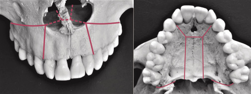
Obr. 1 / Fig. 1
1 Schematické zobrazení chirurgických řezů při H-tomii při pohledu en face a z okluze / Scheme of surgical cuts in H-osteotomy seen from the en face and from the occlusion.
Tab. 1 / Tab. 1
Změny hodnot lineárních charakteristik sledovaného souboru v čase před léčbou (T1), v čase před operací (T2), v čase po ukončení aktivní léčby (T3) a při dlouhodobé kontrole (T4) / Changes in linear parameters of the sample monitored prior to therapy (T1), prior to surgery (T2), after active therapy (T3) and at long-term check-up (T4).
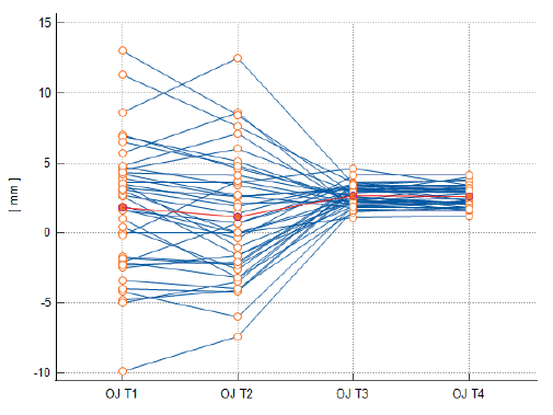
Obr. 2 / Fig. 2
Velikost incizálního schůdku (OJ) v čase před léčbou (T1), v čase před operací (T2), v čase po ukončení aktivní léčby (T3) a při dlouhodobé kontrole (T4). Červeně je označen aritmetický průměr / Overjet size (OJ) prior to therapy (T1), prior to surgery (T2), after active therapy (T3) and at long-term check-up (T4). Arithmetic mean is in red.
Tab. 2 / Tab. 2
Změny charakteristik sledovaného souboru v čase před léčbou (T1), v čase před operací (T2), v čase po ukončení aktivní léčby (T3) a při dlouhodobé kontrole (T4) / Changes prior to therapy (T1), prior to surgery (T2), after active therapy (T3) and at long-term check-up (T4).
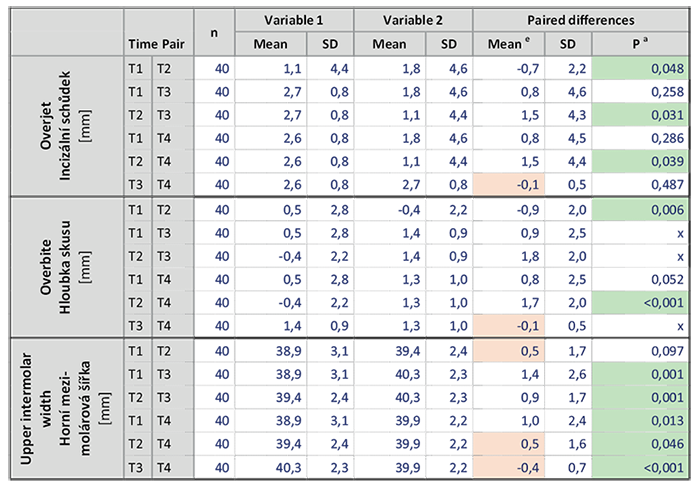
a Paired samples t-test; X = t-test nelze aplikovat (non-normální rozložení v čase T3); e Červeně označené průměrné hodnoty naměřených rozdílů nepřekračují chybu měření dané charakteristiky.
Obr. 3 / Fig. 3
Hloubka skusu (OB) v čase před léčbou (T1), v čase před operací (T2), v čase po ukončení aktivní léčby (T3) a při dlouhodobé kontrole T4). Červeně je označen aritmetický průměr. Záporné hodnoty na ose „y“ značí otevřený skus / Overbite (OB) prior to therapy (T1), prior to surgery (T2), after active therapy (T3) and at long-term check-up (T4). Arithmetic mean is in red. Negative values on y axis mean open bite.
Obr. 4 / Fig. 4
Horní mezimolárová šířka (U6w) v čase před léčbou (T1), v čase před operací (T2), v čase po ukončení aktivní léčby (T3) a při dlouhodobé kontrole (T4). Červeně je označen aritmetický průměr / Upper intermolar width (U6w) prior to therapy (T1), prior to surgery (T2), after active therapy (T3) and at long-term check-up (T4). Arithmetic mean is in red.
Tab. 3 / Tab. 3
Rozdíly v hloubce skusu v čase před léčbou (T1), v čase před operací (T2), v čase po ukončení aktivní léčby (T3) a při dlouhodobé kontrole (T4) / Changes in overbite prior to therapy (T1), prior to surgery (T2), after active therapy (T3) and at long-term check-up (T4).
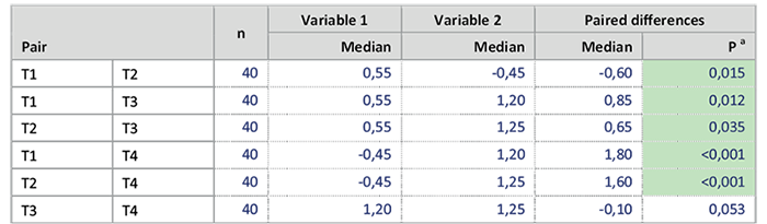
a Wilcoxon test (paired samples)
Tab. 4 / Tab. 4
Korelace změn navozených léčbou v čase T1-T3 a změn, ke kterým došlo v období retence v čase T3-T4 / Correlation of the changes induced by therapy at T1-T3 and the changes that occured during the retention at T3-T4
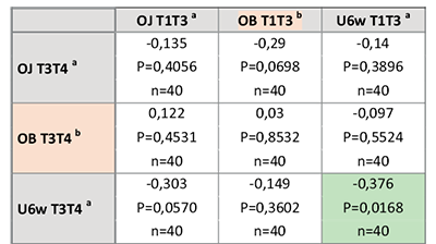
a Pearson correlation coefficient; b Spearman rank correlation coefficient

Literatura / References
1. Proffit WR, Sarver D. Combined surgical and orthodontic treatment. In: Proffit WR, Fields HW, Larson B, Sarver DM. Contemporary orthodontics. 6th ed. Philadelphia: Elsevier Health Sciences; 2018. p. 657–709.
2. Bell WH. Le Fort I Osteotomy. In: Bell WH. Surgical Correction of Dentofacial Deformities: New Concepts Vol III. Philadelphia: WB Saunders Company; 1985. p. 15–44.
3. Posnick JC. Malocclusion after orthodontics and orthognathic surgery: Prevention and treatment pitfalls. In: Posnick JC. Orthognathic surgery - principles and practice. St. Louis, Missouiri: Elsevier Saunders; 2014. p. 543–606. doi: 10.1016/B978-1-4557-2698-1.00017-4
4. Proffit WR, Turvey TA, Phillips C. The hierarchy of stability and predictability in orthognathic surgery with rigid fixation: an update and extension. Head Face Med. 2007;3(1):1–11. doi: 10.1186/1746-160X-3-21
5. Proffit WR, Bailey LTJ, Phillips C, Turvey TA. Long-term stability of surgical open-bite correction by Le Fort I osteotomy. Angle Orthod. 2000;70(2):112–7.
6. Proffit WR, Phillips C, Prewitt JW,Turvey TA. Stability after surgical-orthodontic correction of skeletal Class III malocclusion. 2. Maxillary advancement. Int. J. Adult Orthodont. Orthognathic Surg. 1991;6(2):71–80.
7. Kahnberg KE, Hagberg C. The approach to dentofacial skeletal deformities using a multisegmentation technique. Clin. Plast. Surg. 2007;34(3):477–84. doi: 10.1016/j.cps.2007.05.003
8. Chamberland S, Proffit WR. Short-term and long-term stability of surgically assisted rapid palatal expansion revisited. Amer. J. Orthodont. dentofacial Orthop. 2011;139(6):815–22. doi: 10.1016/j.ajodo.2010.04.032
9. Petr J, Foltán R. Ortodonticko-chirurgická léčba otevřeného skusu z pohledu ortodontisty. Ortodoncie. 2010;19(3):15–26.
10. Esteves LS, Santos JND, Sullivan SM, Martins LMR, Ávila C. Why segment the maxilla between laterals and canines?. Dental Press J. Orthod. 2016;21:110–25. doi: 10.1590/2177-6709.21.1.110-125.sar
11. Shand JM, Heggie AA. Segmental surgery of maxilla. In: Naini FB, Gill DS. Orthognathic surgery: principles, planning and practice. Chichester, West Sussex, UK: John Wiley & Sons; 2017. p. 635–41. doi: 10.1002/9781119004370.ch38
12. Žuffová N. Segmentální osteotomie maxily a její stabilita. Atestační práce. Plzeň; 2023.
13. Kretschmer WB, Baciut G, Baciut M, Zoder W, Wangerin K. Stability of Le Fort I osteotomy in bimaxillary osteotomies: single-piece versus 3-piece maxilla. J. Oral Maxillofac. Surg. 2010;68(2):372–80. doi: 10.1016/j.joms.2009.09.053
14. Haas Jr O, Guijarro-Martínez R, de Sousa Gil AP, da Silva Meirelles L, de Oliveira RB, Hernández-Alfaro F. Stability and surgical complications in segmental Le Fort I osteotomy: a systematic review. Int. J. Oral Maxillofac. Surg. 2017;46(9):1071–87. doi: 10.1016/j.ijom.2017.05.011
15. Arpornmaeklong P, Heggie AA, Shand JM. A comparison of the stability of single-piece and segmental Le Fort I maxillary advancements. J. Craniofac. Surg. 2003;14(1):3–9. doi: 10.1097/00001665-200301000-00002
16. Brandtner C, Hachleitner J, Rippel C, Krenkel C, Gaggl A. Long-term skeletal and dental stability after orthognathic surgery of the maxillo-mandibular complex in Class II patients with transverse discrepancies. J. Cranio-Maxillo-Fac. Surg. 2015;43(8):1516–21. doi: 10.1016/j.jcms.2015.07.007
17. Arnett GW, Trevisiol L, Grendene E, McLaughlin RP, D‘Agostino A. Combined orthodontic and surgical open bite correction. Angle Orthod. 2022;92(2):161–72. doi: 10.2319/101921-779.1
18. Arnett GW, D‘Agostino A, Grendene E, McLaughlin RP, Trevisiol L. Combined orthodontic and surgical open bite correction: Principles for success. Part 2. Angle Orthod. 2022;92(4),431–45. doi: 10.2319/123121-959.1
19. Silva I, Suska F, Cardemil C, Rasmusson L. Stability after maxillary segmentation for correction of anterior open bite: a cohort study of 33 cases. J. Cranio-Maxillo-Fac. Surg. 2013;41(7):154–8. doi: 10.1016/j.jcms.2012.12.003
20. Kamínek M, et al. Ortodoncie. 1st ed. Praha: Galén; 2014.
21. Proffit WR, Fields HW, Nixon WL. Occlusal forces in normal-and long-face adults. J. Dent. Res. 1983;62(5):566–70. doi: 10.1177/00220345830620051201
22. Yao W, Bekmezian S, Hardy D, Kushner HW, Miller AJ, Huang JC, et al. Cone-beam computed tomographic comparison of surgically assisted rapid palatal expansion and multipiece Le Fort I osteotomy. J. Oral Maxillofac. Surg. 2015;73(3):499–508. doi: 10.1016/j.joms.2014.08.024
23. Kretschmer WB, Baciut G, Baciut M, Zoder W, Wangerin K. Transverse stability of 3-piece Le Fort I osteotomies. J. oral maxillofacial Surg. 2011;69(3):861–9. doi: 10.1016/j.joms.2010.05.024
24. Marchetti C, Pironi M, Bianchi A, Musci A. Surgically assisted rapid palatal expansion vs. segmental Le Fort I osteotomy: transverse stability over a 2-year period. J. Cranio-Maxillo-Fac. Surg. 2009;37(2):74–8. doi: 10.1016/j.jcms.2008.08.006
25. Phillips C, Medland WH, Fields Jr HW, Proffit WR, White Jr RP. Stability of surgical maxillary expansion. Int. J. Adult Orthodont. Orthognathic Surg. 1992;7(3):139–46.
26. Reyneke JP, Ferretti C. Surgical correction of skeletal anterior open bite: Segmental maxillary surgery. In: Naini FB, Gill DS. Orthognathic surgery: principles, planning and practice. Chichester, West Sussex, UK: John Wiley & Sons; 2017. p. 480–97. doi: 10.1002/9781119004370.ch28
27. Hoppenreijs TJ, Freihofer HP, Stoelinga PJ, Tuinzing DB. Stability of orthodontic-maxillofacial surgical treatment of anterior open bite deformities. Ned. Tijdschr. Tandheelkd. 2001;108(5):173–8.
28. Hoppenreijs TJ, Van der Linden FPGM, Freihofer HP, Stoelinga PJ, Tuinzing DB, Jacobs BT, et al. Stability of transverse maxillary dental arch dimensions following orthodontic-surgical correction of anterior open bites. 1998. ANTERIOR OPEN BITE DEFORMITY, 99.
29. Foltán R, Petr J. Ortodonticko-chirurgická léčba pacientů s čelistními vadami – základy a nové poznatky. 2022 Jun
23–25; Pec pod Sněžkou, Czechia.
30. Posnick JC, Liu S, Tremont TJ. Long-face dentofacial deformities: occlusion and facial esthetic surgical outcomes. J. Oral Maxillofac. Surg. 2018;76(6):1291–308. doi: 10.1016/j.joms.2017.11.006
31. Posnick JC, Adachie A, Choi E. Segmental maxillary osteotomies in conjunction with bimaxillary orthognathic surgery: indications–safety–outcome. J. Oral Maxillofac. Surg. 2016;74(7):1422–40. doi: 10.1016/j.joms.2016.01.051
32. Ho MW, Boyle MA, Cooper JC, Dodd MD, Richardson D. Surgical complications of segmental Le Fort I osteotomy. Br. J. Oral Maxillofac. Surg. 2011;49(7):562–566. doi: 10.1016/j.bjoms.2010.09.018
33. Bell WH, You ZH, Finn RA, Fields RT. Wound healing after multisegmental Le Fort I osteotomy and transection of the descending palatine vessels. J. Oral Maxillofac. Surg. 1995;53(12):1425–33. doi: 10.1016/0278-2391(95)90670-3
MDDr. Nina Žuffová
stomatologická klinika FN Plzeň
Alej Svobody 80, 304 60 Plzeň