Dentální a skeletální indikátory růstového spurtu v ortodoncii
Dental and Skeletal Indicators of Growth Spurt in Orthodontics




MDDr. Lenka Dostálová, Ph.D.1, MDDr. Iva Voborná, Ph.D.2, MUDr. Eva Sedlatá Jurásková, Ph.D.1, MUDr. Ivana Dubovská, Ph.D.1
1 Ortodontické oddělení, Klinika zubního lékařství UP a FN Olomouc / Department of Orthodontics, Institute of Dentistry and Oral Sciences, University Hospital and Palacky University, Olomouc
2 Protetické oddělení, Klinika zubního lékařství UP a FN Olomouc / Department of Prosthodontics, Institute of Dentistry and Oral Sciences, University Hospital and Palacky University, Olomouc
DOI10.61110/50111
Souhrn
Cíl: Cílem práce bylo zjistit korelaci mezi chronologickým věkem, kostním věkem dle rentgenového snímku ruky a zápěstí a vývojovými stadii zubů a ověřit, zda lze na základě hodnocení vývoje zubů určit fázi růstu jedince.
Materiál a metodika: Do práce bylo zařazeno 362 adolescentů, a to 180 chlapců ve věkovém rozmezí 11,5–15,5 let a 182 dívek ve věkovém rozmezí 10,5–14,5 let. Pro hodnocení zubů na OPG snímku byla využita klasifikace dle Demirjiana, byly hodnoceny zuby 43, 44, 45, 47 a 48 v dolní čelisti a zuby 13, 14, 15, 17 a 18 v horní čelisti. Pro hodnocení rentgenu ruky a zápěstí byla využita klasifikace dle Björka, Grave a Browna.
Výsledky: Byla potvrzena korelace skeletálního a dentálního věku, nejvyšší korelace byla nalezena u dolního stálého špičáku, a to jak u dívek, tak i u chlapců. Stadium S na rentgenu ruky (stadium před maximálním růstovým spurtem) odpovídá fázi, kdy má kořen dolního špičáku paralelní stěny (stadium G dle Demirjiana). Pro klinické využití lze tedy odvodit, že pokud má dolní stálý špičák na OPG snímku paralelní stěny kořenového kanálku, dojde k vrcholu růstového spurtu cca za 0,5 roku až 1 rok. Vrchol růstového spurtu (stadium MP3cap) a následující stadia odpovídají stavu, kdy je apex dolního špičáku uzavřen (stadium H dle Demirjiana), což nám může pomoci při načasování ortodontické léčby.
Závěr: Hodnocení vývojových stadií zubů, a to především dolního stálého špičáku, je využitelné při plánování ortodontické léčby a může nám sloužit jako spolehlivý faktor pro určení stádia růstového spurtu.
(Ortodoncie 2024, 33, č. 2, s. 70–84)
Abstract
Aim: The aim was to confirm the correlation between chronological age, skeletal age according to X-ray picture of the hand and wrist, and the stages of teeth development, and to determine whether the assessment of teeth development can be used to establish the stage of growth of an individual.
Material and method: 362 adolescents were included in the study; 180 boys between the age 11.5–15.5, 182 girls between the age 10.5–14.5. To assess teeth in OPG Demirjian’s classification was used. We assessed teeth 43, 44, 45 and 47 in the mandible, and 13, 14, 15, 17 and 18 in the maxilla. To assess radiographs of the hand and wrist Björk, Grave and Brown classification was applied.
Results: The correlation of skeletal and dental age was proved. The highest correlation was found in lower permanent canine, both in girls and boys. S-stage in radiograph of the hand (the stage prior to the maximum growth spurt) corresponds to the stage of the lower canine root with parallel walls (G-stage according to Demirjian). Thus, in case OPG of lower permanent canine shows parallel walls of the root canal the maximum of growth spurt occurs within 0.5–1 year. The maximum growth spurt (MP3cap stage) and the following stages correspond to the situation when lower canine apex is closed (H-stage according to Demirjian). This can help us in the correct timing of orthodontic treatment.
Conclusion: The assessment of the developmental stages of teeth, namely lower permanent canine, is used in the planning of orthodontic treatment, and can serve as a realiable factor in establishing the stage of the growth spurt.
(Ortodoncie 2024, 33, No. 2, p. 70–84)

Klíčová slova:Růstový spurt, skeletální věk, rentgenový snímek ruky a zápěstí, dentální věk, dolní stálý špičák
Key words: growth spurt, skeletal age, radiograph of the hand and wrist, dental age, lower permanent canine
Úvod
Při plánování ortodontické terapie používáme pro správné načasování léčby určení růstového spurtu. Možnost určení fáze růstu nám pomáhá započít léčbu u většiny ortodontických vad ve správný čas tak, aby bylo dosaženo co nejlepšího funkčního a estetického výsledku v optimálním čase.
Vhodná doba pro ortodontickou léčbu při kompenzační léčbě anomálií typu distookluze je v období maximálního růstového zrychlení, kdy intenzivní růst dolní čelistí pomáhá v úpravě distookluze. Naopak u některých meziookluzí a skeletálních otevřených skusů je lépe začínat léčbu až po maximálním pubertálním růstovém zrychlení, kdy je teprve možné odlišit pacienty vhodné k ortodontické léčbě od těch, kde bude nutné ortodonticko – chirurgické řešení.
V současnosti nejčastěji používáme pro určení růstového spurtu hodnocení rentgenového snímku ruky a distální části předloktí [1], které se stalo zlatým standardem pro určení fáze růstu, nicméně v literatuře lze najít i další metody určování pubertálního růstového zrychlení, které by mohly sloužit jako náhrada nebo alespoň jako adekvátní doplněk při určení této důležité fáze růstu jedince.
Materiál a metodika
Do práce bylo zařazeno 362 adolescentů:
• 180 chlapců ve věkovém rozmezí 11,5–15,5 let,
• 182 dívek ve věkovém rozmezí 10,5–14,5 let.
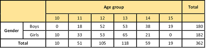
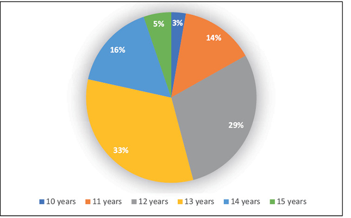
Všichni pacienti byli vybráni ručně z celkového souboru 819 pacientů, u nichž byl zhotoven rentgen ruky a zápěstí a zároveň i ortopantomogram ve stejný den (Tab. 1 a Obr. 1).
Z celkového souboru byli vyřazeni pacienti s agenezí či retencí zubu, pacienti se syndromovým onemocněním, pacienti s rozštěpem a pacienti, u kterých chyběl jeden nebo více hodnocených zubů na OPG snímku (13, 14, 15, 17, 18, 43, 44, 45, 47 a 48) Byly použity jen kvalitní rentgenové snímky. Méně kvalitní rentgenové snímky byly z hodnocení vyřazeny.
Hodnocení rentgenů bylo provedeno vizuálně jejich přímým pozorováním a srovnáváním s tabelárními a grafickými standardy dle dvou níže uvedených metodik.
Všechny sledované hodnoty byly 2× měřeny v rozmezí 2 týdnů stejným hodnotitelem a byla vypočítána chyba a opakovatelnost měření.
Pro učení vývojových stadií zubů 43, 44, 45, 47 a 48 v dolní čelisti a zubů 13, 14, 15, 17 a 18 v horní čelisti dle ortopantomografického snímku byla použita Demirjianova klasifikace (1973) [2], která popisuje 8 stadií označených A–H:
Stadium A je definováno jako počátek mineralizace korunky při horním okraji zubního folikulu ve tvaru obráceného kužele.
Stadium B je charakteristické kompletní mineralizací korunkové části založené na fúzi kalcifikovaných oblastí, čímž vzniká celá okluzní plocha.
Ve stadiu C zpočátku dochází k tvorbě skloviny při cementosklovinné hranici a je již viditelný počátek tvorby dentinu.
Stadium D má korunku již vytvořenou až k cementosklovinné hranici. Dřeňová dutina má trapezoidní tvar.
Stadium E je specifické tím, že délka kořene je menší než délka korunky zubu.
Ve stadiu F je délka kořene stejná nebo větší než korunka zubu, přičemž stěny dřeňové dutiny tvoří rovnoramenný trojúhelník.
Stadium G je charakteristické pro paralelní stěny kořenového kanálku, apex je v této fázi částečně otevřen.
V posledním stadiu H má již kořen ukončený vývoj foramen apicale. Periodontální štěrbina má stejnou šířku na celém povrchu kořene.
Skeletální věk byl určován na základě klasifikace autorů Björk, Grave a Brown (1976) [3] dle rentgenového snímku ruky a distální části předloktí, tato klasifikace byla použita jako zlatý standard pro určení fáze růstu v rámci této experimentální části. Klasifikace zahrnuje 9 stadií:
1. stadium PP2= je označováno, když epifýza proximálního článku 2. prstu dosáhne stejné šířky jako diafýza.
2. stadium MP3= nastává, když epifýza středního článku 3. prstu dosáhne stejné šířky jako diafýza.
3. stadium Pisi je charakteristická osifikací os pisiforme.
4. stadium S značí osifikaci sezamské kůstky ulnárně u metakarpofalangeálního kloubu palce.
5. stadium MP3cap, tzv. čepičkování, označuje stav, kdy epifýza středního článku 3. prstu dostává tvar čepičky.
6. stadium DP3u je v období spojení epifýzy a diafýzy distálního článku 3. prstu.
7. stadium PP3u označuje spojení epifýzy a diafýzy proximálního článku 3. prstu.
8. stadium MP3u je přítomno při spojení epifýzy a diafýzy mediálního článku 3. prstu
9. stadium Ru je hodnoceno jako spojení epifýzy a dialýzy radia.
Statistické zpracování
Naměřené výsledky byly nejprve vloženy do tabulky v programu Excel. Kvalitativní veličiny byly popsány pomocí absolutních a relativních četností (procenta). Kvantitativní proměnné byly prezentovány pomocí průměrů a směrodatných odchylek (SD), minimálních a maximálních hodnot a 95 % intervalů spolehlivosti (95% CI) pro průměr. Všechny testy byly provedeny na hladině statistické významnosti a = 0,05. Statisticky významné výsledky jsou zvýrazněny červenou barvou. Ověření chyby měření a opakovatelnosti měření bylo provedeno pomocí Cohenova kappa koeficientu a procentuální shody. Korelace byla analyzována pomocí Spearmanova korelačního koeficientu.
Ke statistickému zpracování byl použit statistický software IBM SPSS Statistics for Windows, Version 23.0. Armonk, NY: IBM Corp. Statistické zpracování provedla Mgr. Kateřina Langová, Ph.D. na Katedře lékařské biofyziky LF UP a FN Olomouc.
Chyba měření
Všechny hodnoty byly 2× měřeny v rozmezí 2 týdnů a porovnány. Ověření chyby měření a opakovatelnosti bylo provedeno pomocí Cohenova kappa koeficientu a procentuální shody. Měření shody proběhlo jak u hodnocení rentgenu ruky a zápěstí, tak u hodnocení jednotlivých zubů.
Při hodnocení rentgenu ruky se zápěstím a zubů 43, 44, 45, 14, 15, 17 byla shoda perfektní, kappa = 1, shoda byla tedy stoprocentní, což značí velmi dobrou interpretaci a rozlišitelnost jednotlivých stadií v rámci obou metodik. U zubů 47, 48, 13 a 18 byla shoda téměř perfektní, kappa > 0,989 u zubu 47, kappa > 0,991 u zubu 48, kappa > 0,995 u zubu 13 a, kappa > 0,995 u zubu 18.
Výsledky
Průměrný věk v jednotlivých fázích skeletálního zrání
V celém souboru byla nalezena všechna skeletální stadia, kromě 1. stadia (PP2=).
2. stadium (MP3=) skeletálního zrání bylo přítomno pouze u chlapců.
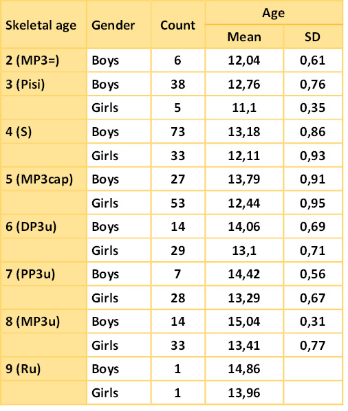
Ve stadiu 3 (Pisi) bylo 38 chlapců ve věku 12,76 ± 0,61 let a 5 dívek ve věku 11,10 ± 0,76 let.
Ve stadiu 4 (S) bylo 73 chlapců ve věku 13,18 ± 0,86 let a 33 dívek ve věku 12,11 ± 0,93 let.
Další stadium, které nás v rámci studie zajímalo, bylo stadium 5 (MP3cap), kde bylo zastoupeno 27 chlapců ve věku 13,79 ± 0,91 let a 53 dívek ve věku 12,44 ± 0,95 let.
Následující stadia 6 (DP3u), 7 (PP3u) a 8 (MP3u) jsou charakteristická zpomalením růstu, počet zhotovených snímků v těchto fázích byl nižší než v předchozích fázích. Ve stadiu 6 (DP3u) bylo 14 chlapců ve věku 14,06 ± 0,69 let a 29 dívek ve věku 13,10 ± 0,71 let.
Stadium 7 (PP3u) bylo zastoupeno 7 chlapci ve věku 14,42 ± 0,56 let a 28 děvčaty ve věku 13,29 ± 0,67 let.
Ve stadiu 8 (MP3u) bylo 14 chlapců ve věku 15,04 ± 0,31 let a 33 dívek ve věku 13,41 ± 0,77 let.
V souboru bylo nalezeno i zastoupení ve stadiu Ru, ale pouze u jednoho chlapce ve věku 14,86 let a jedné dívky ve věku 13.96 let.
Důležité pro plánování ortodontické léčby je stadium S, které předchází růstovému spurtu (stadiu MP3cap). Toto období nastalo u chlapců ve věku 13,18 ± 0,86 let a u dívek ve věku 12,11 ± 0,93 let.
Stadium MP3cap nastalo u chlapců ve věku 13,79 ± 0,91 let a u dívek ve věku 12,44 ± 0,95 let (Tab. 2).
Korelace chronologického a skeletálního věku
Pro ověření korelace chronologického a skeletálního věku byla vytvořena nulová a alternativní hypotéza.
Nulová hypotéza H0: Neexistuje závislost mezi chronologickým a skeletálním věkem.
Alternativní hypotéza HA: Existuje závislost mezi chronologickým a skeletálním věkem.
Na základě výpočtu Spearmanova korelačního koeficientu nulovou hypotézu zamítáme ve prospěch alternativní hypotézy. Bylo prokázáno, že existuje závislost mezi chronologickým a skeletálním věkem.
U chlapců i dívek byla nalezena středně silná korelace mezi chronologickým a skeletálním věkem. U chlapců byla korelace silnější než u dívek, u chlapců měl korelační koeficient hodnotu 0,63, p < 0,0001, zatímco u dívek měl korelační koeficient hodnotu 0,55, p < 0,0001.
Korelace chronologického a dentálního věku
Pro ověření korelace chronologického a dentálního věku byla taktéž vytvořena nulová a alternativní hypotéza.
Nulová hypotéza H0: Neexistuje závislost mezi chronologickým a dentálním věkem.
Alternativní hypotéza HA: Existuje závislost mezi chronologickým a dentálním věkem.
Na základě výpočtu Spearmanova korelačního koeficientu nulovou hypotézu zamítáme ve prospěch alternativní hypotézy. Bylo prokázáno, že existuje závislost mezi chronologickým a dentálním věkem.
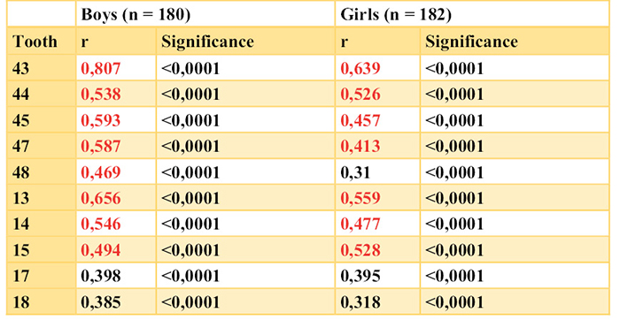
U všech zubů ve vztahu k chronologickému věku byly prokázány statisticky významné korelace, p < 0,0001. U chlapců byly u všech zubů nalezeny středně silné korelace mezi chronologickým a dentálním věkem, u dívek byla nalezen u většiny zubů také středně silné korelace, kromě zubu 45, 48 a 18, kde byly zjištěny slabší korelace. Nejsilnější korelace byla zjištěna u zubu 47 (r = 0,57, p < 0,0001), a u zubu 43 u chlapců (r = 0,55, p < 0,0001), u dívek byl nejsilnější korelační koeficient nalezen u zubu 14 (r = 0,53, p < 0,0001) a u zubu 44 (r = 0,51, p < 0,0001). Zub 43 u dívek měl korelační koeficient k chronologickému věku r = 0,48, p < 0,0001.
Korelace skeletálního věku a vývojových stadií zubů
Nulová hypotéza H0: Neexistuje závislost mezi skeletálním věkem a vývojovými stadii zubů v dolní a horní čelisti.
Alternativní hypotéza HA: Existuje závislost mezi skeletálním věkem a vývojovými stadii zubů v dolní a horní čelisti.
Na základě výpočtu Spearmanova korelačního koeficientu nulovou hypotézu zamítáme ve prospěch alternativní hypotézy. Bylo prokázáno, že existuje závislost mezi skeletálním věkem a vývojovými stadii zubů v dolní a horní čelisti.
U všech zubů byla prokázána statisticky významná korelace mezi vývojovými stadii zubů a skeletálním věkem, p < 0,05. Korelace klesala v tomto pořadí zubů u chlapců: 43, 13, 45, 47, 14, 44, 15, 48, 17, 18. U dívek klesala korelace v tomto pořadí zubů: 43, 13, 15, 44, 14, 45, 47, 17, 18, 48.
U chlapců korelace klesala v dolní čelisti v tomto pořadí zubů: 43, 45, 47, 48, 44.
U chlapců korelace klesala v horní čelisti v tomto pořadí zubů: 13, 14, 15, 17, 18.
U dívek klesala korelace v dolní čelisti v tomto pořadí zubů: 43, 44, 45, 47, 48.
U dívek klesala korelace v horní čelisti v tomto pořadí zubů: 13, 15, 14, 17, 18.
Nejsilnější korelace byla zjištěna u zubu 43 u chlapců (r = 0,81, p < 0,0001), u dívek korelační koeficient u tohoto zubu dosáhl také vysoké hodnoty odpovídající středně silné korelaci (r = 0,64, p < 0,0001). Prognosticky nejzajímavější je tedy zub 43, kde je korelace nejsilnější, zvlášť u chlapců (Tab. 3). V tabulce jsou červeně označeny statisticky významné korelace.
Statisticky významný rozdíl je patrný ještě u zubu 47 (u chlapců: r = 0,59; u dívek r = 0,41). U horních zubů je nejsilnější korelace u zubu 13, r = 0,66 u chlapců a r = 0,56 u dívek. Korelace u horních zubů jsou slabší a nejslabší korelace byla zjištěna u zubu 18 (r = 0,39 u chlapců a r = 0,32 u dívek – jedná se pouze o slabou korelaci).
V rámci korelace dle věkových skupin byla nejvýraznější korelace nalezena u zubu 43 byla u dívek ve věkové skupině 11 let (r = 0,78, p < 0,0001), u chlapců ve věkové skupině 14 let (r = 0,80, p < 0,0001).
Celkově lze říci, že byly v rámci hodnocení tohoto zubu nalezeny statisticky významné korelace u 11letých, 12letých i 13letých dívek i chlapců a také u chlapců ve věku 14 a 15let. Právě u naposledy zmíněných, tedy u 14letých a 15letých chlapců, koreluje skeletální věk nejsilněji s vývojem zubů.
Je zajímavým zjištěním, že u zubu 13 byly statisticky nejsilnější korelace přítomny u 11letých dívek i chlapců, 12letých dívek, 13letých dívek i chlapců a 14letých a 15letých chlapců.
V celkovém souboru všech zubů byly nejslabší korelace u 10letých dívek, žádná nebyla statisticky významná. Dále byla nalezena pouze slabá korelace u 14letých dívek, kde byly ze všech zubů pouze 2 statisticky signifikantní.
U 14letých dívek nebylo možno u zubu 14 spočítat korelační koeficient vzhledem k tomu, že všechna vývojová stadia zubu 14 v dané věkové skupině byla ve stejném vývojovém stadiu (stadium H), v této situaci korelační koeficient není měřitelný.
Distribuce vývojových stadií zubu v jednotlivých fázích skeletálního vývoje
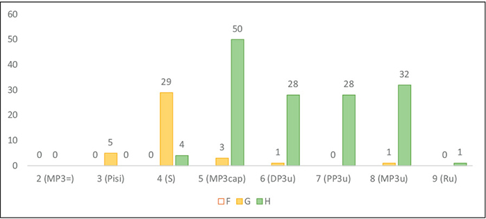
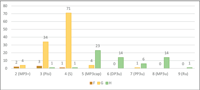
Vzhledem k průkazu nejsilnější korelace mezi dentálním a skeletálním vývojem u zubu 43 jsou níže zpracovány grafy znázorňující zastoupení jednotlivých stadií vývoje dolního špičáku vzhledem ke skeletální fázi vývoje jedince u chlapců a dívek (Obr 2, Obr 3).
Grafy ukazují, že ve skeletální fázi S je u chlapců i dívek nejčastěji zastoupen dolní stálý špičák ve vývojovém stadiu G (stadium paralelity kořene), zatímco ve skeletálních fázích MP3cap a také následných fázích vývoje je již vývoj dolního špičáku ve stadiu H, které je charakteristické uzavřeným apexem zubu.
Diskuse
Pro diagnostiku a naplánování léčby ortodontických anomálií nám může být velmi nápomocná znalost důležitých milníků v rámci růstu a alespoň orientační představa, v jaké konkrétní fázi se daný jedinec nachází. Již v minulosti se mnoho autorů snažilo najít korelaci mezi chronologickým, dentálním a skeletálním věkem jedince a zjistit, zda jsou tyto metody zastupitelné [1].
V této souvislosti je třeba upozornit, že termín zubní věk je často klinicky používán pro počet prořezaných zubů (dočasný, smíšený a stálý chrup). V této studii je dentální věk používán pro hodnocení podle vývojových stadií zubů (dle Demirjiana).
Cílem této práce bylo ověřit vztah mezi chronologickým věkem, kostním věkem dle rentgenu ruky a zápěstí a vývojovými stadii zubů v dolní a horní čelisti na OPG snímku a zjistit, zda lze na základě hodnocení vývojových stadií zubu určit fázi růstu jedince.
V rámci této práce bylo jako zlatý standard použito hodnocení skeletálních stadií na rentgenu ruky a distální části předloktí dle Björka, Grave a Browna [3]. Použití tohoto hodnocení patří mezi mezinárodně uznávané a dostupné metody k určení růstové fáze jedince a spolehlivost této metody byla prokázána v mnoha studiích [4–9].
V experimentální části této práce byla nalezena korelace mezi chronologickým a skeletálním věkem, chronologickým a dentálním věkem a mezi skeletálním a dentálním věkem navzájem. Tyto výsledky odpovídají studiím zahraničních i českých autorů [1, 4, 6, 10–13]. Je však důležité si uvědomit, že chronologický věk jako samotný faktor nelze spolehlivě použít k predikci růstové fáze. Konkrétně to lze ukázat na skeletálním stadiu MP3cap, tedy vrcholu růstového spurtu, které v této studii nastává u dívek v průměrném věku 12,44 let, u chlapců ve věku 13,79 let. Tento výsledek se shoduje s výsledky studie autorů Björk, Grave a Browna [3], původními autory metodiky, která byla v této studii použita. Z výsledků jejich výzkumu vyplývá, že vrchol růstového spurtu nastal u dívek průměrně ve 12,4 letech a u chlapců ve 14 letech [3]. Podobné výsledky byly nalezeny i ve studii Hagga a Tarangera [14], kde byl vrchol růstového spurtu zaznamenán u dívek průměrně ve 12,42 letech a u chlapců ve 14,62 letech. Velmi podobných výsledků bylo také dosaženo v rámci studie Fishmana [5], v jehož studii byl růstový spurt ve 12,1 letech u dívek a v 13,8 letech u chlapců. Také nedávná studie Motghara z roku 2016 [15] zaznamenala stadium MP3cap v průměrném věku 12,01 u dívek a 14,08 u chlapců. Nicméně pro určení růstového spurtu kalendářní věk nelze použít, jedná se pouze o orientační údaj. V rámci našeho zkoumaného souboru stadium MP3cap totiž nastalo u dívek nejdříve v 10,59 letech, nejpozději v 14,32 letech. U chlapců stadium MP3cap nastalo nejdříve v 12,45 letech, nejpozději v 15,41 letech. Stejně je tomu i u stadia S, tedy stadia skeletálního věku před vrcholem růstového spurtu – u dívek bylo rozpětí této fáze od 10,61 až 14,07 let, u chlapců 11,59 až 15,05 let. Tyto výsledky ukazují, že kalendářní věk není spolehlivým ukazatelem procesu dospívání.
Zajímavé je také srovnání se zahraničními studiemi v rámci rozdílů mezi pohlavími. Na základě výsledků této studie bylo prokázáno, že růstový spurt přichází u chlapců cca o 1,35 roku později než u dívek. Tento časový údaj odpovídá zjištěním mnoha studií, že u chlapců dochází ke skeletálnímu dozrávání později než u dívek [5, 6, 16, 17]. Nejčastěji se v literatuře uvádí orientační údaj, že pubertální růstové zrychlení u chlapců nastává asi o 2 roky později než u dívek [18].
V souladu s dostupnou literaturou byla také nalezena korelace mezi skeletálním a dentálním věkem [15,19–24]. U všech zubů zkoumaných v rámci této studie byla prokázána statisticky významná korelace mezi vývojovými stadii zubů a skeletálním věkem, p < 0,05. Korelace klesala v tomto pořadí zubů u chlapců: 43, 13, 45, 47, 14, 44, 15, 48, 17, 18. U dívek klesala korelace v tomto pořadí zubů: 43, 13, 15, 44, 14, 45, 47, 17, 18, 48. Tyto výsledky tedy jasně ukazují nejsilnější korelaci mezi dolním stálým špičákem, a to jak u dívek, tak i u chlapců, což odpovídá zjištěním studie Coutinho z USA [17]. Nejslabší korelace byla v této studii nalezena u horních a dolních třetích molárů a u druhého moláru, což může být způsobeno špatnou čitelností této oblasti na OPG snímku a také celkovou variabilitou.
Překvapivé je ale porovnání se studií Uysala a kol. [20], u kterých byla u obou pohlaví nalezena nejsilnější korelace u druhého moláru. Ve studii Krailassiri a kol. [21] byla nejsilnější korelace u druhého premoláru. Obě zmíněné studie používaly stejnou metodiku jako jsme použili i v této studii, přičemž však hodnotily pouze zuby dolní čelisti. Nesoulad výsledků je zřejmě dán populačními rozdíly, protože studie byly prováděny na turecké a thajské populaci.
Optimální načasování ortodontické léčby bylo v minulosti zájmem mnoha autorů. Již původní studie Björka [7] a na něj navazujících autorů [3, 7] vypozorovaly souvislost mezi skeletálním zráním a tzv. PHV (peak high velocity), tedy maximálním růstovým spurtem. Je možno zmínit, že někteří autoři zdokumentovali výraznější skeletální efekt snímacích aparátů během období maximálního růstu oproti období před maximálním růstem. Patří k nim například McNamara a kol. [25], který publikoval studii dětí léčených Fränkelovým aparátem nebo Hägg a Pancherz při léčbě pacientů Herbstovým aparátem [26]. Podobné zjištění publikoval kolektiv autorů a Malmgren, který studoval skeletální efekt modifikovaných aktivátorů v kombinaci s headgearem [27]. Kopecky a kolektiv [28] zjistili, že skeletální korekci při použití krčního headgearu je optimální začínat před pubertálním zrychlením růstu a pokračovat v léčbě během růstového spurtu s ohledem na závažnost skeletálního podkladu anomálie. Stejného principu je využíváno také při léčbě fixními aparáty. Lze tedy shrnout, že pro ortodontické plánování léčby je tedy především důležitá predikce růstového spurtu, protože u velké části ortodontických anomálií je vhodná doba pro léčbu v období maximálního růstového zrychlení. Björk [4] doporučuje zahájení ortodontické léčby využívající růst ve fázi S a zakončení před nástupem DP3u.
V rámci srovnání vývojových stadií dolního stálého špičáku s konkrétními stadii kostního zrání byla v roce 1979 ve studii Chertkow a Fatti [29] a v roce 1993 ve studii Coutinho [17] prokázána souvislost. Coutinho definovala základní pravidla použitelná pro možnost orientačního určení fáze růstu. Stadium S na rentgenu ruky (stadium před maximálním růstovým spurtem) dle zmíněné studie odpovídá fázi, kdy má kořen dolního špičáku paralelní stěny (stadium G dle Demirjiana). Tuto korelaci potvrdila i studie Gupty. Chady a Sharmy [30] a tato souvislost byla potvrzena i v naší studii – u chlapců je tomu tak v 97 %, u 71 chlapců ze 73. U dívek byl tento výsledek nalezen v 88 %, tedy u 29 dívek z 33. Interval spolehlivosti byl v obou případech vysoký. Přesto tento výsledek nelze používat jednoznačně, protože stadium G dolního špičáku bylo často nalezeno i ve fázi Pisi (89 % u chlapců, 100 % u dívek), také bylo nalezeno v několika procentech případů i ve stadiu MP3cap (15 % u chlapců, 6 % u dívek). Tyto výsledky lze však použít pro předpoklad, že stadium G se vždy vyskytuje před vrcholem růstového spurtu, což je pro klinické využití důležité zjištění.
Možnost využití tohoto zjištění je také důležitá pro predikci maximálního růstového spurtu, což nám může být velice nápomocné při plánování započetí ortodontické léčby. Obecně k tomuto účelu slouží skeletální stadium S, které dle Björka nastupuje obvykle 0,5 až 1 rok před maximálním růstovým spurtem [4]. Například Onat a kol. [9] uvádí, že ulnární sezamoid (stádium S) je radiograficky viditelný nejčastěji cca 0,5 roku před objevením pubického ochlupení a 0,75 roku před iniciací růstového spurtu. Autoři Coutinho Bushang a Miranda v roce 1993 [17] na základě výzkumu závislosti vývoje dolního špičáku a kostního zrání ruky u 200 chlapců a 215 dívek prokázali, že stadium G, které koinciduje s erupcí špičáku, nastává průměrně 1 rok před PHV u chlapců a 5 měsíců před PHV u dívek, což se orientačně shoduje s výsledky Björka. Toto bylo potvrzeno také ve studii Litte a Vaughana [31], v jejichž výzkumu stadium G nastalo průměrně 1,3 roku před PHV u chlapců a 0,4 roku před PHV u dívek. Pro klinické využití tedy při nalezení stádia G (stadium paralelity kořenového kanálku) u dolního stálého špičáku můžeme predikovat, že je jedinec s velkou pravděpodobností ve stadiu S dle rentgenu ruky a tím pádem k vrcholu růstového spurtu daného jedince by mohlo dojít orientačně za 6 až 12 měsíců.
Podobně byla hledána souvislost mezi ukončeným vývojem kořene špičáku a vrcholem růstového spurtu. Dle některých studií [20, 21] bylo prokázáno, že vrchol růstového spurtu (stadium MP3cap) odpovídá stavu, kdy je apex dolního špičáku uzavřen (stadium H). Dle výsledků prezentované studie lze říci, že i tato souvislost byla potvrzena – u chlapců je tomu tak v 85 %, tedy u 23 chlapců z 27. U dívek došlo k potvrzení v 94 %, konkrétně u 50 dívek z 53. Interval spolehlivosti byl v obou případech vysoký. Samozřejmě je nutno vzít také v potaz, že stadium H bylo nalezeno také v několika případech i ve stadiích před růstovým spurtem, nicméně toto zastoupení bylo pouze v malém počtu případů (ve stadiu S byl apex špičáku uzavřen v 1 % u chlapců a ve 12 % u dívek). Pro klinické využití lze tedy říci, že pokud je dolní špičák na OPG snímku ve stadiu H, je vysoká pravděpodobnost, že je jedinec na vrcholu růstového spurtu nebo již růstový spurt proběhl.
Zajímavá je zjištěná vysoká spolehlivost při interpretaci jednotlivých stadií vývoje kořene zubů a vývoje osifikace na rentgenu ruky jedním hodnotitelem, tzv. intraobservační spolehlivost. Výsledky naší studie korelují s výsledky španělské studie z roku 2020 [32], kde byla v daných věkových skupinách prokázána 99% intraobservační spolehlivost, v rámci interobservační spolehlivosti při klasifikaci více hodnotiteli byla nalezena 98% spolehlivost, což patří k velmi nadstandardním výsledkům a poukazuje na to, že jednotlivá stadia jsou dobře rozlišitelná jedním i více hodnotiteli.
Závěr
Z našeho výzkumu vyplývají tyto závěry pro praxi:
1. Byla potvrzena korelace skeletálního a dentálního věku, nejvyšší korelace byla nalezena u dolního stálého špičáku, a to především u chlapců, u dívek byla korelace středně silná. Hodnocení vývojových stadií dolního stálého špičáku nám tedy může sloužit jako spolehlivý faktor pro určení růstového spurtu.
2. Stadium S na rentgenu ruky (stadium před maximálním růstovým spurtem) a případně i předchozí skeletální stadium ve většině případů odpovídá fázi, kdy má kořen dolního špičáku paralelní stěny (stadium G dle Demirjiana). Pro klinické využití lze tedy odvodit, že pokud má dolní stálý špičák na OPG snímku paralelní stěny kořenového kanálku, tak s velkou pravděpodobností dojde k vrcholu růstového spurtu za 0,5 až 1 rok.
3. Vrchol růstového spurtu (stadium MP3cap) a následující stadia odpovídají stavu, kdy je apex dolního špičáku uzavřen (stadium H dle Demirjiana), což nám může pomoci při načasování ortodontické léčby.
4. Závěrem lze říci, že hodnocení vývojových stadií zubů, a to především dolního stálého špičáku, výrazně koreluje se skeletálním věkem. Tato metoda je tedy využitelná při plánování ortodontické léčby a může pomoci při načasování zhotovení rentgenu ruky a distální části předloktí ve správném období, čímž snížíme radiologickou zátěž pacienta. Stále ovšem platí, že hodnocení rentgenu ruky a distální části předloktí je považováno za nejspolehlivější metodu pro určení fáze růstu jedince. Klinicky nejvhodnější metodou je komplexní hodnocení všech projevů vývoje a růstu.
Tento výzkum nebyl financován z grantových projektů, veřejných ani komerčních zdrojů ani ze zdrojů neziskových organizací.
Autoři nemají komerční, vlastnické nebo finanční zájmy na produktech nebo společnostech popsaných v tomto článku.
Introduction
To time and plan orthodontic therapy we need to determine the stage of pubertal growth spurt. It helps us to start the treatment of orthodontic anomalies in time in order to achieve the best functional and esthetic results at the optimum time.
The appropriate time to start orthodontic treatment in compensation therapy of anomalies (such as distoocclusion) is at the maximum growth spurt when the intensive growth of the mandible helps to adjust distoocclusion. On the contrary, in some mesioocclusions and skeletal open bites it is better to start the treatment after the maximum growth spurt when it is possible to distinguish between the patients appropriate for orthodontic treatment and those who need orthodontic-surgical intervention.
Nowadays we determine the growth spurt according to the assessment of x-ray of the hand and distal part of forearm [1] that is considered the golden standard method. However, we can find other methods that may contribute to the assessment of this important growth stage.
Material and method
The study included 362 adolescents:
• 180 boys, within the interval 11.5–15.5 yrs
• 182 girls, within the interval 10.5–14.5 yrs
The patients were selected manually from the sample of 819 patients with radiograph of the hand and wrist and OPG taken on the same day (Table 1, Fig. 1)
Patients with missing or impacted teeth, syndromes, clefts, and with missing one or more teeth assessed in OPG (13, 14, 15, 17, 18, 43, 44, 45, 47, 48) were excluded from the study. We worked only with high quality x-rays.
OPGs were assessed visually, i.e. by direct inspection and comparison with tabular and graphic standards using the methods given below.
All parameters were measured twice within the interval of two weeks by the same evaluator, error of measurement and repeatability of measurement were calculated.
To identify developmental stage of teeth 43, 44, 45, 47 and 48 in the mandible, and 13, 14, 15, 17 and 18 in the maxilla we used the classification by Demirjian (1973) [2] working with 8 stages, A–H:
Stage A: the beginning of mineralization of the crown at the superior level of dental folicle in the form of an inverted cone.
Stage B: completed mineralization of the crown, fusion of calcified points resulting in a regularly outlined occlusal surface.
Stage C: enamel formation at cemento-enamel junction; dentine begins to form.
Stage D: crown formation is complete, extending down to the cemento-enamel junction; pulp chamber takes on a form of trapezoid.
Stage E: the root length is less than the crown height.
Stage F: the root length is the same or longer than the crown; the walls of pulp chamber form an isosceles triangle.
Stage G: the walls of root canal are parallel, apex is partially open.
Stage H:the development of foramen apicale of the root is completed – the apical end of the root is completely closed; the periodontal membrane has a uniform width around the tooth apex.
Skeletal age was determined with classification of Björk, Grave and Brown (1976) [3] using the x-ray of the hand and distal part of forearm. The classification was used as the golden standard to determine the growth stage in the experimental part of this work. The classification includes 9 stages of bone maturation:
1st stage PP2 – the width of epiphysis and the width of diaphysis of the proximal part of second finger are identical
2nd stage MP3 – the width of epiphysis and the width of diaphysis of the middle phalanx of third finger are identical
3rd stage Pisi – it is characterized by os pisiforme ossification
4th stage S – in metacarpophalangeal joint of the thumb sesamoid bone is ossificated ulnarly
5th stage MP3cap – the so-called capping – epiphysis of the middle phalanx of third finger takes a form of a cap
6th stage DP3u – epiphysis and diaphysis of distal phalanx of third finger are joined
7th stage PP3u – epiphysis and diaphysis of proximal phalanx of third finger are joined
8th stage MP3u – epiphysis and diaphysis of medial phalanx of third finger are joined
9th stage Ru – epiphysis and diaphysis of radius are joined
Statistical data processing
The measured values were represented in the Excel table. Qualitatives were described with absolute and relative frequency (per cents). Quantitatives were represented with mean values and standard deviations (SD), minimum and maximum values, and 95% confidence intervals (95% CI) for mean values. All tests were performed at the level of statistical significance a = 0.05. Statistically significant results are marked in red. Error of measurement and repeatability of measurement were verified with Cohen’s kappa coefficient and concordance rate. The correlation was analyzed with Spearman’s coefficient of correlation.
The data were processed with statistical software IBM SPSS Statistics for Windows, Version 23.0, Armonk, NY:IBM Corp. The work was performed by Mgr. Kateřina Langová, Ph.D., Department of Medical Biophysics, Faculty of Medicine and Dentistry in Olomouc.
Error of measurement
All values were measured twice within two weeks and compared. Error of measurement and repeatability of measurement were assessed with Cohen’s kappa coefficient and concordance rate. Measurement of concordance was performed both in the assessment of x-ray of the hand and wrist, and in the assessment of individual teeth.
In the assessment of radiograph of the hand and wrist and teeth 43, 44, 45, 14, 15, 17 the concordance was perfect, kappa = 1, i.e.100%. This suggests a very good interpretation and discriminability of individual stages in both methods of assessment used. In teeth 47, 48, 13 and 18 the concordance was almost perfect, kappa > 0.989 for 47, kappa > 0.991 for 48, kappa > 0.995 for 13, and kappa > 0.995 for 18.
Results
Mean age in individual stages of growth
All skeletal stages, except stage 1 (PP2=) were present in the whole sample. Stage 2 (MP3=) was found only in boys.
Stage 3 (Pisi) included 38 boys of the age 12.76 ±0.61 yrs, and 5 girls of the age 11.10 ±0.76 yrs. Stage 4 (S): 73 boys of the age 13.18 ±0.86 yrs, 33 girls of the age 12.11 ±0.93 yrs. Stage 5 (MP3cap): 27 boys of the age 13.79 ±0.91, 53 girls of the age 12.44 ±0.95 yrs.
Stages 6 (DP3u), 7 (PP3u) and 8 (MP3u) are characterized by slower growth, there were less OPGs than in previous stages. Stage 6 (DP3u): 14 boys of the age 14.06 ±0.69 yrs, 29 girls of the age 13.10 ±0.71 yrs. Stage 7 (PP3u): 7 boys of the age 14.42 ±0.56 yrs, 28 girls of the age 13.29 ±0.67 yrs. Stage 8 (MP3u): 14 boys of the age 15.04 ±0.31 yrs, 33 girls of the age 13.41 ±0.77 yrs.
Stage 9 (Ru) was found in only 1 boy (14.86 yrs) and 1 girl (13.96 yrs).
Stage S, that precedes the growth spurt (i.e. MP3cap) is important for the planning of orthodontic treatment. This stage occured in boys at the age 13.18 ± 0.86 yrs, and in girls at the age 12.11 ± 0.93 yrs.
Stage MPScap started in boys at the age 13.79 ± 0.91, in girls at the age 12.44 ± 0.95 yrs (Tab. 2).
Correlation of chronological and skeletal age
To confirm the correlation of chronological and skeletal age zero and alternative hypotheses were formulated.
Zero hypothesis H0 : There is no correlation between chronological and skeletal age.
Alternative hypothesis HA: There exists correlation between chronological and skeletal age.
Based on Spearman’s correlation coefficient the zero hypothesis is rejected. The correlation between chronological and skeletal age was proved.
Both in boys and girls moderate correlation between chronological and skeletal age was found. In boys the correlation was stronger; in boys the correlation coeficient was 0.63, p < 0.0001, in girls 0.55, p < 0.0001.
Correlation of chronological and dental age
To confirm the correlation of chronological and dental age zero and alternative hypotheses were formulated.
Zero hypothesis H0: There is no correlation between chronological and dental age.
Alternative hypothesis HA: There exists correlation between chronological and dental age.
Based on Spearman’s correlation coefficient the zero hypothesis is rejected. The correlation between chronological and dental age was proved.
Statistically significant correlations were confirmed in all teeth related to chronological age, p < 0.0001. In boys in all teeth moderate correlations between chronological and dental age were found; in girls in most teeth moderate correlations were found as well, except teeth 45,48 and 18 (weak correlations). The strongest correlation was found in the tooth 47 (r = 0.57, p < 0.0001) and 43 (r = 0.55, p < 0.0001) in boys, and in the tooth 14 (r=0.53, p < 0.0001) and 44 (r = 0.51, p < 0.0001) in girls. In girls the tooth 43 had correlation coefficient to chronological age r = 0.48, p < 0.0001.
Correlation of skeletal age and developmental stages of teeth
Zero hypothesis H0: There is no correlation between skeletal age and stages of teeth development in the mandible and in the maxilla.
Alternative hypothesis HA: There exists correlation between skeletal age and stages of teeth development in the mandible and in the maxilla.
Based on Spearman’s correlation coefficient the zero hypothesis is rejected. The correlation between skeletal age and stages of teeth development was proved.
Statistically significant correlation between developmental stages of teeth and skeletal age was proved in all teeth, p < 0.05. The correlation decreased in boys in the following order: 43, 13, 45, 47, 14, 44, 15, 48, 17, 18. The correlation decreased in girls in the following order: 43, 13, 15, 44, 14, 45, 47, 17, 18, 48.
In boys, in the mandible the correlation decreased in the following order: 43, 45, 47, 48, 44.
In boys, in the maxilla the correlation decreased in the following order: 13, 14, 15, 17, 18.
In girls, in the mandible the correlation decreased in the following order: 43, 44, 45, 47, 48.
In girls, in the maxilla the correlation decreased in the following order: 13, 15, 14, 17, 18.
The strongest correlation was found in the tooth 43 in boys (r = 0.81, p < 0.0001), in girls the CC of the tooth 43 was moderate (r = 0,64, p < 0.0001). Thus, the tooth 43 is very interesting, the correlation is the strongest in boys (Tab. 3). Statistically significant correlations are marked in red.
A statistically significant difference is found also in the tooth 47 (boys: r = 0.59, girls: r = 0.41). The strongest correlation within the upper teeth is found in the tooth 13 (boys: r = 0.66, girls: r = 0.56). In the maxilla the correlation is weaker, the weakest correlation was found in the tooth 18 (boys: r = 0.39, girls: r = 0.32).
According to age groups, the strongest correlation was found for the tooth 43 in girls of the age 11 years (r = 0.78, p < 0.0001), and in boys of the age 14 years (r = 0.80, p < 0.0001).
We can say that for this tooth statistically significant correlations were found both in girls and boys of the age 11, 12 and 13 years, and in boys of the age 14 and 15 years. In boys of the age 14 and 15 the correlation between skeletal age and teeth development is strongest.
It is interesting that for the tooth 13 statistically strongest correlations were found in both girls and boys of the age of 11 years, girls of 12 years, both girls and boys of the age 13 years, and in boys of the age 14 and 15.
The weakest correlations were found in girls of the age of 10; however, none was statistically significant. A weak correlation was found in girls of the age 14 – only 2 teeth were statistically significant.
In the group of girls of the age 14 we could not set the correlation coefficient for the tooth 14 because all developmental stages of the tooth 14 at the given age group were identical (Stage H), and in such a case the correlation coefficient cannot be measured.
Distribution of tooth developmental stages in individual stages of skeletal development
The strongest correlation between dental and skeletal development was found for the tooth 43 (Fig. 2, Fig 3).
The charts show individual stages of development of lower canine at G-stage (root parallelism). At skeletal stages MPScap and subsequent ones the development of lower canine is at H-stage, i.e. tooth apex is closed.
Discussion
To be aware of and to know significant stages of growth and potential stage of development of an individual can help in diagnostics and in the planning of orthodontic anomalies treatment. A number of authors tried to find the correlation between chronological, dental and skeletal age, and to determine whether the methods can replace each other [1].
The term dental age is often related to the number of erupted teeth (deciduous, mixed and permanent dentition). In our study the dental age is classified according to developmental stages of teeth (according to Demirjian).
The aim of our work was to confirm correlations between chronological, skeletal (in x-ray of the hand and wrist), and developmental stages of lower and upper teeth in OPG, and to establish whether we can determine the stage of growth of an individual according to the developmental stages of a tooth.
The method of golden standard to assesss skeletal age in radiograph of the hand and wrist is the classification by Björk, Grave and Brown [3]. This classification is widely used and its reliability was confirmed by many studies [4–9].
The experimental part of our work confirmed correlations between chronological and skeletal age, between chronological and dental age, and between skeletal and dental age. This corresponds to conclusions of a number of works published by foreign as well as Czech authors [1, 4, 6, 10–13]. However, we should be aware of the fact that chronological age itself cannot be used to reliably predict the growth stage. This can be shown on skeletal stage MP3cap, i.e. maximum growth spurt, which occurs (in our study) in girls at the mean age of 12.44 years, and in boys at the mean age of 13. 79. This corresponds to the results of Björk, Grave and Brown [3], i.e. the authors whose classification was used in our work. Their results suggest that maximum growth spurt starts in girls at the mean age of 12.4 years and in boys at the mean age of 14 [3]. Similar results are given in the study of Hagg and Taranger [14] (for girls at the age of 12.42 years, boys at the age of 14.62 years). Very similar results are also brought by the study of Fishman [5] (for girls at the age of 12.1 years, for boys at the age of 13.8 years). The recent study by Mothgar [15] recorded MP3cap stage at the age of 12.01 (girls) and 14.08 (boys). Nevertheless, chronological age cannot be used to determine the growth spurt, it is only an indicative date. In our sample of patients MP3cap stage occured in girls at the age of 12.45 at the earliest, and at 15.41 years at the latest. In boys it was at the age of 12.45 years, and at the age of 15.41, respectively. The same holds true for S stage, i.e. the stage prior to the maximum growth spurt - in girls it was within the interval of 10.61–14.07 years, in boys 11.59–15.05 years. Therefore, we can see that the chronological age is not a reliable indicator of the process of adolescence.
Comparison with studies published abroad dealing with differences between sexes is also interesting. The results of our study confirmed that the growth spurt starts in boys approx.by 1.35 years later than in girls. This agrees with a number of studies proving that skeletal maturation in boys starts later than in girls [5, 6, 16, 17]. The literature published reports that the growth spurt in boys occurs about 2 years later [18].
We also confirmed the correlation between skeletal and dental age, which agrees with conclusions of the publications available [15, 19-24]. In all teeth assessed in our study the statistically significant correlation between developmental stages of teeth and skeletal age was found, p<0. 05. In boys, the correlation decreased in the following order: 43, 13, 45, 47, 14, 44, 15, 48, 17, 18. In girls, the correlation decreased in the following order: 43, 13, 15, 44, 14, 45, 47, 17, 18, 48. The results show the strongest correlation in lower permanent canine, both in girls and boys. This is in agreement with the results cited in the study by Coutinho [17]. The weakest correlation was found in upper and lower third molars and second molar, which may be also due to more difficult interpretation of the area in OPG as well as to variability of the area.
Uysal et al. [20] found the strongest correlation for second molar in both sexes which is rather surprising. Krailassiri et al. [21] gave the strongest correlation for second premolar. Both studies used the same methodology as our work. However, they assessed only the mandibular teeth. The different results may be due to different populations, as the authors worked with Turkish and Thai populations.
A number of authors dealt with optimum timing of orthodontic treatment. The early studies of Björk [7] and other authors [3, 7] found the relationship between skeletal age and the so-called PHV (peak high velocity). i.e. the maximum growth spurt. Some authors documented more distinct skeletal effect of removable appliances during the maximum growth compared to the period before the maximum growth. We can cite e.g. McNamara et al. [25] who dealt with children treated with Fränkel appliance, or Hägg and Pancherz who used Herbst appliance [26]. Similar results were published by Malmgren et al. [27] who focused on skeletal effect of modified appliances combined with headgear [27]. Kopecký et al. [28] recommended that the skeletal correction using cervical headgear should start before the adolescent growth and the treatment should continue during the growth spurt with regard to the severity of skeletal basis of anomaly. The same principle is used in the treatment with fixed appliances. Therefore, we can sum up that in orthodontic treatment planning it is important to predict the growth spurt. Björk [4] recommends to start orthodontic treatment using growth at S stage and to complete the treatment before DP3u stage.
The works by Chertkow and Fatti [29] in 1979, and Coutinho [17] in 1993 compared the stages of development of lower permanent canine and individual stages of skeletal age. The correlation was proved. Coutinho defined basic rules to be used to estimate the growth stage. S stage in x-ray of the hand (taken prior to the maximum growth spurt) corresponds to the stage with lower canine root parallelism (G stage according to Demirjian). The correlation was confired by Gupta, Chad and Sharm [30], and also by our study – 97% in boys (71 out of 73); 88% in girls (29 out of 33). Credibility interval is rather high in both cases. The result is not clear-cut, as G stage of lower canine was often found also at Pisi stage (89% in boys, 100% in girls), as well as in MP3cap stage (15% in boys, 6% in girls). However, the results can be used to support the fact that G stage always precedes the maximum growth spurt. This can be very well used in clinical practice.
This finding is also important in prediction of the maximum growth spurt, i.e. in deciding when to start orthodontic treatment. In general, skeletal stage S serves well to this purpose. According to Björk the stage usually starts 0.5–1 year prior to the maximum growth spurt [4]. Onat et al. [9] report that ulnar sesamoide (stage S) is evident in radiographs about 0.5 year before the occurrence of pubic hair and 0.75 year prior to the maximum growth spurt. In 1993 Miranda [17] studied the correlation between lower canine development and skeletal maturity of hand in 200 boys and 215 girls. The study proved that stage G, which coincides with canine eruption, starts 1 year prior to PHV in boys, and 5 months prior to PHV in girls. This corresponds to the results cited by Björk. The fact was also proved by the study of Litte and Vaughan [31] – stage G started 1.3 years prior to PHV in boys, and 0.4 year prior to PHV in girls. Stage G in lower permanent canine thus predicts that an individual is at stage S (based on x-ray of the hand), and, therefore, their maximum growth spurt should occur within approximately 6 to 12 months.
The correlation between the completed development of canine root and the maximum growth spurt was also studied. Several works [20,21] proved that the maximum growth spurt (stage MP3cap) corresponds to the situation when lower canine apex is closed (stage H). Our study confirmed the results – in 85% of boys (i.e. in 23 boys out of 27); in 94% of girls (i.e. in 50 girls out of 53). In both cases credibility interval was high. However, we should be aware of the fact that stage H was found also in stages prior to the growth spurt. Nevertheless, this was found only in a small number of cases (at stage S canine apex was closed in 1% of boys, and in 12% of girls). We can sum up that in case lower canine is at stage H in OPG, it is highly probable that an individual is at the peak of the growth spurt or the growth spurt is already finished.
The high reliability of interpretation of individual stages of roots development and ossification in x-ray of the hand by one and the same evaluator (the so-called interrater or interobserver reliability) is interesting. Our results correspond to those given by the Spanish study of 2020 [32], where in the given age groups 99% intraobserver reliability was proved. In the classification performed by more evaluators the interobserver reliability reached 98% which si above standard results. It confirms the fact that individual stages are well distinguished by both one evaluator or more evaluators.
Conclusion
Our study brought the following results applicable in clinical practice:
1. The correlation between skeletal and dental age was confirmed. The highest correlation was found in lower permanent canine (in boys, in particular; in girls there was a medium correlation). Thus the assessment of developmental stages of lower permanent canine may serve as a reliable factor in determining the growth spurt.
2. Stage S in x-ray of the hand (stage prior to the maximum growth spurt) and possibly previous skeletal stage mostly correspond to the stage when lower canine root has parallel walls (stage G according to Demirjian). Therefore, in case lower permanent canine has parralel walls of the root canal in OPG, the maximum growth spurt probably occurs within 0.5 to 1 year.
3. The peak of the growth spurt (stage MP3cap) and the subsequent stages correspond to the situation where lower canine apex is closed (stage H according to Demirjian). This may help in accurate timing of orthodontic treatment.
4. The assessment of developmental stages of teeth, lower permanent canine in particular, correlates significantly with skeletal age. Therefore, the method can be used in the planning of orthodontic treatment, and can help in correct timing of radiograph of the hand and wrist. The radiological load of patients is thus reduced. However, the interpretation of radiograph of the hand and distal part of forearm is still considered the most reliable method to determine the growth stage of an individual. The complex evaluation of all manifestations of development and growth is the most appropriate for clinical practice.
This study was not financed from grant projects or other public or commercial sources.
Authors have no commercial, proprietary or financial interests in products or companies mentioned in the article.
Tab. 1 / Tab. 1
Soubor pacientů zařazených do studie / The sample of patients included in the study
Obr. 1 / Fig. 1
Procentuální zastoupení chlapců a dívek v jednotlivých věkových skupinách / Proportion of boys and girls in individual age groups
Tab. 2 / Tab. 2
Průměrný věk v jednotlivých stadiích kostního zrání u dívek a u chlapců / Mean age at individual stages of skeletal growth in girls and in boys
Tab. 3 / Tab 3
Korelační koeficienty jednotlivých zubů horní a dolní čelisti a skeletálních fází / Correlation coefficients of individual upper and lower teeth
Obr. 2 / Fig. 2
Vývojová stadia zubu 43 vzhledem ke skeletální fázi vývoje jedince u chlapců / Developmental stages of the tooth 43 related to skeletal development stage in boys
Obr. 3 / Fig. 3
Vývojová stadia zubu 43 vzhledem ke skeletální fázi vývoje jedince u dívek / Developmental stages of the tooth 43 related to skeletal development stage in girls

Literatura / References
1. Krásničanová H, Zemková D. Růst a biologický věk. Čs Pediatr 1991;46:525–30.
2. Demirjian A, Goldstein H, Tanner JM. A new system of dental age assessment. Hum Biol. 1973;211–27.
3. Grave K, Brown T. Skeletal ossification and the adolescent growth spurt. Amer. J. Orthodont. 1976;69(6):611–9. doi: 10.1016/0002-9416(76)90143-3
4. Björk A. Timing of interceptive orthodontic measures based on stages of maturation. Trans. eur. orthodont. Soc. 1972;61–74.
5. Fishman LS. Chronological versus skeletal age, an evaluation of craniofacial growth. Angle Orthodont. 1979;49(3):181–9.
6. Greulich WW, Pyle SI. Radiographic atlas of skeletal development of the hand and wrist. Stanford university press; 1959. doi: 10.1097/00000441-195909000-00030
7. Helm S, Siersbaek-Nielsen S, Skieller V, Björk A. Skeletal maturation of the hand in relation to maximum puberal growth in body height. Tandlaegebladet. 1971;75(12):1223–34.
8. Bala M, Pathak A, Jain R. Assessment of skeletal age using MP3 and hand-wrist radiographs and its correlation with dental and chronological ages in children. J. Indian. Soc. pedod. prev. Dent. 2010;28(2):95–9. doi: 10.4103/0970-4388.66746
9. Onat T, Numan-Cebeci E. Sesamoid Bones of the Hand: Relationships to Growth, Skeletal and Sexual Development in Girls. Hum. Biol. 1976;48(4):659–76.
10. Tanner JM. 1 Normal growth and techniques of growth assessment. Clin. endocrinol. Metab. 1986;15(3):411–51. doi: 10.1016/S0300-595X(86)80005-6
11. Tanner J, Whitehouse R, Marshall W, Carter B. Prediction of adult height from height, bone age, and occurrence of menarche, at ages 4 to 16 with allowance for midparent height. Arch., DiS. Child. 1975;50(1):14–26. doi: 10.1136/adc.50.1.14
12. Fishman LS. Maturational patterns and prediction during adolescence. Angle Orthodont. 1987;57(3):178–93.
13. Björk A, Helm S. Prediction of the age of maximum puberal growth in body height. Angle Orthodont. 1967;37(2):134–43.
14. Hägg U, Taranger J. Maturation indicators and the pubertal growth spurt. Amer. J. Orthodont. 1982;82(4):299–309. doi: 10.1016/0002-9416(82)90464-X
15. Motghare PC, Bedia AS, Degwekar SS, Indurkar AD, Bedia S. Correlation of calcification of permanent mandibular canine, mandibular premolars, and permanent mandibular first and second molars with skeletal maturity in Indian population. J. forensic. dent. Sci. 2016;8(2):67. doi: 10.4103/0975-1475.186370
16. Hägg U, Taranger J. Skeletal stages of the hand and wrist as indicators of the pubertal growth spurt. Acta odontol. Scand. 1980;38(3):187–200. doi: 10.3109/00016358009004719
17. Coutinho S, Buschang PH, Miranda F. Relationships between mandibular canine calcification stages and skeletal maturity. Amer. J. Orthodont. dentofacial. Orthop. 1993;104(3):262–8. doi: 10.1016/S0889-5406(05)81728-7
18. Taranger J, Hägg U. The timing and duration of adolescent growth. Acta Odontol. Scand. 1980;38(1):57–67. doi: 10.3109/00016358008997719
19. Demirjian A, Buschang PH, Tanguay R, Patterson DK. Interrelationships among measures of somatic, skeletal, dental, and sexual maturity. Amer. J. Orthodont. 1985;88(5):433–8. doi: 10.1016/0002-9416(85)90070-3
20. Uysal T, Sari Z, Ramoglu SI, Basciftci FA. Relationships between dental and skeletal maturity in Turkish subjects. Angle Orthodont. 2004;74(5):657–64.
21. Krailassiri S, Anuwongnukroh N, Dechkunakorn S. Relationships between dental calcification stages and skeletal maturity indicators in Thai individuals. Angle Orthodont. 2002;72(2):155–66.
22. Jourieh A, Khan H, Mheissen S, Assali M, Alam MK. The Correlation between dental stages and skeletal maturity stages. BioMed res. Int. 2021;2021. doi: 10.1155/2021/9986498
23. Bittencourt MV, Cericato G, Franco A, Girão R, Lima APB, Paranhos L. Accuracy of dental development for estimating the pubertal growth spurt in comparison to skeletal development: a systematic review and meta-analysis. Dentomaxillofac. Radiol. 2018;47(4):20170362. doi: doi.org/10.1259/dmfr.20170362
24. Lecca-Morales RM, Carruitero MJ. Relationship between dental calcification and skeletal maturation in a Peruvian sample. Dent. Press J. Orthodont. 2017;22:89–96. doi: 10.1590/2177-6709.22.3.089-096.oar
25. McNamara Jr JA, Bookstein FL, Shaughnessy TG. Skeletal and dental changes following functional regulator therapy on Class II patients. Amer. J. Orthodont. 1985;88(2):91–110. doi: 10.1016/0002-9416(85)90233-7
26. Pancherz H, Hägg U. Dentofacial orthopedics in relation to somatic maturation: An analysis of 70 consecutive cases treated with the Herbst appliance. Amer. J. Orthodont. 1985;88(4):273–87. doi: 10.1016/0002-9416(85)90126-5
27. Malmgren O, Ömblus J, Hägg U, Pancherz H. Treatment with an orthopedic appliance system in relation to treatment intensity and growth periods A study of initial effects. Amer. J. Orthodont. dentofacial. Orthop. 1987;91(2):143–51.doi: 10.1016/0889-5406(87)90472-0
28. Kopecky GR, Fishman LS. Timing of cervical headgear treatment based on skeletal maturation. Amer. J. Orthodont. dentofacial. Orthop. 1993;104(2):162–9. doi: 10.1016/S0889-5406(05)81006-6
29. Chertkow S, Fatti P. The relationship between tooth mineralization and early radiographic evidence of the ulnar sesamoid. Angle Orthodont. 1979 Oct;49(4):282–8.
30. Gupta B, Anegundi R, Sudha P. Comparison of dental age of Hubli Dharwad children by Moore’s method with the skeletal age and chronological age. Intern. J. dent. Sci. 2008;6(1):11–4. doi: 10.5580/2344
31. Demirjian A, Buschang PH, Tanguay R, Patterson DK. Interrelationships among measures of somatic, skeletal, dental, and sexual maturity. Amer. J. Orthodont. 1985;88(5):433–8. doi: 10.1016/0002-9416(85)90070-3
32. Paz Cortés MM, Rojo R, Alía García E, Mourelle Martínez MR. Accuracy assessment of dental age estimation with the Willems, Demirjian and Nolla methods in Spanish children: Comparative cross-sectional study. BMC Pediatr. 2020;20(1):1–9. doi: 10.1186/s12887-020-02247-x
MDDr. Lenka Dostálová, Ph.D.
Klinika zubního lékařství FNOL a UPOL
Palackého 12, 772 00 Olomouc