Srovnání změn v dolním zubním oblouku u pacientů léčených konvenčními
zámky a samoligujícími zámky
Changes in the lower dental arch in patients treated with conventional
brackets and self-ligating brackets – a comparison



MDDr. Aneta Hrubá, MUDr. Hana Tycová, doc. MUDr. Josef Kučera, Ph.D.
Ortodontické oddělení, Stomatologická klinika 1. LF UK a VFN, Praha / Department of Orthodontics, Clinic of Stomatology, 1st Medical Faculty of Charles University and University Hospital, Prague
DOI10.61110/50119
Souhrn
Cíl: Cílem studie bylo posoudit změny v dolní čelisti pacientů léčených konvenčními zámky ve srovnání s pacienty léčenými samoligujícími zámky. Zaměřili jsme se na srovnání míry transverzálních změn mezi oběma systémy a také na délku léčby.
Materiál a metodika: Studijní soubor zahrnoval 43 pacientů, z toho 11 mužů a 32 žen. Průměrný věk byl 22,2 let. Pacienti zařazení do studie měli kompletní stálý chrup, byli léčeni neextrakčně a moláry byly v I. Angleově třídě. Pacienti byli rozděleni do dvou skupin – první skupina byla léčena konvenčními zámky, druhá samoligujícími zámky. K měření byly použity skeny sádrových modelů zhotovených před léčbou (T1) a po sejmutí fixního aparátu (T2), u každého pacienta byla zaznamenána délka léčby v měsících. Srovnávali jsme změny v mezišpičákové a mezimolárové vzdálenosti, a to jak na zubech, tak v příslušných bodech na linii WALA na čelistní kosti.
Výsledky: V oblasti prvních molárů dolní čelisti byla změna mezimolárové šíře signifikantní u obou skupin mezi časy T2 a T1. Tato změna byla u skupiny se samoligujícími zámky signifikantně větší než u skupiny s konvenčními zámky. Změna mezišpičákové šíře mezi časy T2 a T1 byla statisticky významná pouze u skupiny se samoligujícími zámky jen v měření na hrotech špičáků, v měření v bodech FA (nejvíce prominující bod uprostřed podélné osy klinické korunky) nebyla prokázaná signifikantní změna. V oblasti WALA byla signifikantní změna mezi časy T2 a T1 jen u skupiny se samoligujícími zámky, a to jen u prvních dolních molárů (klinicky však i tato změna byla malá). Změna vzdálenosti WALA-FA straně u prvních dolních molárů vlevo i vpravo mezi časy T2 a T1 byla statisticky významná u obou skupin a signifikantně větší u skupiny se samoligujícími zámky. Rozdíl v délce léčby mezi oběma skupinami nebyl statisticky významný.
Závěr: U pacientů léčených samoligujícími zámky dochází k větší expanzi v prvních molárech a ve špičácích. Dále u těchto pacientů pozorujeme větší vestibulární sklon špičáků a prvních molárů ve porovnání s pacienty léčenými konvenčními zámky. U pacientů léčených samoligujícími zámky nebyla ortodontická léčba rychlejší než za použití konvenčních zámků. WALA se jeví jako klinicky relativně stabilní struktura.
(Ortodoncie 2024, 33, č. 2, s. 86–103
Abstract
Aim: The aim was to assess the changes in the mandible in patients treated with conventional brackets compared to those treated with self-ligating brackets. In particular, we focused on the degree of transversal changes and the length of therapy.
Material and method: The sample included 43 patients; 11 men and 32 women. The mean age was 22.2 years. The patients had complete permanent dentition, were treated without extractions, molars in Angle Class I. The subjects were divided into two groups – conventional brackets, self-ligating brackets. Measurements were performed in scans of cast models made prior to therapy (T1), after removal of fixed appliance (T2), the length of treatment was given in months. The changes in intercanine and intermolar distance were compared in teeth and in relevant points on WALA ridge of the mandible.
Results: In lower first molars the change in intermolar width was significant in both groups both at T1 and T2. The change was significantly bigger in the group with self-ligating brackets. The change in intercanine width between T2 and T1 was statistically significant only in the group with self-ligating brackets when measured on canine tips. Measurements at FA points (the most prominent point in the middle of longitudinal axis of the clinical crown) did not prove significant change. At WALA the change between T2 and T1 was significant only in the group with self-ligating brackets and only in first lower molars (clinically the change was small). The change in the distance WALA-FA in first lower molars on the left and on the right between T2 and T1 was statistically significant in both groups, however, it was significantly bigger in the group with self-ligating brackets. The difference in the length of therapy was not statistically significant.
Conclusion: In patients with self-ligating brackets there is a greater expansion in first molars and canines. We can also observe more prominent vestibular inclination of canines and first molars. In patients with self-ligating brackets the orthodontic treatment was not shorter. WALA appears to be a clinically relatively stable structure.
(Ortodoncie 2024, 33, No. 2, p. 86–103)

Klíčová slova: WALA, expanze, samoligující zámky
Key words: WALA, expansion, self-ligating brackets
Úvod
Vytvoření estetické, funkční a stabilní okluze je základním cílem ortodontické léčby. K dosažení tohoto cíle musíme při plánování ortodontické terapie brát v úvahu mnoho faktorů. Pro pacienty je zejména důležitá rychlost ortodontické léčby. Je nepochybně žádoucí, aby byl pacient léčen fixními aparáty co nejkratší možnou dobu, protože přítomnost fixního ortodontického aparátu v dutině ústní je spojena s vyšší akumulací plaku a obtížnější zubní hygienou, která může zvýšit riziko výskytu zubního kazu nebo parodontálního onemocnění [1, 2]. Rychlost ortodontické léčby určuje mnoho faktorů: povaha a rozsah ortodontické vady, individuální anatomické dispozice pacienta na straně jedné, nebo typ použitého materiálu na straně druhé, mimo jiné také velikost drážky ortodontických zámků či výběr a sekvence ortodontických oblouků [3, 4]. V tomto ohledu hraje nezanedbatelný vliv především tření vznikající mezi zámkem a obloukem [5]. Na velikost tření mají vliv fyzikální vlastnosti použitého ortodontického oblouku, materiál ortodontického zámku a způsob připevnění oblouku do zámku [6, 7]. U konvenčních zámků, u nichž jsou používány elastické ligatury, je tření zvýšeno ligaturovým materiálem [8, 9]. Ve snaze snížit tření ortodontických zámků bylo vyvinuto mnoho typů samoligujících zámků, které nevyžadují použití elastických nebo kovových ligatur. Především pasivní samoligující zámky se vyznačují výrazně redukovaným třením oproti zámkům konvenčním [10, 11]. Podle Damona lze použitím samoligujících zámků s nízkým třením a aplikací velice lehkých sil významně redukovat dobu léčby a zmenšit počet kontrol, v laterálních úsecích chrupu lze také dosáhnout větší expanze než při použití konvenčních zámků [12, 13]. Na druhou stranu příliš rychlý pohyb zubů a použití nevhodně velkých sil může také vést k nežádoucím vedlejším efektům, jakými jsou diskomfort pacienta [14,15] nebo apikální resorpce kořenů zubů [16, 17]. Nadměrná expanze zubních oblouků může také způsobit nežádoucí změny tvaru zubních oblouků, které byly popsány jako dlouhodobě nestabilní [18, 19].
Zubní oblouky, kostní podklad a okolní měkké tkáně by měly být ve vzájemné harmonii. K dosažení stabilní okluze je důležité umístit zuby do správné polohy v alveolárním výběžku a respektovat jeho limity [20]. Individualizace ortodontických oblouků dle unikátní anatomie daného pacienta se proto jeví jako základní předpoklad ke zlepšení stabilního výsledku ortodontické terapie [21, 22, 23]. Pro tyto účely byla Andrewsem jako jedna z možných referenčních struktur navržena struktura WALA [24], která představuje nejvíce prominující pruh keratinizované gingivy těsně nad mukogingivální hranicí. Následně se objevilo hned několik studií o využití WALA k individualizaci ortodontických oblouků [21, 25, 26, 27]. Výhodou WALA je její snadná klinická identifikace a neměnnost v čase. Tato struktura naznačuje, kde se nachází bazální kost, a tak v podstatě označuje fyziologickou hranici pro ortodontický pohyb zubů [28]. Andrews popisuje také vzdálenost jednotlivých zubů v dolní čelisti vzhledem k WALA, kdy je dolní zubní oblouk v ideální pozici vzhledem k apikální bázi dolní čelisti [24]. Díky tomu lze také v případě transverzální diskrepance mezi horním a dolním zubním obloukem poměrně snadno klinicky ověřit, jestli nejsou v dolní čelisti přítomny kompenzační lingvální sklony postranních zubů [29].
Cílem studie bylo porovnat změny, které nastanou na dolním zubním oblouku po ortodontické léčbě konvenčními zámky a samoligujícími zámky. U vybrané skupiny neextrakčních pacientů jsme se zaměřili na transverzální rozměry dolního oblouku, zejména pak na rozsah expanze v oblasti molárů a špičáků. Dalším cílem pak bylo ověřit platnost teze Andrewse, že struktura WALA je stabilní a neměnnou strukturou a zda není ovlivněna léčbou fixními aparáty. Byla také porovnávána průměrná doba terapie v obou skupinách.
Materiál
Do této retrospektivní studie bylo zařazeno celkem 43 pacientů (11 mužů, 32 žen). Soubor pacientů byl shromážděn z Ortodontického oddělení 1. LF UK a VFN v Praze a z privátní praxe MUDr. Darji Kadlecové v Praze. Vzhledem k tomu, že všechna měření probíhala pouze na skenech sádrových modelů, nebyl u této studie vyžadován souhlas etické komise. Do studie byli zařazeni pacienti splňující následující kritéria:
1. Artikulace I. třída Angleova v molárech
2. Neextrakční terapie
3. Kompletně prořezaná dentice v rozsahu 37-47
4. V dolním zubním oblouku nejsou přítomny ageneze, retinované, semiretinované ani nadpočetné zuby
5. V rámci terapie nebyla použita žádná z metod rychlé nebo pomalé maxilární expanze (hyrax, bihelix) nebo extraorální tah
6. Pacienti bez závažné skeletální vady, rozštěpu nebo syndromového onemocnění
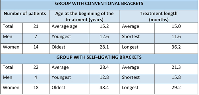
Všichni pacienti byli bílé rasy a ve věkovém rozmezí 12–41 let (průměr 22,2 let). Délka léčby fixním aparátem probíhala v rozmezí 11–36 měsíců (průměr 21,2 měsíců). Pacienti byli rozděleni do dvou skupin podle typu použitých zámků (Tab. 1 a 2).
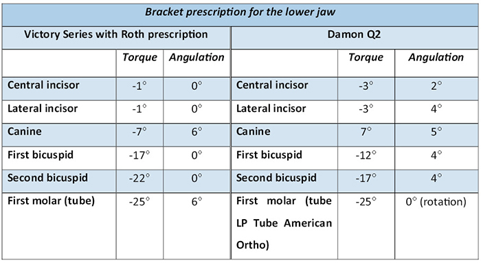
1. Skupina s konvenčními zámky (Tab. 1) byla léčena konvenčními zámky Victory Series (3M Unitek, USA) s preskripcí dle Rotha (Tab. 2) a s kombinovanou drážkou – pro frontální úsek (v rozsahu 3-3) drážka 0,018“, laterálně drážka 0,022“. Byla použita sekvence ortodontických oblouků Nitinol SuperElastic (3M Unitek, USA) ve tvaru OrthoForm III Ovoid v pořadí 0,014“, 0,016×0,022“ a 0,017×0,025“, následně pak ocelové oblouky (3M Unitek, USA) ve tvaru Orthoform III Ovoid 0,017×0,025“ [1].
2. Skupina se samoligujícími zámky (Tab. 1) byla léčena samoligujícimi zámky Damon Q2 (Ormco, Glendora, California, USA) s preskripcí Damon a drážkou 0,022“ (Tab. 2). V této skupině byla používána následující sekvence ortodontických oblouků: Damon Copper Ni-Ti (Ormco, Glendora, California, USA) v pořadí 0,014“, 0,014×0,025“ a 0,018×0,025“, dále pak ocelové oblouky 0,017×0,025“ od téže firmy.
Metodika
U všech pacientů byly zhotoveny skeny sádrových modelů ve dvou časech: před začátkem léčby fixním aparátem (T1) a těsně po sejmutí fixního aparátu (T2). Všechny skeny byly zhotoveny na přístroji 3Shape TRIOS (3Shape A/S, Dánsko). Následně byly v programu 3Shape Ortho Analyzer (3Shape A/S) zhotoveny virtuální modely, na nichž byla provedena analýza.
Zhotovení virtuálního modelu
Na virtuálním modelu byly v programu Ortho Analyzer skeny připraveny k analýze následujícím způsobem. Nejprve byla určena okluzní rovina, která byla definována dolním řezákovým bodem a meziobukálními hrbolky prvních molárů. Dále byl izolován sken dolní čelisti a zhotovena báze vhodné velikosti. V dolním zubním oblouku pak byla změřena meziodistální šířka všech zubů. Na skenech bylo měřeno 8 lineárních parametrů (Obr. 1-4), z dokumentace byl také zaznamenán údaj o délce léčby v měsících jako rozdíl mezi časy T2 (sejmutí fixního aparátu) a T1 (vstupní vyšetření) jako parametr T.
Přehled měřených parametrů
C – mezišpičáková šíře, vzdálenost hrotů pravého a levého dolního špičáku (v případě abraze hrotů špičáků byly určeny body v geometrickém středu plošek).
M6 – mezimolárová šíře, vzdálenost mezi hroty meziobukálních hrbolků prvních dolních molárů na pravé a levé straně (v případě abraze hrbolků byly určeny body v geometrickém středu plošek).
FA-C – mezišpičáková šíře, vzdálenost pravého a levého dolního špičáku v bodech FA, které jsou definovány jako nejvíc prominující bod uprostřed podélné osy klinické korunky.
FA-M6 – mezimolárová šíře, vzdálenost pravého a levého prvního dolního moláru v bodech FA, které jsou definovány jako nejvíc prominující bod uprostřed dlouhé osy korunky procházející meziobukálním hrbolkem.
WALA-C – šířka dolní čelisti v bodech WALA v oblasti dolních špičáků, které jsou definovány na pruhu WALA jako průsečík kolmice spuštěné na linii WALA ze špičákového bodu FA.
WALA-M6 – šířka dolní čelisti v bodech WALA v oblasti dolních prvních molárů, které jsou definovány na pruhu WALA jako průsečík kolmice spuštěné na linii WALA z bodu FA prvního dolního moláru.
WALA-FA P – vzdálenost WALA-M6 a FA bodu u prvního dolního moláru vpravo.
WALA-FA L – vzdálenost WALA-M6 a FA bodu u prvního dolního moláru vlevo.
Změna mezimolárové a mezišpičákové šířky byla hodnocena v oblasti FA bodů a v oblasti vrcholků hrbolků zubů (Obr. 1–3). Bod FA je definován jako nejvíc prominující bod v polovině podélné osy klinické korunky, vrcholky hrbolků zubů byly určeny jako geometrický střed meziálních hrbolků prvních molárů a hrotů špičáků. U prvních molárů byly vzhledem k větší přesnosti body FA umístěny na střed dlouhé osy procházející meziobukálním hrbolkem.
Šířka bazální kosti byla měřena v bodech WALA v oblasti špičáků a prvních molárů (Obr. 3). Pruh WALA je definován jako nejvíce prominující pruh keratinizované gingivy těsně nad mukogingivální hranicí [24]. Body WALA k měření byly označeny přímo pod daným bodem FA kolmicí od bodu FA k pruhu WALA. Následně byla také změřena horizontální vzdálenost obou bodů WALA.
K změření vzdálenosti WALA-FA u prvního moláru (Obr. 4) byla použita rovina vedoucí přes vrcholek meziobukálního hrbolku a vrcholek meziolingválního hrbolku. Tato rovina byla paralelní s dlouhou osou zubu a následně byla změřena vzdálenost v bodě FA na této rovině (nejvíc prominující bod na bukální straně korunky) a příslušném bodě WALA. Toto měření bylo provedeno zvlášť pro pravou a zvlášť pro levou stranu.
Statistické zpracování
Naměřené výsledky byly vloženy do tabulky v programu Excel (Microsoft Office, 2022) a následně zpracovány metodami popisné statistiky. Kvantitativní data byla vyjádřena pomocí průměru, směrodatné odchylky (SD), mediánu, minimální a maximální hodnoty. Shapiro-Wilkovými testy normality bylo prokázáno, že všechny parametry mají normální distribuci, kromě parametru označujícího rozdíl FA-C v čase T2-T1 ve skupině se samoligujícími zámky. Pro ověření hypotéz byly použity parametrické metody. Pro porovnání závislých vzorků párový t-test, pro nezávislé vzorky dvouvýběrový t-test. U veličiny FA-C, která neměla normální distribuci, byly výsledky parametrických testů validovány i pomocí příslušných neparametrických metod (Wilcoxonovým párovým testem a Mannovým-Whitneyovým U-testem). Všechny testy byly provedeny na hladině signifikance 0,05.
Ke statistickému zpracování byl použit statistický software IBM SPSS Statistics for Windows, Version 23.0. Armonk, NY: IBM Corp. A program MedCalc v18.2 (MedCalc Software, Ostend, Belgium). Statistika byla zpracována Mgr. Kateřinou Langovou, Ph.D.
Chyba měření
Reprodukovatelnost měření byla ověřena opakovaným měřením na 10 náhodně vybraných pacientech, a to 1 měsíc po prvním měření. Pro hodnocení chyby měření byla použita Dahlbergova formule (D), relativní Dalbergova chyba (RDE) a míra absolutní shody mezi prvním a kontrolním měřením byla ověřena také výpočtem koeficientu ICC (Intraclass correlation coefficient). Relativní Dahlbergova chyba se pohybovala na velmi nízkých hladinách mezi 0,33 % (pro parametr M6-M6) a 2,14 % (pro FA WALA/P). Hodnoty koeficientu ICC byly zjištěny v rozmezí 0,970 až 0,995, což představuje perfektní shodu. Výsledky hodnocení chyby měření jsou přehledně zpracovány v tabulce (Tab. 3). Ke grafickému znázornění chyby byly použity také Bland-Altmanovy grafy.
Výsledky
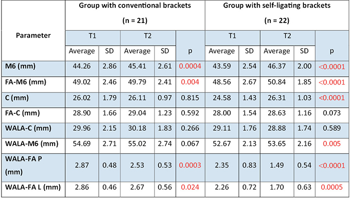
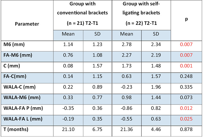
U parametru mezimolárové šíře měřené na meziobukálních hrbolcích dolních prvních molárů (M6) byla u obou skupin zaznamenána signifikantní změna mezi časy T2 a T1 (Tab. 4). U skupiny se samoligujícími zámky došlo k větší expanzi oblouku v oblasti dolních prvních molárů (průměrně o 2,78 mm) než u pacientů skupiny s konvenčními zámky (průměrně o 1,14 mm). Tento rozdíl v mezihrbolkové vzdálenosti prvních molárů (M6 v čase T2-T1) mezi oběma skupinami je statisticky významný, p = 0,007 (Tab. 5). U parametru mezimolárové šíře měřené na FA bodech (FA-M6) byla u obou skupin zaznamenána signifikantní změna mezi časy T2 a T1 (Tab. 4). U pacientů skupiny se samoligujícími zámky došlo k větší expanzi oblouku v oblasti prvních molárů v bodech FA (průměrně o 2,27 mm) než u pacientů skupiny s konvenčními zámky (průměrně o 0,76 mm). Rozdíl mezi oběma skupinami (FA-M6 v čase T2-T1) je statisticky významný, p = 0,007 (Tab. 5).
U parametru mezišpičákové šíře měřené na hrotech dolních špičáků byla u skupiny se samoligujícími zámky zaznamenána signifikantní změna mezi časy T2 a T1 (p < 0,0001), u skupin s konvenčními zámky změna signifikantní nebyla (p = 0,815) (Tab. 4). U pacientů skupiny se samoligujícími zámky došlo k větší expanzi oblouku v oblasti dolních špičáků (průměrně o 1,73 mm) než u pacientů skupin s konvenčními zámky (průměrně o 0,08 mm). Tento rozdí v mezišpičákové vzdálenosti (parametr v čase T2-T1) mezi oběma skupinami je statisticky významný, p = 0,001 (Tab. 5). U parametru mezišpičákové šíře měřen v FA bodech (FA-C) nebyla ani u jedné skupiny zaznamenána signifikantní změna mezi časy T2 a T1 (pro skupin s konvenčními zámky p = 0,592, pro skupinu se samoligujícími zámky p = 1,16) (Tab. 4). U pacientů skupiny se samoligujícími zámky došlo k větší expanzi oblouku v oblasti dolních špičáků v bodech FA (průměrně o 0,63 mm) než u pacientů skupin s konvenčními zámky (průměrně o 0,14 mm). Rozdíl mezi oběma skupinami (FA- v čase T2-T1) není statisticky významný, p = 0,248 (Tab. 5).
U parametru šířky čelisti v bodech WALA u dolních špičáků (WALA-C) nebyla u obou skupin zaznamenána signifikantní změna mezi časy T2 a T1 (Tab. 4). U pacientů skupiny s konvenčními zámky došlo k větší změně šířky čelisti v bodech WALA u dolních špičáků (průměrně o 0,22 mm) než u pacientů skupiny se samoligujícími zámky (průměrně o –0,23 mm). Rozdíl mezi oběma skupinami (WALA- v čase T2-T1) není statisticky významný, p = 0,335 (Tab. 5). U parametru šířky čelist v bodech WALA u dolních prvních molárů (WALA-M6) byla u skupiny se samoligujícími zámky zaznamenána signifikantní změna mezi časy T2 a T1 (p = 0,005), u skupiny s konvenčními zámky změna signifikantní nebyla (p = 0,067) (Tab. 4). U pacientů skupiny se samoligujícími zámky došlo k větší změně šířky čelisti v bodech WALA u prvních dolních molárů (průměrně o 0,98 mm) než u pacientů skupiny s konvenčními zámky (průměrně o 0,33 mm). Rozdíl mezi oběma skupinami (WALA-M v čase T2-T1) není statisticky významný, p = 0,073 (Tab. 5).
U parametru změny vzdálenosti WALA-FA u dolních molárů na pravé straně (WALA-FA P) byla u obou skupin zaznamenána signifikantní změna mezi časy T2 a T1 (Tab. 4). U pacientů skupiny se samoligujícími zámky došlo k výraznější redukci této vzdálenosti (průměrně o –0,86 mm) než u pacientů skupiny s konvenčními zámky (průměrně o –0,35 mm). Tento rozdíl ve vzdálenosti WALA-FA u dolních molárů na pravé straně (WALA-FA v čase T2-T1) mezi oběma skupinami je statisticky významný, p = 0,012 (Tab. 5). U parametru změny vzdálenosti WALA-FA u dolních molárů na levé straně (WALA-FA L) byla u obou skupin zaznamenána signifikantní změna mezi časy T2 a T1 (Tab. 4). U pacientů skupiny se samoligujícími zámky došlo k výraznější redukci této vzdálenosti (průměrně o –0,55 mm) než u pacientů skupiny s konvenčními zámky (průměrně o –0,19 mm). Tento rozdíl ve vzdálenosti WALA-FA u dolních molárů na levé straně (WALA-FA v čase T2-T1) mezi oběma skupinami je statisticky významný, p = 0,025 (Tab. 5).
U pacientů skupin s konvenčními zámky trvala léčba kratší dobu (21,10 měsíců) než u pacientů skupiny se samoligujícími zámky (21,36 měsíců) (Tab. 5). Tento rozdíl však není statisticky významný, p = 0,878.
Diskuse
Cílem této studie bylo porovnat změny na dolním zubním oblouku po ortodontické léčbě pomocí konvenčních zámků (skupiny s konvenčními zámky) a samoligujících zámků (samoligující Damon skupina) a zjistit, zda jsou tyto rozdíly statisticky signifikantní. Hlavní pozornost byla věnována dentoalveolárním změnám na dolním zubním oblouku po léčbě fixním ortodontickým aparátem se zaměřením zejména na změny mezimolárové a mezišpičákové šířky. Tyto změny nebyly posuzovány jen na vrcholcích hrbolků, ale také v bodech FA, které představují bukálně nejvíc prominující bod uprostřed podélné osy klinické korunky. Vzdálenost v bodech FA reprezentuje šířku oblouku, ve které lepíme ortodontické zámky – místo, kterým probíhají ortodontické oblouky, tedy místo přenosu ortodontické síly na zub. Dále jsme posuzovali změny na čelistní kosti v bodech WALA a také změnu vzdálenosti v bodech FA a příslušném bodě WALA u prvních molárů. Zároveň jsme porovnávali také délku ortodontické léčby u obou skupin.
Prvním sledovaným parametrem byla změna mezimolárové šířky. U obou skupin byla zaznamenána signifikantní změna mezi časy T2 a T1 a to jak u mezimolárové šíře měřené na meziobukálních hrbolcích, tak v bodech FA. U skupiny se samoligujícími zámky došlo k statisticky významně větší expanzi v oblasti prvních molár v obou měřených parametrech. Zatímco Kamínek [18] nedoporučuje žádnou expanzi dolního zubního oblouku v transverzálním směru, (s výjimkou napřímení lingválních sklonů dolních laterálních zubů), je dle Proffita [19] expanze v oblasti prvních molárů do 3 mm stabilní. Ani jedna skupina v naši studii nepřesáhla expanzi nad 3 mm
V souladu s našimi výsledky Pandis et al. [30] zaznamenali při srovnání míry expanze po ortodontické léčbě u pacientů léčených samoligujícími a konvenčními zámky (měřené u prvních molárů v centrální rýze) statisticky významně větší expanzi v oblasti prvních molárů u samoligujících zámků než u skupin s konvenčními zámky. Ve studii použili samoligující Damon 2 zámky (Ormco, Glendora, Calif) a konvenční zámky (Microarch, GAC, Central Islip, NY). Závěry studií Jianga et al. [31], Fleminga et al. [32] nebo Yu et al. [33] prezentují také podobné výsledky při srovnání pacientů léčených samoligujícími a konvenčními zámky u neextrakčních případů. Naopak Almeida et al. [34] při měření mezimolárové vzdálenosti na meziobukálních hrbolcích nenašli statisticky významný rozdíl v míře expanze mezi skupinou pacientů léčených pomocí samoligujících a konvenčních zámků. K podobným závěrům dospěli i Scott et al. [35]. Soubor jejich pacientů však tvořili pacienti po extrakcích prvních premolárů. Zdařilová [36] po nivelizační fázi dokonce zaznamenala u skupin s konvenčními zámky (zámky Omniarch, GAC) statisticky významně větší změnu mezimolárové šířky ve srovnání se skupinou se samoligujícímí zámky (In-Ovation R, GAC). Objektivní srovnání výsledků mezi studiemi je však poměrně obtížné vzhledem k tomu, že studie se liší v použití různých samoligujících a konvenčních zámků, preskripcí zámků, odlišnou metodikou použitou pro měření vzdáleností a také sekvencí použitých drátů.
Dále byla hodnocena a porovnávána změna mezišpičákové vzdálenosti. Změna mezišpičákové šířky na hrotech špičáků mezi časy T2 a T1 byla signifikantní jen u skupiny se samoligujícími zámky. U parametru mezišpičákové šířky v bodech FA nebyla mezi časy T2 a T1 zaznamenána signifikantní změna ani u jedné skupiny. Při srovnání obou skupin navzájem, došlo u skupiny se samoligujícícmi zámky ke statisticky významně větší změně u mezišpičákové šířky měřené na hrotech špičáků, v bodech FA změna signifikantní mezi dvěma skupinami nebyla.
Tento nález je možné interpretovat tak, že spíše, než k výrazné expanzi, došlo pouze k napřímení lingválního sklonu špičáků. Na změnu mezišpičákové šířky má vliv více faktorů, například preskripce zámků a použitá sekvence ortodontických oblouků a jejich rozměry. Zámky Damon Q2 mají u špičákových zámků kladnou torzi 7°, zatímco zámky Victory series s preskripcí dle Rotha použité u skupiny s konvenčními zámky mají u dolních špičáků torzi –7°. Se změnou torze však může dojít i ke změně mezišpičákové šíře, a to zejména tehdy, je-li měřena na hrotech špičáků
Mnoho autorů se shoduje na tom, že mezišpičáková vzdálenost by se neměla ortodontickou léčbou zvětšovat, přičemž jako maximální přijatelná expanze v mezišpičákové vzdálenosti je uváděna změna do 1 mm [19, 37]. Větší změny jsou považovány za nestabilní s větší tendencí k recidivě. V žádné aktuálně dostupné studii však nenajdeme statisticky významný rozdíl v stabilitě výsledku ortodontické léčby mezi pacienty léčenými samoligujícími a konvenčními zámky [38, 33, 39, 34]. V dostupných studiích srovnávajících změnu mezišpičákové vzdálenosti mezi pacienty léčenými samoligujícími a konvenčními zámky nebyl prokázán statisticky významný rozdíl mezi oběma skupinami [30, 36, 32, 33, 35]. Ve všech těchto studiích byly konfrontovány změny mezišpičákové vzdálenosti na hrotech špičáků, přičemž v žádné studii se zatím neměřila změna mezišpičákové vzdálenosti v bodech FA. Výsledky dostupných studií jsou tedy v souladu s výsledky naší studie.
Dalším naším cílem bylo ověřit tezi Andrewse, že struktura WALA je stabilní. Protože odráží průběh apikální báze, nemělo by během léčby docházet k její změně. Byly zkoumány změny šíře čelisti v bodech WALA a to v oblasti dolních špičáků a prvních dolních molárů. Změna šířky čelisti v bodech WALA mezi časy T2 a T1 byla signifikantní pouze v měření u prvních molárů u skupiny se samoligujícími zámky. Při srovnání mezi oběma skupinami však nebyl nalezen statisticky významný rozdíl. Nutno poznamenat, že změna u skupiny se samoligujícími zámky byla sice statisticky významná, přesto byl rozdíl v hodnotách klinicky poměrně malý (0,98 mm). Dalším faktorem však mohla být i větší expanze v oblasti prvních molárů během léčby, mezimolárová šíře se průměrně zvětšila o 2,78 mm (parametr M6), respektive o 2,27 mm (parametr FA-M6).
Výsledky našich měření jsou v souladu se závěry studií Estevese et al. [23], kteří zaznamenali u skupiny se samoligujícími zámky statisticky významně větší expanzi v mezišpičákové i mezimolárové šířce jak v FA bodech, tak na hrbolcích a v bodech WALA. Výjimkou byla pouze změna mezimolárové šířky na meziobukálních hrbolcích u prvních molárů a změna rozměru WALA u špičáků, které byly statisticky nevýznamné. Esteves et al. také konstatují, že dentální expanze u pacientů léčených samoligujícím (Damon) systémem přesahovala limit daný rozměrem apikální báze měřeným ve WALA bodech. To by znamenalo, že se zuby u skupiny se samoligujícími zámky posunuly až na bukální kraj alveolární kosti. Arnold et. al [40] však neprokázali rozdíl ve stavu parodontu mezi pacienty léčenými konvenčními a samoligujícími zámky. Někteří autoři dokonce považují samoligující zámky za metodu volby u dospělých pacientů s již existujícím parodontálním onemocněním [41]. Mavreas [41] se domnívá, že právě použití lehkých sil spojených s minimálním třením představuje pro pacientův parodont menší zátěž.
Další srovnávaný parametr byla změna vzdálenosti bodů FA na prvním moláru a příslušného WALA bodu u prvního moláru. U obou skupin došlo během léčby k signifikantní změně této vzdálenosti jak na levé, tak na pravé straně. U skupiny se samoligujícími zámky došlo na levé i na pravé straně ke statisticky významně větší změně než u skupiny s konvenčními zámky. U skupiny se samoligujícími zámky byla průměrná hodnota vzdálenosti WALA-FA po léčbě 1,49 mm vpravo a 1,70 mm vlevo, u skupiny s konvenčními zámky byla průměrná hodnota vzdálenosti WALA-FA po léčbě 2,53 mm vpravo a 2,67 mm vlevo. Vzdálenost WALA-FA dle Andrewse progresivně stoupá od řezáků k molárům od 0 mm až po 2,5 mm u druhého moláru [42, 43, 44]. V oblasti prvního moláru by měla být tato vzdálenost při správném sklonu moláru 2 mm. Výsledná vzdálenost WALA-FA se u obou skupin v naší studii nepatrně liší od hodnot, které uvádí Andrews. Když vezmeme v potaz změnu mezimolárové šíře v bodech FA a na hrbolcích a také změnu WALA-FA, lze z těchto výsledků vyvodit, že u prvních molárů došlo kromě expanze i k určité míře napřímení. U obou skupin totiž došlo k větší změně mezimolárové šíře na hrbolcích než v bodech FA a u obou skupin došlo k statisticky významnému zmenšení vzdálenosti WALA-FA oboustranně. Lze tedy konstatovat, že u skupiny se samoligujícími zámky došlo k výraznějšímu napřímení lingválního sklonu dolních prvních molárů, a to i přesto, že kanyly použité u obou skupin měly shodnou torzi –25°.
Nakonec jsme porovnávali délku léčby u obou skupin. Délka léčby u obou byla téměř shodná, přičemž skupina s konvenčními zámky měla v průměru nepatrně kratší délku léčby (21,10 měsíců) než skupina se samoligujícími zámky (21,36 měsíců). Tento rozdíl však nebyl statisticky významný. V naší studii, stejně jako v řadě jiných [30, 36, 45, 46, 47], tedy nebylo prokázáno tvrzení Damona, že za použití samoligujících zámků dochází k urychlení ortodontické léčby. V jiných studiích se naopak uvádí, že léčba samoligujícimi zámky je rychlejší [48, 49]. Vliv na objektivitu realizovaných šetření má nepochybně typ a závažnost vady vybraných pacientů, některé studie posuzují celkovou délku léčby, jiné pouze nivelizační fázi [30, 36]. Vzorek pacientů v naší studii měl před léčbou Angleovu I. třídu a neextrakční terapii. Je možné, že by se u pacientů se závažnější vadou léčba pomocí samoligujících zámků mohla prokázat jako rychlejší. Pandis et al. [30] ve své studii rozdělili pacienty do dvou skupin dle závažnosti stěsnání zubů. Celkově nezpozorovali statisticky významný rozdíl v rychlosti léčby mezi pacienty léčenými samoligujícími a konvenčními zámky, avšak u skupiny s mírným stěsnáním se projevila signifikantně rychlejší (2,7×) nivelizace u skupiny se samoligujícími zámky. U skupiny s větším stěsnáním rozdíl v rychlosti nivelizace nepozorovali. Velkým limitem jejich studie bylo, že posuzovali pouze rychlost nivelizace dolních frontálních zubů. Studie Harradine et al. [48] zahrnovala poměrně komplexní vzorek pacientů se skusem ve všech třídách dle Anglea, také pacienty s agenezemi, extrakcemi nebo dystopickými špičáky. U pacientů léčených samoligujícimi zámky pozorovali signifikantně kratší dobu léčby (průměrně o 4 měsíce). Podobný vzorek pacientů a obdobné výsledky se ukázaly také ve studii Ebertinga et al. [49]. DiBiase et al. [45] a Songra et al. [50] srovnávali rychlost léčby u pacientů s extrakcemi premolárů. Ani v jedné z těchto studií nebyla prokázána u skupiny se samoligujícími zámky rychlejší léčba. Lze tedy konstatovat, že co se týče urychlení léčby za použití samoligujících zámků, se závěry studií značně rozcházejí.
Naše měření potvrzují, že WALA je stabilní struktura jen málo ovlivnitelná ortodontickou léčbou. V případě, že expanze v dolní čelisti přesahuje limity apikální báze čelisti, lze však pozorovat změny i v rozměrech čelisti v oblasti WALA. Určitě by bylo přínosné, měření zopakovat s časovým odstupem v retenční fázi léčby, aby se ukázala stabilita výsledku léčby u pacientů z obou skupin.
Limitace studie
Pacienti zahrnutí do naší studie měli poměrně velký věkový rozptyl – nejmladšímu bylo 12 let, nejstaršímu 48 let. Skupina s konvenčními zámky měla průměrný věk 15,0 let, zatímco u skupiny se samoligujícími zámky byl průměrný věk 28,4 let. Všichni pacienti však měli plně prořezanou stálou dentici v rozsahu 37–47. Podle Bishary [51] se zubní oblouk po prořezání stálé dentice v transversálním rozměru významně nemění, větší věkový rozptyl by tedy neměl mít na přesnost této studie žádný vliv. Stejně tak by věk pacientů neměl ovlivnit ani délku ortodontické léčby, a to za předpokladu, že pacienti mají plně prořezanou stálou dentici [52]. K měření jsme zvolili 3D modely místo sádrových a tento způsob se ukazuje jako přesný a spolehlivý [53, 54, 55]. Uvádí se, že tloušťka měkkých tkání může být ovlivněna kompresí při otiskování. Tyto body by byly nejpřesněji určeny a změřeny dle CBCT [56], což však má v ortodoncii stále omezené indikace a nemělo by to být součástí rutinní dokumentace ortodontického pacienta [57]. Moura Neto ve své studii [58] porovnával přesnost měření rozměrů WALA ridge na modelech a CBCT. Prokázal, že oba výše zmíněné způsoby jsou srovnatelně přesné, bez statisticky významné odchylky.
Závěr
• U pacientů léčených samoligujícími zámky dochází ke statistiky významně větší expanzi v prvních molárech než u pacientů léčených konvenčními zámky.
• U pacientů léčených konvenčními zámky nedochází při léčbě ke změně mezišpičákové šířky.
• U pacientů léčených samoligujícími zámky dochází k zvětšení mezišpičákové šířky v oblasti hrotů špičáků, v oblasti bodů FA nebyla změna statisticky významná, jedná se tedy spíše o napřímení lingválního sklonu špičáků než o expanzi.
• U pacientů léčených samoligujícími zámky dochází ke statistiky významně větší expanzi ve špičácích (měřeno na hrotech špičáků) než u pacientů léčených konvenčními zámky.
• U pacientů léčených samoligující zámky dochází k většímu napřímení lingválního sklonu prvních molárů než u pacientů léčených konvenčními zámky.
• U pacientů léčených samoligujícími zámky nebyla ortodontická léčba rychlejší než při použití konvenčních zámků.
Autoři nemají komerční, vlastnické nebo finanční zájmy na produktech nebo společnostech popsaných v tomto článku.
Introduction
The goal of orthodontic treatment is an esthetic, functional and stable occlusion. To achieve the goal, we have to consider a number of factors. The patients appreciate the pace of orthodontic treatment. The treatment with fixed appliance should be as short as possible as the appliance is associated with higher accumulation of plaque and difficult dental hygiene which may result in caries or diseases of periodontium [1, 2]. The speed of orthodontic treatment is affected by a number of factors: the kind and extent of orthodontic disturbance, individual anatomical dispositions of a patient, the kind of materials used, orthodontic bracket slot, or selection and sequence of orthodontic archwires [3, 4]. An important role is played by the friction occuring between the bracket and the wire [5]. The friction is affected by physical characteristics of orthodontic archwire, material of an orthodontic bracket, and adjustment of the archwire in the bracket [6, 7]. In conventional brackets using elastic ligatures the friction is increased by the material of the ligature [8, 9]. To reduce the friction of orthodontic brackets a lot of self-ligating brackets have been developed. These brackets do not require elastic or metal ligatures. Passive self-ligating brackets in particular, show significantly reduced friction compared to the conventional ones [10, 11]. According to Damon the use of self-ligating brackets with low frictional force and application of light forces can significantly reduce the length of therapy as well as number of check-ups, in lateral segments of dentition a greater expansion is achieved than in conventional brackets [12, 13]. On the other hand, too rapid movement of teeth and the use of inappropriate forces may lead to undesirable adverse effects, such as patients’ discomfort [14, 15] or apical resorption of teeth roots [16, 17]. Excessive expansion of dental arches may also cause undesirable changes in the shape of dental arches that were described as unstable [18, 19].
Dental arches, skeletal basis and adjacent soft tissues should be in harmony. To achieve stable occlusion, it is important to place the teeth in the correct position in the alveolar process, and to respect its limits [20]. Orthodontic archwires adapted individually according to the unique anatomy of each patient is thus the basic precondition of improved and stable result of the therapy [21, 22, 23]. Therefore, Andrews suggested the structure WALA as one of possible reference structures [24]. WALA is the most prominent band of ceratinized gingiva immediately above mucogingival junction of the mandible. Soon after the WALA introduction several studies were published on the use of WALA for individualization of orthodontic archwires [21, 25, 26, 27]. The advantage of WALA is in its easy clinical identification and stability over time. This structure suggests the location of base bone, and thus it indicates physiological limits of orthodontic movement [28]. Andrews also described the distance of individual teeth in the maxilla related to WALA, when the lower dental arch is in ideal position in relation to apical base of the mandible [24]. Therefore, even in case of transversal discrepancy between upper and lower dental arches we can relatively easily verify whether there are present compensating lingual inclinations of lateral teeth in the mandible [29].
The aim of the study was to compare the changes in the lower dental arch following orthodontic therapy with conventional and self-ligating brackets. In the group of non-extraction patients, we focused on transversal dimensions of the lower dental arch, the extent of expansion within the area of molars and canines in particular. We also verified Andrewsʼ proposition of WALA as a stable and constant structure, and possible impacts of fixed appliances on the structure. The length of therapy in both groups was also compared.
Material
The retrospective study included 43 patients (11 men, 32 women). The sample of patients comes from the Department of Orthodontics of 1st Medical Faculty Charles University and University Hospital in Prague and from private practice of MUDr. Darja Kadlecová in Prague. All measurements were performed in scans of plaster models, therefore, the approval of Ethical commission was not required. The subjects met the following inclusion criteria:
1. Articulation – Angle Class I in molars
2. Non-extraction therapy
3. Erupted permanent dentition within 37-47
4. No missing, impacted or semiimpacted teeth, no supernumerary teeth in the lower dental arch
5. No method of rapid or slow maxillary expansion (hyrax, bihelix) or extraoral traction was applied during the therapy
6. No severe skeletal disorder, cleft or syndrome disease
All patients were Caucassians, the age interval 12–41 years (average 22.2 years). The treatment with fixed appliance took 11–36 months (average 21.2 months). The patients were divided to two groups based on the kind of brackets used (Tab. 1, Tab. 2).
1. Conventional brackets group (Tab. 1) was treated with conventional brackets Victory Series (3M Unitek, USA), prescription according to Roth (Tab. 2), with a combined slot – front segment (within 3-3) 0.018“, lateral 0.022“. The sequence of orthodontic wires Nitinol SuperElastic (3M Unitek, USA) was used, in the shape OrthoForm III Ovoid in the sequence 0.014“, 0.016“ and 0.017×0.025“, followed by steel wires Unitek stainless steel archwire (3M Unitek, USA) in the shape Orthoform III Ovoid 0.017×0.025“ [1].
2. Self-ligating brackets group (Tab. 1) was treated with self-ligating brackets Damon Q2 (Ormco, Glendora, California, USA) with Damon prescription and the slot 0.022 (Tab. 2). The following sequence of orthodontic wires was used: Damon Copper Ni-Ti (Ormco, Glendora, California, USA) in the order 0.014“, 0.014×0.025“and 0.018×0.025“, and stainless steel wires 0.017×0.025“ from the same company.
Method
In all patients scans of plaster models were made at two times: prior to treatment with fixed appliance (T1), immediately after removal of fixed appliance (T2). Scans were made using 3Shape TRIOS (3Shape A/S, Denmark). In 3Shape Ortho Analyzer (3Shape A/S, Denmark) virtual models were made.
Virtual model
First the occlusal plane was determined – defined by the lower incisal point and mesiobuccal cusps of first molars. Then a scan of the mandible was separated and an appropriate base was made. In the lower dental arch mesiodistal width of all teeth was measured. 8 linear parameters were measured (Fig.1-4); using documentation the length of treatment in months was recorded – the difference between T2 (removal) and T1 (initial assessment) = parameter T.
Survey of the parameters measured
C – intercanine width, distance between tips of right and left lower canines (in case of canine tips abrasion the points were in the geometrical centre of surfaces).
M6 – intermolar width, distance between tips of mesiobuccal cusps of first lower molars on the right and on the left (in case of abrasion the points were in the geometrical centre of surfaces).
FA-C – intercanine width, distance of right and left lower canine at points FA which are defined as the most prominent points in the middle of the longitudinal axis of the clinical crown
FA-M6 – intermolar width, distance of right and left first lower molar at points FA, which are defined as the most prominent point in the middle of the long axis of the crown running through mesiobuccal cusp
WALA-C – the mandible width at points WALA in the area of lower canines that are defined on the WALA band as the crossection of the perpendicular running to WALA line from the canine point FA
WALA-M6 – the mandible width at WALA points in the area of lower first molars that are defined on the WALA band as the crossection of the perpendicular running to WALA line from the first lower molar point FA
WALA-FA P – distance between WALA-M6 and FA in first lower molar on the right
WALA-FA L - distance between WALA-M6 and FA in first lower molar on the left
The change in intermolar and intercanine width was assessed at FA points and at tips of cusps (Fig. 1–3). Point FA is the most prominent point in the middle of longitudinal axis of the clinical crown, tips of teeth cusps are defined as the geometrical centre of mesial cusps of first molars and tips of canines. To be more precise, in first molars FA points were put in the middle of the long axis running through mesiobuccal cusp.
The width of base bone was measured in WALA points in the area of canines and first molars (Fig. 3). WALA band is defined as the most prominent band of ceratinized gingiva just above mucogingival junction (24). WALA points were marked just below the given point FA with the perpendicular from the point FA to WALA band. Then the horizontal distance of both
points of WALA was measured.
To measure the distance of WALA in first molar (Fig.4) the plane running over the tip of mesiobuccal cusp and the tip of mesiolingual cusp was used. The plane runs parallel with the long axis of the tooth. The distance was measured in the point FA (the most prominent point on the buccal side of the crown) and in the relevant point of WALA. The measurement was performed separately for the right side and for the left side.
Statistical data processing
The values obtained were represented in the Excel table (Microsoft Office, 2022) and subsequently they were processed with methods of descriptive statistics. Quantitative data were expressed as mean values, standard deviation (SD), median, minimum and maximum values. Shapiro-Wilk tests of normality proved that all the parameters have normal distribution, except the parameter for the difference FA-C at time T2-T1 in the group with self-ligating brackets. To verify hypotheses parametric methods were applied. To compare dependent samples we used the pair t-test, for independent samples we used two-sample t-test. In the quantity FA-C that did not have normal distribution the results of parametric tests were validated also with the help of relevantt non-parametric methods (Wilcoxon pair test and Mann-Whitney U-test). All the tests were performed at the level of significance 0.05.
For data processing we used the statistical software IBM SPSS Statistics for Windows, Version 23.0, Armonk, NY: IBM Corp., and MedCals v18.2 (MedCalc Software, Ostend, Belgium). The statistical part of the study was carried out by Mgr.Kateřina Langová, Ph.D.
Error of measurement
Reproducibility of measurement was verified with repeated measurements of 10 randomly chosen subjects 1 month after the first ones. To determine the error of measurement we used Dahlberg’s formula (D), relative Dahlberg’s error (RDE), and the degree of absolute concordance between the first and control measurements was verified with intraclass correlation coefficient (ICC). Relative Dahlberg’s error was in very low levels between 0.33% (in the parameter M6-M6) and 2.14% (in the parameter FA WALA/P). ICC values were within the interval 0.970 and 0.995 which means perfect concordance. The results are represented in Table 3. To represent the error of measurement we also used Bland-Altman plots.
Results
In the parameter of intermolar width measured in mesiobuccals cusps of lower first molars (M6) the significant change was recorded in both groups between T2 and T1 (Tab. 4). In the group with self-ligating brackets there was a greater expansion of the dental arch in the area of lower first molars (by 2.78 mm on the average) than in patients with conventional brackets (by 1.14 mm on the average). The difference in intercuspal distance of first molars (M6 at T2-T1) between the groups is statistically significant, p = 0.007 (Tab. 5). In the parameter of intermolar width measured in FA points (FA-M6) there was significant change between T2 and T1 in both groups (Tab. 4). In the group with self-ligating brackets there was a greater expansion in the area of first molars at points FA (by 2.27 mm on the average) than in patients with conventional brackets (by 0.76 mm on the average). The difference between the two groups (FA-M6 at T2-T1) is statistically significant, p = 0.007 (Tab. 5).
In the parameter of intercanine width measured on the tips of lower canines the significant change between T2 and T1 (p < 0.0001) was found in the group with self-ligating brackets; in the group with conventional brackets the change was not significant (p = 0.815) (Tab. 4). In patients with self-ligating brackets there was a greater expansion of the dental arch in the area of lower canines (by 1.73 mm on the average) than in patient with conventional brackets (by 0.08 mm on the average). The difference in the intercanine distance (parameter C at T2-T1) between the two groups is statistically significant, p = 0.0001 (Tab. 5). In the parameter of intercanine width measured in FA points (FA-C) the change between T2 and T1 was not statistically significant in any group (conventional group p = 0.592, self-ligating group p = 1.16) (Tab. 4). In the group with self-ligating brackets there was a greater expansion of the dental arch in the area of lower canines at points FA (by 0.63 mm on the average) than in patients with conventional brackets (by 0.14 mm on the average). The difference between the two groups (FA-C at T2-T1) is not statistically significant, p = 0.248 (tab. 5).
In the parameter of the mandible width at points WALA in lower canines (WALA-C) there was not found significant change between T2 and T1 in both groups (Tab. 4). In the group with conventional brackets there was a more significant change in the width of the mandible at WALA in lower canines (by 0.22 mm on the average) than in patients with self-ligating brackets (by –0.23 on the average). The difference between the two groups (WALA-C at T2-T1) is not statistically significant, p = 0.335 (Tab. 5). In the parameter of the mandible width at points WALA in lower first molars (WALA-M6) there was significant change between T2 and T1 in the group with self-ligating brackets (p = 0.005); the change was not statistically significant in the group with conventional brackets (p = 0.067) (Tab. 4). In the group with self-ligating bracketds there was a greater change in the width at points WALA in first lower molars (by 0.98 mm on the average), than in patients with conventional brackets (by 0.33 mm on the average). The difference between the two groups (WALA-M6 at T2-T1) is not statistically significant, p = 0.073 (Tab. 5).
In the parameter change in the distance WALA-FA in lower molars on the right (WALA-FA P) the significant change between T2 and T1 was found in both groups (Tab. 4). In the group with self-ligating brackets the distance was more distinctly reduced (by = 0.88 mm on the average) than in patients with conventional brackets (by = 0.35 mm on the average). This difference between the two groups is statistically significant, p = 0.012 (Tab. 5). In the parameter change in the distance WALA-FA in lower molars on the left (WALA-FAL) the significant change between T2 and T1 was found in both groups (Tab. 4). In the group with self-ligating brackets the reduction was more distinct (by –0.55 mm on the average) than in patients with conventional brackets (by –0.19 on the average). This difference between the two groups is statistically significant, p = 0.025 (Tab. 5).
In the group with conventional brackets the therapy took 21.10 months, in the group with self-ligating brackets 21.36 months (Tab. 5). However, this difference is not statistically significant, p = 0.878.
Discussion
The aim of our study was to compare changes in the lower dental arch following orthodontic therapy with conventional brackets (the group with conventional brackets) and self-ligating brackets (the group with self-ligating brackets), and to find whether the differences are statistically significant. We focused on the dentoalveolar changes in the lower dental arch after the treatment with fixed orthodontic appliance, in particular on the changes in intermolar and intercanine width. The changes were not assessed only in tips of cusps but also at points FA, that are the buccally most prominent point in the middle of longitudinal axis of the clinical crown. The distance in points FA represents the width of the arch where we bond orthodontic brackets – the place through which orthodontic arches run, i.e. the place where orthodontic forces act upon the tooth. We also assessed the chages in the mandible bone at points WALA, and the change of distance at points FA and the given point WALA in first molars. The length of orthodontic therapy in the two groups was compared.
The first parameter assessed was the change in intermolar width. In both groups a significant change between T2 and T1 was recorded – in intermolar width measured in mesiobuccal cusps, and in points FA. In the group with self-ligating brackets there was a significantly greater expansion in the area of first molars in both parameters measured. Kamínek [18] does not recommend any transversal expansion of the lower dental arch (with the exception of straightening of lingual inclinations of lower lateral teeth). However, Proffit [19] states that the expansion up to 3 mm within first molars is stable. None of the groups in our study extended expansion over 3 mm.
Similarly to our results, Pandis et al. [30] found a significantly greater expansion in the area of first molars in self-ligating brackets than in conventional ones. They worked with self-ligating brackets Damon 2 (Ormco, Glendora, California) and conventional brackets (Microarch, GAC, Central Islip, NY). Virtually the same results are reported also by Jiang et al. [31], Fleing et al. [32] or Yu et al. [33]. On the contrary, Almeida et al. [34] measured intermolar distance in mesiobuccal cusps, and they did not find statistically significant difference in the degree of expansion between self-ligating and conventional brackets. Similar results are given also by Scott et al. [35]. However, their subjects were patients after extraction of first premolars. Zdařilová [36] after the nivelization stage even recorded statistically significant change in intermolar width in patients with conventional brackets (Omniarch, GAC) compared to the patients treated with self-ligating brackets (In-Ovation R, GAC). However, an objective comparison of the results of individual studies is relatively difficult as the authors work with different self-ligating and conventional brackets, prescriptions, different methods of measurement, and sequence of the wires used.
Further we assessed and compared the change in intercanine distance. The change measured in tips of canines between T2 and T1 was statistically significant only in the group with self-ligating brackets. In the parameter intercanine width at points FA there was not recorded any significant change in any group. When we compared the two groups, there was statistically significant change in intercanine width measured in canine tips in the group with self-ligating brackets. We found no statistically significant change in points FA.
It appears that lingual inclination of canines was corrected, and we should not speak about significant expansion. The change of intercanine width is the result of more factors, e.g. brackets prescription, sequence of orthodontic brackets, dimensions of the brackets. Brackets Damon Q2 in canines have positive torque 7°, while brackets Victory series with Roth’s prescription (used in the group with conventional brackets) have negative torque –7°. However, the change in torque may result is the change of intercanine width, especially when measured in canine tips.
A number of authors agree on that the intercanine distance should not increase due to orthodontic treatment; they see the maximum acceptable expansion up to 1 mm [19, 37]. More profound changes are considered unstable with a tendency to relapse. However, in the works available we did not find a statistically significant difference in the stability of the orthodontic treatment results between patients with self-ligating and conventional brackets [38, 33, 39, 34]. In the works comparing the change in intercanine distance between patients with self-ligating and conventional brackets there was not proved statistically significant difference between the two groups [30, 36, 32, 33, 35].
In all the works mentioned above the intercanine distances measured in tips of canines were compared. The parameter has not been measured in FA points yet. The results of the published studies agree with our conclusions.
Another objective was to verify Andrews’ statement that WALA ridge is stable. As the structure reflects the apical base, it should not change during the therapy. We studied changes in the width of the mandible at WALA points in the are of lower canines and lower first molars. The change between T2 and T1 was significant only in first molars in the group with self-ligating brackets. However, the comparison between the two groups did not reveal a statistically significant difference. We have to note that though the change in the group with self-ligating brackets was statistically significant, from the clinical viewpoint the difference in the values was relatively low (0.98 mm). The greater expansion in the area of first molars during the therapy may have contributed to the extension of intermolar width by 2.78 mm (parameter M6), or by 2.27 mm respectively (parameter FA-M6).
Our results agree with the conclusion of Esteves et al. [23], who reported a significantly greater expansion in both intercanine and intermolar width at FA points, cusps and WALA points in the group with self-ligating brackets. The only exception was the change in intermolar width at mesiobuccal cusps of first molars and the change in WALA in canines – the changes were not statistically significant. Esteves et al. also concluded that dental expansion in patients treated with self-ligating (Damon) system exceeded the limit represented by the dimension of the apical base measured at WALA points. This could mean that the teeth in the group with self-ligating brackets moved as far as to the buccal edge of the alveolar bone. Nevertheless, Arnold et al. [40] did not prove the difference in the condition of periodontium between the patients with conventional and self-ligating brackets. Some authors even consider self-ligating brackets as the method of choice in adults with diseased periodontium [41]. Mavreas [41] presumes that the use of light forces associated with minimum friction brings about lower load for a patient’s periodontium.
The change in the distance of FA points in first molar and the relevant WALA point in first molar was another parameter compared. There was recorded statistically significant change in both groups on the left as well as on the right side. In the group with self-ligating brackets the change was significantly greater on the left as well as on the right side than in patients with conventional brackets. In the group with self-ligating brackets the mean parameter WALA-FA after the therapy was 1.49 mm on the right, and 1.70 mm on the left side; in the group with conventional brackets it was 2.53 mm on the right, and 2.67 mm on the left side. According to Andrews the WALA-FA distance increases from incisors to molars by 0 mm to 2.5 mm in second molar [42, 43, 44]. The parameter in the area of correctly inclined first molar should be 2 mm. The WALA-FA distance measured in our study slightly differs from the values given by Andrews. When we consider the change in intermolar width at points FA and cusps, and also the change in WALA-FA, we can conclude that in first molars the expansion was accompanied with some straightening. In both groups there was recorded a greater change in intermolar width at cusps than at FA points, and WALA-FA parameter was significantly reduced on both sides. Therefore, we can conclude that in the group with self-ligating brackets the lingual inclination of lower first molars was more distinctly corrected, even though tubes applied in both groups had the same torque –25°.
Finally, the length of therapy was compared. It was almost the same; the therapy took slightly shorter time (21.10 months) in the group with conventional brackets than in patients with self-ligating brackets (21.36 months). However, the difference is not statistically significant. In our study, as well as in number of other works [30, 36, 45, 46, 47] the Damon’s thesis about more rapid treatment with self-ligating brackets was not confirmed. However, some authors state that the therapy using self-ligating brackets takes less time [48, 49]. But we should also consider the kind and severity of the anomaly, some studies assess the overall treatment, other only the phase of nivelization [30, 36]. Our patients had Angle Class I and non-extraction therapy prior to the orthodontic treatment. It is possible that in a patient with a more severe anomaly the treatment with self-ligating brackets cold take less time. Pandis et al. [30] worked with two groups of patients divided subclassified according to the severity of crowding. They did not prove a statistically significant difference between patients with self-ligating and conventional brackets. However, in the group with moderate crowding the nivelization phase in patients with self-ligating brackets was significantly shorter (2.7×). The study was nevertheless limited, as they assessed only the speed of nivelization in lower front segment. Harradine et al. [48] worked with a relatively complex sample of patients with all Angle classes, as well as with patients with missing teeth, extractions or dystopic canines. They recorded significantly shorter time of therapy in patients with self-ligating brackets (by 4 months on the average). Ebertinger et al. [49] worked with a similar sample of patients and arrived at similar conclusions. DiBiase et al. [45] and Songr et al. [50] compared the length of therapy in patients with extracted premolars. The authors did not record shorter time of therapy in patients with self-ligating brackets. Therefore, we can conclude that different authors arrived at different results.
Our measurements confirm that WALA is a stable structure that is not much affected by orthodontic treatment. In case the expansion in the mandible exceeds the limits of apical base we can see also changes in the WALA area. It would be helpful to repeat the measurement in the retention phase of the treatment in order to prove the stability of the therapy results in patients of both groups.
Study limitations
The age of patients included in our study was relatively dispersed – the youngest one was 12 years old, the oldest 48 years old. The mean age in the group with conventioal brackets was 15.0 yrs, in the group with self-ligated brackets 28.4 yrs. Nevertheless, all patients had fully erupted permanent dentition of 37–47. According to Bishara [51] after eruption of permanent dentition the dental arch does not change significantly in transversal dimension, therefore the age dispersal should not affect the results of the study in term of its accuracy. The age of patients should not affect the length of orthodontic treatment in case the patients have fully erupted permanent dentition [52]. We chose 3D models instead of casts and, therefore, the measurements were accurate and reliable [53, 54, 55]. However, the thickness of soft tissues may be affected by compression during impression. These points were precisely identified and measured in CBCT [56]. The method has still restricted indications in orthodontics and should not become a routine part of the documentation of orthodontic patients [57]. Moura Neto [58] compared the accuracy of measurement of WALA ridge in models and in CBCT. He proved that both techniques are comparably accurate, without a statistically significant deviation.
Conclusion
• In patients with self-ligating brackets there is a significantly greater expansion in first molars compared to those with conventional brackets.
• In patients with conventional brackets the treatment does not result in the change of intercanine width.
• In patients with self-ligating brackets the intercanine width increases in the area of canine tips. The change in the area of FA points was not statistically significant, i.e. it is rather straightening of lingual inclination of canines, not expansion.
• In patients with self-ligating brackets there is observed a significantly greater expansiom in canines (measured in canine tips) than in those treated with conventional brackets.
• In patients with self-ligating brackets the lingual inclination of first molars is straightened more than in those treated with conventional brackets.
• The length of orthodontic therapy was comparable in both groups of patients.
Authors have no commercial, proprietary or financial interests in products or companies mentioned in the article.
Tab. 1 / Tab. 1
Charakteristika skupin / Characteristics of both groups.
Tab. 2 / Tab. 2
Preskripce pro dolní čelist pro obě skupiny / Lower jaw bracket prescription for both groups
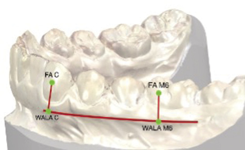
Obr. 1 / Fig. 1
Znázornění bodů FA a WALA u špičáků a prvních molárů /Representation of FA and WALA points of canines and first molars
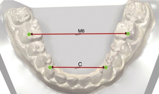
Obr. 2 / Fig. 2
Měření vzdálenosti prvních molárů a špičáků ve vrcholcích hrbolků / Intermolar and intercanine distance on cusp tips
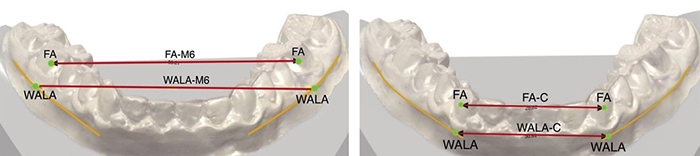
Obr. 3 / Fig. 3
>Měření vzdálenosti v bodech FA a WALA u špičáků a prvních molárů / Intermolar and intercanine distance measured at FA and WALA points
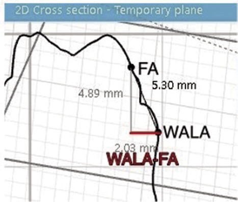
Obr. 4 / Fig. 4
Měření vzdálenosti WALA-FA u prvních molárů / Measurement of WALA-FA distance on the first molar
Tab.3 / Tab. 3
Chyba měření / Measurement error
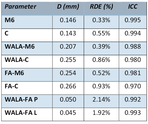
D - Dahlbergova chyba, Dahlberg formula; RDE - relativní chyba dle Dahlberga, relative Dahlberg error; ICC - koeficient vnitrotřídní korelace, intraclass correlation coefficient
Tab.4 / Tab. 4
Srovnání měřených parametrů v časech T1 a T2 u obou skupin / Comparison of measurements between times T2 and T1 in both groups
Tab. 5 / Tab. 5
Změna měřených parametrů v čase T2-T1 / Change in measured parameters in time T2-T1

Literatura / References
1. Talic NF. Adverse effects of orthodontic treatment: A clinical perspective. Saudi Dent J. 2011;23(2):55–9. doi: 10.1016/j.sdentj.2011.01.003
2. Alfuriji S, Alhazmi N, Alhamlan N, Al-Ehaideb A, Alruwaithi M, Alkatheeri N, Geevarghese A. The effect of orthodontic therapy on periodontal health: a review of the literature. Int. J. Dent. 2014;2014:585048. doi: 10.1155/2014/585048
3. Cobb NW 3rd, Kula KS, Phillips C, Proffit WR. Efficiency of multi-strand steel, superelastic Ni-Ti and ion-implanted Ni-Ti archwires for initial alignment. Clin. Orthodont. Res. 1998;1(1):12–9. doi: 10.1111/ocr.1998.1.1.12
4. Evans TJ, Jones ML, Newcombe RG. Clinical comparison and performance perspective of three aligning arch wires. Amer. J. Orthodont. dentofacial Orthop.1998;114(1):32–9. doi: 10.1016/S0889-5406(98)70234-3
5. Tidy DC. Frictional forces in fixed appliances. Amer. J. Orthodont. dentofacial Orthop.1989;96(3):249–54 doi: 10.1016/0889-5406(89)90462-9
6. Ireland AJ, Sherriff M, McDonald F. Effect of bracket and wire composition on frictional forces. Eur. J. Orthodont. 1991;13(4):322–8. doi: 10.1093/ejo/13.4.322
7. Taylor NG, Ison K. Frictional resistance between orthodontic brackets and archwires in the buccal segments. Angle Orthodont. 1996;66(3):215–22.
8. Michelberger DJ, Eadie RL, Faulkner MG, Glover KE, Prasad NG, Major PW. The friction and wear patterns of orthodontic brackets and archwires in the dry state. Amer. J. Orthodont. dentofacial Orthop. 2000;118(6):662–74. doi: 10.1067/mod.2000.105529
9. Schumacher HA, Bourauel C, Drescher D. The influence of bracket design on frictional losses in the bracket/arch wire system. J. orofacial Orthop. 1999;60(5):335–47. doi: 10.1007/BF01301246
10. Henao SP, Kusy RP. Evaluation of the frictional resistance of conventional and self-ligating bracket designs using standardized archwires and dental typodonts. Angle Orthodont. 2004;74(2):202–11
11. Khambay B, Millett D, McHugh S. Evaluation of methods of archwire ligation on frictional resistance. Eur. J. Orthodont. 2004;26(3):327–32. doi: 10.1093/ejo/26.3.327
12. Damon D. Damon System: The Workbook. Orange County, CA: Ormco; 2005.
13. Maijer R, Smith DC. Time savings with self-ligating brackets. J. clin. Orthod. 1990 Jan; Ngan P, Kess B, Wilson S. Perception of discomfort by patients undergoing orthodontic treatment. Amer. J. Orthodont. dentofacial Orthop.1989;96(1):47–53. doi: 10.1016/0889-5406(89)90228-X
14. Ngan P, Kess B, Wilson S. Perception of discomfort by patients undergoing orthodontic treatment. Amer. J. Orthodont. dentofacial Orthop. 1989;96(1):47–53. doi: 10.1016/0304-3940(89)90241-3
15. Wilson S, Ngan P, Kess B. Time course of the discomfort in young patients undergoing orthodontic treatment. Pediatr. Dent. 1989;11(2):107–10.
16. Linge L, Linge BO. Patient characteristics and treatment variables associated with apical root resorption during orthodontic treatment. Amer. J. Orthodont. dentofacial Orthop. 1991;99(1):35–43. doi: 10.1016/S0889-5406(05)81678-6
17. Remington DN, Joondeph DR, Artun J, Riedel RA, Chapko MK. Long-term evaluation of root resorption occurring during orthodontic treatment. Amer. J. Orthodont. dentofacial Orthop.1989;96(1):43–6. doi: 10.1016/0889-5406(89)90227-8
18. Kamínek M. Ortodoncie. 1. ed. Praha: Galén; 2014.
19. Proffit WR, Fields HW. Contemporary orthodontics, 3rd ed. St Louis: Mosby Yearbook; 2000.
20. Wennström JL, Lindhe J, Sinclair F, Thilander B. Some periodontal tissue reactions to orthodontic tooth movement in monkeys. J. clin. Periodont. 1987;14(3):121–9. doi: 10.1111/j.1600-051X.1987.tb00954.x
21. Ronay V, Miner RM, Will LA, Arai K. Mandibular arch form: the relationship between dental and basal anatomy. Amer. J. Orthodont. dentofacial Orthop. 2008;134(3):430–8. doi: 10.1016/j.ajodo.2006.10.040
22. Triviño T, Siqueira DF, Scanavini MA. A new concept of mandibular dental arch forms with normal occlusion. Amer. J. Orthodont. dentofacial Orthop. 2008;133(1):10.e15–22. doi: 10.1016/j.ajodo.2007.07.014
23. Esteves T, Salvatore Freitas KM, Vaz de Lima D, Cotrin P, Cançado RH, Valarelli FP, et al. Comparison of WALA ridge and dental arch dimensions changes after orthodontic treatment using a passive self-ligating system or conventional fixed appliance. Indian J. dent. Res. 2019;30(3):386–392. doi: 10.4103/ijdr.IJDR_361_18
24. Andrews LF. The Six Elements of Orofacial Harmony. Andrews J. Orthodont. Orofac. Harmony. 2000; 1:13–22.
25. Ball RL, Miner RM, Will LA, Arai K. Comparison of dental and apical base arch forms in Class II Division 1 and Class I malocclusions. Amer. J. Orthodont. dentofacial Orthop. 2010;138(1):41–50. doi: 10.1016/j.ajodo.2008.11.026
26. Conti MF, Vedovello M, Vedovello SAS, Valdrighi HC, Kuramae M. Longitudinal evaluation of dental arches individualized by the WALA ridge method. Dental Press J. Orthodont. 2011;16(2):65–74. doi: 10.1590/S2176-94512011000200009
27. Gupta D, Miner RM, Arai K, Will LA. Comparison of the mandibular dental and basal arch forms in adults and children with Class I and Class II malocclusions. Amer. J. Orthodont. dentofacial Orthop. 2010;138(1):10.e1-8; discussion 10–1. doi: 10.1016/j.ajodo.2010.01.024
28. Lundström AF. Malocclusion of the teeth regarded as a problem in connection with the apical base. Int. J. Orthod. oral Surg. Radiogr. 1925;11(12): 1109–1133. doi: 10.1016/S0099-6963(25)80052-8
29. Sampermans G. Difficult orthodontic cases. AO webseminar. s. l; American Orthodontics; Oct 30;2020.
30. Pandis N, Polychronopoulou A, Eliades T. Self-ligating vs conventional brackets in the treatment of mandibular crowding: a prospective clinical trial of treatment duration and dental effects. Amer. J. Orthodont. dentofacial Orthop. 2007;132(2):208–15. doi: 10.1016/j.ajodo.2006.01.030
31. Jiang RP, Fu MK. Non-extraction treatment with self-ligating and conventional brackets. Zhonghua Kou Qiang Yi Xue Za Zhi. 2008;43(8):459–63.
32. Fleming PS, DiBiase AT, Sarri G, Lee RT. Comparison of mandibular arch changes during alignment and leveling with 2 preadjusted edgewise appliances. Amer. J. Orthodont. dentofacial Orthop. 2009;136(3):340–7. doi: 10.1016/j.ajodo.2007.08.030
33. Yu Z, Jiaqiang L, Weiting C, Wang Y, Zhen M, Ni Z. Stability of treatment with self-ligating brackets and conventional brackets in adolescents: a long-term follow-up retrospective study. Head Face Med. 2014;20;10:41. doi: 10.1186/1746-160X-10-41
34. Almeida MR, Futagami C, Conti AC, Oltramari-Navarro PV, Navarro Rde L. Dentoalveolar mandibular changes with self-ligating versus conventional bracket systems: A CBCT and dental cast study. Dental Press J. Orthodont. 2015;20(3):50–7. doi: 10.1590/2176-9451.20.3.050-057.oar
35. Scott P, DiBiase AT, Sherriff M, Cobourne MT. Alignment efficiency of Damon3 self-ligating and conventional orthodontic bracket systems: a randomized clinical trial. Amer. J. Orthodont. dentofacial Orthop. 2008;134(4):470.e1–8. doi: 10.1016/j.ajodo.2008.04.018
36. Zdařilová H. Léčba samoligujícími zámky. Atestační práce. Stomatologická klinika, Fakultní nemocnice u sv. Anny v Brně, Lékařská fakulta Masarykovy univerzity; Brno : 2010.
37. Alexander RG. The Alexander discipline: Long-term stability. Hanover Park : Quintessence books; 2011.
38. Ab Rahman N, Wey MC, Othman SA. Mandibular arch orthodontic treatment stability using passive self-ligating and conventional systems in adults: A randomized controlled trial. Korean J. Orthodont. 2017;47(1):11–20. doi: 10.4041/kjod.2017.47.1.11
39. Atik E, Taner T. Stability comparison of two different dentoalveolar expansion treatment protocols. Dental Press J. Orthodont. 2017;22(5):75–82. doi: 10.1590/2177-6709.22.5.075-082.oar
40. Arnold S, Koletsi D, Patcas R, Eliades T. The effect of bracket ligation on the periodontal status of adolescents undergoing orthodontic treatment. A systematic review and meta-analysis. J. Dent. 2016;54:13–24. doi: 10.1016/j.jdent.2016.08.006
41. Mavreas D. Self-Ligation and the Periodontally Compromised Patient: A Different Perspective. Semin. Orthodont. 2008; 14(1):36–45. doi: 10.1053/j.sodo.2007.12.004
42. Conti MD, Filho MV, Amélia S, Vedovello SAS, Valdrighi HC, Kuramae M. Longitudinal evaluation of dental arches individualized by the WALA ridge method. Dental Press J. Orthodont. 2011; 16(2):65–74. doi: 10.1590/S2176-94512011000200009
43. Andrews LF, Andrews WA. Syllabus of Andrews philosophy and techniques. San Diego: Lawrence F. Andrews Foundation, 1999.
44. Triviño T, Siqueira DF, Andrews WA. Evaluation of distances between the mandibular teeth and the alveolar process in Brazilians with normal occlusion. Amer. J. Orthodont. dentofacial Orthop. 2010;137(3):308.e1-4; discussion 308–9. doi: 10.1016/j.ajodo.2009.09.017
45. DiBiase AT, Nasr IH, Scott P, Cobourne MT. Duration of treatment and occlusal outcome using Damon3 self-ligated and conventional orthodontic bracket systems in extraction patients: a prospective randomized clinical trial. Amer. J. Orthodont. dentofacial Orthop. 2011;139(2):e111–6. doi: 10.1016/j.ajodo.2010.07.020
46. Torres CB, Cabrilla MCP, Quintanilla DS. Comparative assessment of the effectiveness of dental alignment between low friction conventional ligated and self-closing brackets on the maxillary arch in 18 patients. Proceedings of the European Orthodontic Society; Amsterdam. London: European Orthodontic Society. 2005:294.
47. Hamilton R, Goonewardene MS, Murray K. Comparison of active self-ligating brackets and conventional pre-adjusted brackets. Aust. Orthodont. J. 2008;24(2):102–9. doi: 10.2478/aoj-2008-0015
48. Harradine NW. Self-ligating brackets and treatment efficiency. Clin. Orthodont. Res. 2001;4(4):220–7. doi: 10.1034/j.1600-0544.2001.40406.x
49. Eberting JJ, Straja SR, Tuncay OC. Treatment time, outcome, and patient satisfaction comparisons of Damon and conventional brackets. Clin. Orthodont. Res. 2001;4(4):228–34. doi: 10.1034/j.1600-0544.2001.40407.x
50. Songra G, Clover M, Atack NE, Ewings P, Sherriff M, Sandy JR, Ireland AJ. Comparative assessment of alignment efficiency and space closure of active and passive self-ligating vs conventional appliances in adolescents: a single-center randomized controlled trial. Amer. J. Orthodont. dentofacial Orthop. 2014;145(5):569–78. doi: 10.1016/j.ajodo.2013.12.024
51. Bishara SE, Jakobsen JR, Treder J, Nowak A. Arch width changes from 6 weeks to 45 years of age. Amer. J. Orthodont. dentofacial Orthop. 1997;111(4):401–9. doi: 10.1016/S0889-5406(97)80022-4
52. Mavreas D, Athanasiou AE. Factors affecting the duration of orthodontic treatment: a systematic review. Eur. J. Orthodont. 2008;30(4):386–95. doi: 10.1093/ejo/cjn018
53. Quimby ML, Vig KW, Rashid RG, Firestone AR. The accuracy and reliability of measurements made on computer-based digital models. Angle Orthodont. 2004;74(3):298–303.
54. Zilberman O, Huggare JA, Parikakis KA. Evaluation of the validity of tooth size and arch width measurements using conventional and three-dimensional virtual orthodontic models. Angle Orthodont. 2003;73(3):301–6.
55. Costalos PA, Sarraf K, Cangialosi TJ, Efstratiadis S. Evaluation of the accuracy of digital model analysis for the American Board of Orthodontics objective grading system for dental casts. Amer. J. Orthodont. dentofacial Orthop. 2005;128(5):624–9. doi: 10.1016/j.ajodo.2004.08.017
56. Suk KE, Park JH, Bayome M, Nam YO, Sameshima GT, Kook YA. Comparison between dental and basal arch forms in normal occlusion and Class III malocclusions utilizing cone-beam computed tomography. Korean J. Orthod. 2013;43(1):15–22. doi: 10.4041/kjod.2013.43.1.15
57. Kapila S, Conley RS, Harrell WE Jr. The current status of cone beam computed tomography imaging in orthodontics. Dentomaxillofac. Radiol. 2011;40(1):24–34. doi: 10.1259/dmfr/12615645
58. Moura Neto G. A comparative assessment of the WALA ridge in dissected mandibles and cast models as well as its measurement in occlusal radiographies and tomographies. Dizertační práce. Universidad de Sao Paulo; Sao Paulo, Brazil: 2010.
MDDr. Aneta Hrubá
Stomatologická klinika, 1. LF UK a VFN
Kateřinská 32, 128 01, Praha 2