Replantace retinovaného ankylotického horního středního řezáku s dokončeným vývojem. Kazuistika.
Replantation of impacted ankylosed upper central incisor with completed development. Case report.



MDDr. Kristina Vaidišová, MUDr. et MDDr. Jiří Genčur, MUDr. Hana Böhmová
Ortodontické oddělení Stomatologické kliniky LF a FN Plzeň / Orthodontic Department, Clinic of Dentistry, Medical Faculty and University Hospital Plzeň
DOI10.61110/50599
Souhrn
Úvod: Ankylóza je definována jako spojení mineralizovaného povrchu zubu (cementu nebo dentinu) s alveolární kostí. Aktivní řazení ankylotických zubů nebývá úspěšné. Cílem sdělení je prezentovat případ zařazení retinovaného ankylotického řezáku chirurgicky pomocí replantace včetně přilehlé kostní lamely.
Materiál a metodika: Pacientka (13 let 7 měsíců) byla delegována na ortodontické oddělení Stomatologické kliniky FN Plzeň z důvodu retence zubu 21. V 9 letech byly u pacientky extrahovány dva nadpočetné zuby v oblasti premaxily. Na našem oddělení proběhla ortodontická terapie s cílem aktivního zařazení retinovaného zubu 21 a následně replantací ankylotického zubu 21 včetně části vestibulární kompakty do správné pozice.
Závěr: Replantací retinovaného ankylotického zubu 21 do korektní pozice se podařilo u rostoucí pacientky dosáhnout uspokojivé estetické rehabilitace. Zároveň se podařilo zmírnit defekt alveolární kosti, který by ztěžoval případné řešení v případě, že bychom se rozhodli retinovaný ankylotický zub extrahovat.
(Ortodoncie 2026, 35, č. 1, s. 42–55)
Abstract
Introduction: Ankylosis is defined as the fusion of the mineralized tooth surface (cementum or dentin) with the alveolar bone. Active orthodontic alignment of ankylosed teeth is typically unsuccessful. The aim of this report is to present a case of surgical replantation of an unerupted ankylosed incisor, together with the adjacent bone lamella.
Material and Methods: A female patient (aged 13 years and 7 months) was referred to the Orthodontic Department of the Dental Clinic at the University Hospital Plzeň due to the impaction of tooth 21. At the age of 9, two supernumerary teeth in the premaxillary region had been extracted. Orthodontic therapy was performed at our department with the goal of active alignment of the unerupted tooth 21, followed by the surgical replantation of the ankylosed tooth 21, including a portion of the vestibular cortical bone, into the correct position.
Conclusion: By replanting the impacted ankylosed tooth 21 into the correct position, satisfactory aesthetic rehabilitation was achieved in a growing patient. Simultaneously, the alveolar bone defect was mitigated, which would have complicated potential treatment had the decision been made to extract the impacted ankylosed tooth.
(Ortodoncie 2026, 35, No. 1, p. 42–55)

Klíčová slova: ankylóza, replantace, retinovaný řezák, endodontické ošetření.
Key words: ankylosis, replantation, impacted incisor, endodontic management.
Úvod
Ankylóza je definována jako spojení mineralizovaného povrchu zubu (cementu nebo dentinu) s alveolární kostí. V případě horních řezáků dochází ke vzniku ankylózy nejčastěji v důsledku poškození integrity periodontálních vláken následkem úrazu. Nejrizikovější mechanismy úrazu pro vznik ankylózy jsou intruzivní luxace a avulze s následnou replantací [1]. Další faktory, které mohou vést ke vzniku ankylózy, jsou endokrinní či metabolická onemocnění, periapikální infekce či předchozí chirurgický zákrok v dané oblasti [2, 3].
Předpokládá se, že vzniku ankylózy předchází zánět v oblasti periodoncia, kdy osteoklasty resorbují i část povrchu kořene. Následné reparativní pochody mohou vést ke spojení alveolární kosti s povrchem kořene [4].
Pokud dojde ke vzniku ankylózy v raném věku, bývá řešení náročnější, neboť s vertikálním růstem alveolárních výběžků dochází k reinkluzi, která se klinicky projeví infraokluzí ankylotického zubu [5].
Při diagnostice ankylózy vycházíme z anamnézy a klinického vyšetření. Anamnesticky bývá pozitivní úrazová anamnéza, klinicky u ankylotických zubů bývá při poklepu přítomný „zvonivý, kovový“ zvuk. Studie ovšem udávají, že aby perkusní test byl klinicky pozitivní, musí být ankylózou postiženo alespoň 20 % povrchu kořene [2, 5]. Pokud dojde ke vzniku ankylózy u rostoucího jedince, začne se zub vlivem růstu dostávat vůči sousedním zubům do infraokluze, která se s dalším růstem zvýrazňuje. Rentgenologicky na intraorálním rentgenovém snímku zpravidla nebývá ankylóza průkazná. Někdy můžeme detekovat narušení průběhu periodontální štěrbiny [6]. Pokud je ovšem okrsek ankylózy malý nebo lokalizovaný na bukální nebo palatinální straně zubu, nebude narušení periodontální štěrbiny na 2D snímcích patrné. Ani na CBCT snímcích nemusí být možné spolehlivě detekovat vymizení periodontální štěrbiny vzhledem k její malé šířce okolo 0,2 mm. Můžeme tak většinou pouze vyslovit podezření na ankylózu.
Další metodou v diagnostice ankylózy může být digitální zvuková analýza, přičemž ankylotické zuby vykazují větší podíl signální energie ve vysokofrekvenčních pásmech. Periotestem (Siemens/Medizintechnik-Gulden, Bensheim, Germany) může být naopak ověřena mobilita zubu, ankylotické zuby vykazují nižší hodnoty než okolní zdravé zuby [7].
Pokud jde o retinovaný zub, podezření na ankylózu vyslovujeme, pokud sledujeme zub v čase, ten neprořezává, neposouvá se k vrcholu alveolu i když jeho kořen se dále vyvíjí a nejsou patrné jiné příčiny retence jako překážka, nedostatek místa či špatný erupční směr.
V případě ortodontické terapie na ankylózu pomýšlíme, pokud daný zub nereaguje na aktivní tah, a naopak pozorujeme nežádoucí pohyb okolních zubů směrem k ankylotickému zubu, deformaci okluzální roviny a otevírání skusu
Kazuistika:
Dívka (13 let 7 měsíců) byla v červnu 2022 delegována na ortodontické oddělení Stomatologické kliniky FN Plzeň z důvodu retence zubu 21 v nepříznivé poloze. Dle lékařské zprávy byly u dívky v roce 2018 extrahovány dva nadpočetné zuby v dané oblasti. Od té doby byl pouze observován stav. Byla zhotovena kompletní ortodontická dokumentace včetně CBCT snímku.
Osobní anamnéza neodhalila žádná celková onemocnění či alergie, pacientka byla bez trvalé medikace. Úraz či zánětlivé onemocnění ve frontálním úseku horní čelisti v dočasném chrupu rodiče neudávali.
Extraorálně byl obličej symetrický, s přímým až lehce konvexním profilem a nízkou linií úsměvu.
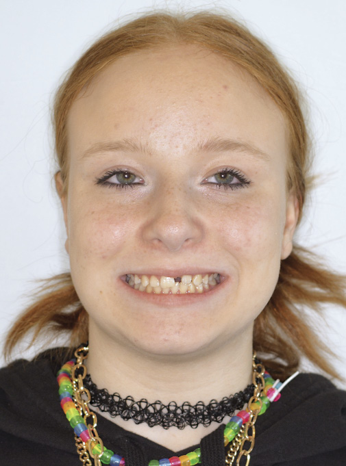
Pacientka měla založený kompletní stálý chrup v rozsahu 17-27 a 47-37, přičemž zub 21 byl retinovaný v nepříznivé poloze (obr. 1, 2). Chrup byl intaktní, hygiena dobrá.
Intraorálně byla v molárech artikulaceAngle I, ve špičácích byla Angle II o 3 mm. Incizální schůdek byl 2,5 mm, hloubka skusu 40 %. Byl patrný posun horního středu doleva, zuby 11 a 22 byly meziálně skloněny do mezery, přičemž zub 22 byl v infraokluzi vůči zubu 12. Zub 11 měl meziodistálně 8 mm, zatímco mezera pro zub 21 byla 3,5 mm. V horním zubním oblouku byla tremata 14-13, 12-11 a 22-23 o celkové velikosti 2,5 mm.
Dle CBCT byly založeny všechny stálé zuby, včetně třetích molárů. Retinovaný zub 21 měl dokončený vývoj kořene, byl ve vertikální poloze, distorotován o 90°, incizální hrana směřovala do poloviny délky kořene zubu 11, se kterým byla téměř v kontaktu, toho času bez známek resorpce na kořeni zubu 11. Apex zubu 21 dosahoval nad apex zubu 22 (obr. 3).
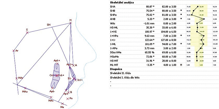
Analýza profilového snímku ukázala hraniční II. skeletální třídu – ANB 5,3°, WITS 1 mm. Úhel mandibulární linie byl 35°, tedy průměrný. Sklon horního řezáku k linii SN byl 101°, tedy v dobrém osovém postavení, dolní řezáky byly vykloněny k mandibulární linii rovněž 101°, tedy v mírné protruzi (obr. 4a, 4b).
Léčebný plán I
Vzhledem k tomu že překážky v erupci, dva přespočetné zuby, byly odstraněny už v 9 letech a s ohledem na polohu zubu 21 ve 13 letech bylo vysloveno podezření na možnou ankylózu. Pacientce byly navrženy dvě varianty léčby. Varianta A se snahou o aktivní řazení retinovaného zubu 21 k fixnímu ortodontickému aparátu a varianta B s extrakcí retinovaného zubu 21, úpravou postavení okolních zubů a náhradou chybějícího zubu 21 adhezivním můstkem jako dlouhodobě provizorní řešení. Při tomto řešení by ale velmi pravděpodobně došlo k prohloubení kostního defektu, který by bylo nutné a náročné rekonstruovat.
Součástí obou variant léčby byl i návrh terapie v dolním zubním oblouku.
Pacientka i její rodiče byli motivováni a souhlasili s variantou řazení retinovaného zubu.
Byl stanoven následující léčebný plán:
1. Nivelizace v horním zubním oblouku fixním ortodontickým aparátem, otevření prostoru pro řazení zubu 21 využitím drobných tremat, napřímením zubů 11, 22 a mírnou protruzí horních řezáků, korekce postavení střední čáry.
2. Chirurgická expozice korunky zubu 21, nalepení attachmentu na vestibulární plochu a aktivní stahování k ortodontickému aparátu uzavřenou metodou. Po erupci 21 jeho derotace a zařazení do oblouku.
3. Dle průběhu léčby v horním zubním oblouku nasazení fixního aparátu v dolní čelisti, nivelizace zubního oblouku, úprava hloubky skusu a vzájemné artikulace obou zubních oblouků.
Průběh terapie
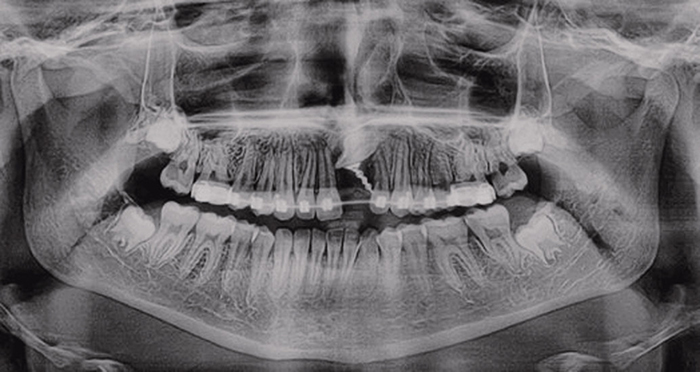
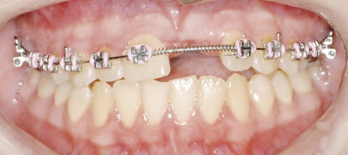
V září 2022 byl nasazen fixní ortodontický aparát v rozsahu 16-26 s použitím zámků Discovery Smart MBT a šířkou drážky 0.022“ (Dentaurum, Ispringen, Německo). Místo pro zub 21 bylo získáno využitím tremat v horním zubním oblouku, úpravou sklonu horních řezáků a protruzí. Po získání dostatečného místa následovala chirurgická expozice s lepením attachmentu k aktivnímu tahu na vestibulární plochu zubu 21 poblíž incizální hrany. Peroperační zkouška poklepem na zub 21 byla neprůkazná. Během zákroku byla rovněž snesena kost ve směru plánovaného pohybu zubu a ortodontický attachment byl vyveden skrze mukoperiostální lalok ve směru plánovaného tahu. Elastickým vláknem byl veden tah distokaudálně k oblouku blízko zámku zubu 22. Výměna elastického vlákna probíhala každé tři až čtyři týdny. Kontrolní OPG zhotovený po třech měsících aktivního tahu (obr. 5), ovšem neukázalo žádnou změnu polohy retinovaného zubu. Bylo rozhodnuto změnit směr aktivního tahu z distokaudálního na více kaudální směr z obavy kolize v oblasti apexů kořenů zubů 21 a 22. Bohužel ani za další tři měsíce aktivního tahu nebyla klinicky patrná žádná změna, naopak bylo pozorováno sklánění okluzální roviny s tendencí k otevřenému skusu (obr. 6a a 6 b).
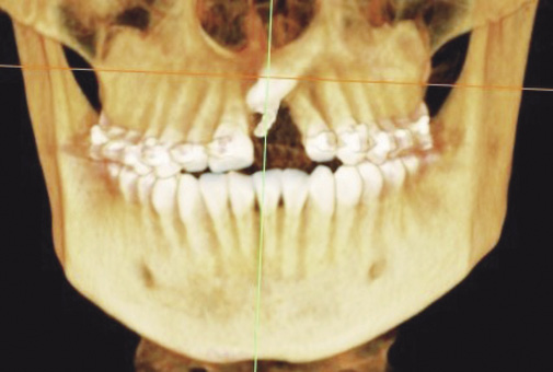
Bylo zhotoveno CBCT, které potvrdilo, že nedochází k pohybu retinovaného zubu a tendence sklánět okluzální rovinu nás utvrdila v našem podezření na ankylózu zubu 21. Byl aktualizován léčebný plán.
Léčebný plán II
Byly zvažovány následující možnosti změny léčebného plánu:
1. Extrakce a náhrada zubu implantátem v dospělosti, přičemž by stav vyžadoval rozsáhlou augmentaci tvrdých i měkkých tkání.
2. Extrakce a mezializace zubů II. kvadrantu - náročná a ne zcela vhodná varianta vzhledem k artikulaci v I. třídě Angle a rozsáhlému defektu vestibulární kompakty v oblasti zubu 21. Tato varianta by rovněž vyžadovala úpravu tvaru zubů 22, 23 a 24.
3. Subapikální osteotomie následovaná distrakční osteogenezí – tato varianta nebyla možná z důvodu šikmé polohy zubu.
4. Ostektomie bločku kosti se zubem 21 a přesunutí do požadované pozice. Fixace ve vyhovující pozici a po zhojení ortodontická úprava postavení okolních zubů.
Po konzultaci s pacientkou a rodiči bylo rozhodnuto pro ostektomii bločku kosti se zubem 21 a jeho přesunutí do požadované pozice. Provedení zákroku bylo naplánováno v celkové anestezii.
Průběh terapie
V lednu 2024 proběhla v celkové anestezii modifikovaná replantace zubu 21 s kostním bločkem (obr. 7 až 10). Vzhledem ke komplexnosti potřebné preparace se však podařilo z původně plánovaného bločku ponechat pouze ventrální část na vestibulární ploše kořene zubu 21 (obr. 8). Po dobu přípravy lůžka byl zub 21 uchován ve fyziologickém roztoku. Po vložení do lůžka byl zub 21 fixován k okolním zubům flow kompozitem, zároveň byl na zub 21 pasivně nalepen ortodontický zámek a zub byl fixován retenční ligaturou k ostatním zubům a drátěnou ligaturou k ocelovému oblouku (obr. 9 a 10). Pacientce byla doporučena kašovitá strava a šetřící režim.
3 týdny po chirurgickém zákroku proběhlo endodontické ošetření s plněním MTA, neboť zub měl již dokončený vývoj kořene (obr. 12).
6 týdnů po zákroku byla odstraněna fixace flow kompozitem, zub byl pevný, sondáž do 3 mm, pouze bodově meziovestibulárně byla sondáž 3,5 mm. Pacientka byla bez obtíží.
Pacientka odmítla léčbu v dolním zubním oblouku, přestože by pomohla vylepšit artikulaci, požadovala pouze doléčení v horním zubním oblouku. Osm měsíců po zákroku proběhlo sejmutí fixního ortodontického aparátu a recontouring dostavbou incizálních hran 11-22 pomocí fotokompozita z volné ruky (obr. 13 a 14). Jako retence byl zhotoven horní fixní retainer v rozsahu 12-22 a horní foliový aparát. Na intraorálním rentgenovém snímku zhotoveném 1 rok po replantaci (obr. 15) lze pozorovat obnovení kostní struktury v apikální oblasti a mezi zuby 21 a 22, meziálně u zubu 21 přetrvává defekt, nicméně zub je klinicky klidný, pevný a pacientka je bez obtíží. Došlo také k eliminaci černého trojúhelníku mezi zuby 11-21. Nadále jsou v plánu pravidelné kontroly v rámci retenčního protokolu. Lze očekávat, že se replantovaný zub vlivem zbytkového růstu začne dostávat do infraokluze a zároveň je důležitá monitorace z důvodu zvýšeného rizika vzniku resorptivních procesů.
Diskuse
Možnosti řešení ankylotických horních řezáků:
Stanovení léčebného plánu by mělo vycházet z celkového stavu pacienta, jeho motivace, úrovně postižení, stavu ankylotického zubu a případně přidružené ortodontické vady.
Obecnými možnostmi jsou:
1. Extrakce ankylotického zubu a protetická náhrada. Extrakce ankylotického zubu je spojena s rozsáhlou ztrátou alveolární kosti, zejména tenké bukální kompakty a výsledný defekt může kompromitovat estetiku protetické rekonstrukce [4].
2. Extrakce ankylotického zubu a uzávěr mezery ortodontickým posunem [8].
3. Chirurgická subluxace ankylotického zubu a ortodontická repozice. Tato metoda je vhodná pokud ankylóza postihuje pouze malý okrsek povrchu kořene a není výrazný vertikální defekt tvrdých a měkkých tkání. Zároveň je zde riziko vzniku nové ankylózy a zastavení pohybu zubu během léčby [4].
4. Chirurgická subapikální osteotomie následovaná distrakční osteogenezí – tato metoda manipulace novotvořenou kostí může pomoci obnovit vertikální rozměr alveolárního výběžku i měkkých tkání. K distrakci můžeme použít intraorální distraktor (vhodný u velmi těžkých postižení) nebo ortodontický aparát (vhodný spíše u menších či středně těžkých postižení) [4,9–11]. Distrakční osteogeneze byla prvně popsána v ortopedii Ilizarovem v padesátých letech 20. století a dnes je v ortodoncii využívána hlavně při řešení výrazných skeletálních malformit, nejčastěji u syndromových pacientů.
Proces distrakční osteogeneze lze rozdělit na 3 po sobě následující období: období latence, distrakce a konsolidace. Obdobím latence nazýváme čas mezi chirurgickým zákrokem subapikální osteotomie a začátkem distrakce, je časem formace svalku. Zpravidla bývá kolem 1 týdne. Období distrakce je pak obdobím pohybu segmentu, protokoly ohledně rychlosti distrakce se však značně liší. Období konsolidace je pak obdobím remodelace a zrání vytvořené kosti poté, co byla skončena distrakce [12].
Nevýhodou užití distraktoru je, že vektor distrakce nelze stanovit přesně ve všech třech rozměrech. Tento nedostatek pak bývá korigován pomocí „floating bone“ konceptu, kdy se postavení upravuje při vyjmutí distraktoru manuláně tlakem prstů na měkkou nově vytvořenou tkáň [13], pomocí ortodontického aparátu nebo kombinací obou metod.
5. Dekoronace – metoda dekoronace ankylotického zubu bývá doporučena u zubů s pokračující náhradovou resorpcí, kdy prognóza zubu není příznivá. Cílem dekoronace je zachování objemu alveolární kosti pro další protetickou náhradu zubu.
Dekoronace se provádí pod úrovní cementosklovinné hranice, 1 mm pod úrovní alveolární kosti, přičemž všechny kořenové výplně musí být ze zubu odstraněny. Na závěr je indukováno krvácení z periapikální oblasti za účelem podpoření formace koagula uvnitř kořenového kanálku. Následně je mobilizovaný mukoperiostální lalok hermeticky přišit zpět [14,15].
6. Autotransplantace – nejvhodnějšími zuby pro náhradu horních řezáků pomocí autotransplantace bývají premoláry ve vývojovém stádiu 3-5 podle Moorreese [1,16]. Úspěšnost této metody je v tomto období velmi vysoká a lze jí dosáhnout estetického výsledku s dobrou dlouhodobou prognózou [17].
Případ prezentované pacientky byl poměrně komplikovaný a žádná z výše zmíněných možností terapie se nezdála být vhodná. Extrakci a protetické náhradě jsme se chtěli z důvodu věku pacientky vyhnout, zejména proto, že již při prvotním vyšetření byl patrný defekt alveolární kosti v oblasti retinovaného zubu a bylo pravděpodobné, že extrakcí by se ještě zvětšil. Extrakce a uzávěr poextrakční mezery mezializací zubů druhého kvadrantu by byla náročná a vyžadovala by recontouring zubů 22, 23 a 24. Rovněž artikulace v I.tř. Angle v moláru a defekt alveolární kosti v místě zubu 21 hovořily proti této variantě. Chirurgická subluxace s ortodontickou repozicí se z důvodu polohy zubu a možných komplikací jevila jako příliš riziková s malou pravděpodobností úspěchu. Chirurgická subapikální osteotomie následovaná distrakční osteogenezí také nebyla možná – šikmá poloha zubu neumožňovala stanovení přijatelného vektoru distrakce. Ani dekoronace by v tomto případě nepřinesla žádný benefit, neboť kořen zubu 21 se nacházel vysoko nad apexem zubu 22 a nepřispěl by tak k žádnému významnému udržení objemu alveolární kosti do období, kdy by přicházela v úvahu implantace. Autotransplantaci jsme u této pacientky rovněž zamítli – její premoláry již v době iniciálního vyšetření měly vývojové stádium dle Moorreese 5 až 6 a autotransplantace premoláru s již dokončeným vývojem kořene do oblasti tak velkého defektu se nám jevila jako příliš riziková. Proto bylo u pacientky přistoupeno k poměrně netypickému řešení replantace ankylotického zubu včetně lamely přilehlé alveolární kosti.
Přestože výsledek má několik estetických nedostatků – došlo k diskoloraci autotransplantovaného zubu v krčkové oblasti (pravděpodobně vlivem plnění MTA) a je nedokonalá růžovobílá estetika horního frontálního úseku, lze tento případ považovat, co se vytvořené kosti týká, za poměrně úspěšný.
U rostoucí pacientky se podařilo i přes nepříznivou prognózu zachovat vlastní zub, díky nízké linii úsměvu nejsou estetické nedostatky na první pohled patrné a pacientka je s výsledkem spokojena.
Závěr
Replantací ankylotického zubu včetně přilehlé části alveolární kosti se podařilo dosáhnout uspokojivé estetické rehabilitace u rostoucí pacientky bez nutnosti protetického řešení. Na místě je ale i nadále opatrnost a dlouhodobé sledování – lze očekávat, že se postupem času replantovaný zub začne dostávat do infraokluze a důležitá je i monitorace z důvodu zvýšeného rizika vzniku resorptivních procesů. O těchto skutečnostech jsou pacientka i rodiče poučeni. Avšak i v případě, že dojde k selhání, bude výchozí stav pro náhradu zubu příznivější než pokud by byl zub extrahován na začátku léčby, jelikož se podařilo alespoň částečně obnovit rozměr alveolární kosti v dané oblasti a nebyla by pravděpodobně nutná tak rozsáhlá augmentace.
Autoři nemají komerční, vlastnické nebo finanční zájmy na produktech nebo společnostech popsaných v tomto článku.
Introduction
Ankylosis is defined as the fusion of the mineralized tooth surface (cementum or dentin) with the alveolar bone. In the case of upper incisors, ankylosis most commonly occurs due to damage to the integrity of the periodontal ligament following a traumatic injury. The highest-risk injury mechanisms for the development of ankylosis are intrusive luxation and avulsion followed by replantation [1]. Other factors that may lead to the development of ankylosis include endocrine or metabolic diseases, periapical infection, or previous surgical intervention in the affected area [2, 3].
It is assumed that the development of ankylosis is preceded by inflammation in the periodontal region, during which osteoclasts resorb a portion of the root surface. Subsequent reparative processes can lead to the fusion of the alveolar bone with the root surface [4].
If ankylosis occurs at an early age, management is typically more challenging because the vertical growth of the alveolar processes leads to secondary impaction (re-inclusion), which manifests clinically as infraocclusion of the ankylosed tooth [5].
The diagnosis of ankylosis is based on medical history and clinical examination. There is typically a positive history of trauma; clinically, ankylosed teeth often exhibit a ‚dull, metallic‘ sound upon percussion. However, studies indicate that at least 20% of the root surface must be affected by ankylosis for the percussion test to be clinically positive [2, 5]. If ankylosis occurs in a growing individual, the tooth begins to move into infraocclusion relative to adjacent teeth due to growth, which becomes more pronounced as growth continues.
Radiographically, ankylosis is generally not conclusive on intraoral X-rays. Occasionally, a disruption in the continuity of the periodontal ligament space can be detected [6]. However, if the area of ankylosis is small or located on the buccal or palatal side of the tooth, the disruption of the periodontal ligament space will not be visible on 2D images. Even on CBCT scans, it may not be possible to reliably detect the disappearance of the periodontal ligament space due to its narrow width of approximately 0.2 mm. Therefore, we can usually only express a suspicion of ankylosis.
Another method for diagnosing ankylosis is digital sound analysis, where ankylosed teeth show a higher proportion of signal energy in high-frequency bands. Conversely, tooth mobility can be verified using the Periotest (Siemens/Medizintechnik-Gulden, Bensheim, Germany); ankylosed teeth exhibit lower values than surrounding healthy teeth [7].
In the case of an impacted tooth, ankylosis is suspected if the tooth is monitored over time and fails to erupt or move toward the alveolar crest, even though its root continues to develop and no other causes of impaction-such as an obstruction, lack of space, or incorrect eruption path-are apparent.
In orthodontic therapy, ankylosis is considered if the tooth fails to respond to active traction, and instead, undesirable movement of adjacent teeth toward the ankylosed tooth, deformation of the occlusal plane, and development of an open bite are observed.
Case Report:
A female patient (aged 13 years and 7 months) was referred in June 2022 to the Orthodontic Department of the Dental Clinic at the University Hospital Plzeň due to the impaction of tooth 21 in an unfavorable position. According to the medical report, two supernumerary teeth had been extracted in the affected area in 2018. Since then, the condition had only been observed. Complete orthodontic records, including a CBCT scan, were obtained.
The medical history revealed no systemic diseases or allergies; the patient was not on any permanent medication. The parents reported no history of trauma or inflammatory disease in the maxillary anterior segment of the primary dentition.
Extraorally, the face was symmetrical, with a straight to slightly convex profile and a low smile line.
The patient presented with a complete permanent dentition ranging from 17-27 and 47-37, with tooth 21 impacted in an unfavorable position (Fig. 1, 2). The dentition was intact, and oral hygiene was good.
Intraorally, the molar relationship was Angle Class I, while the canine relationship was Angle Class II with a 3 mm discrepancy. The overjet was 2.5 mm, and the overbite was 40%. A deviation of the maxillary midline to the left was evident; teeth 11 and 22 were mesially inclined into the space, with tooth 22 in infraocclusion relative to tooth 12. Tooth 11 had a mesiodistal width of 8 mm, while the space for tooth 21 was 3.5 mm. Spacing of 2.5 mm in total was present in the maxillary arch between teeth 14-13, 12-11, and 22-23.
According to the CBCT, all permanent teeth, including third molars, were present. The impacted tooth 21 had completed root development and was in a vertical position, distorotated by 90°. The incisal edge was directed toward the mid-root level of tooth 11, with which it was almost in contact; at that time, no signs of resorption on the root of tooth 11 were present. The apex of tooth 21 reached above the apex of tooth 22 (Fig. 3).
Cephalometric analysis showed a borderline skeletal Class II relationship (ANB 5.3°, WITS 1 mm). The mandibular plane angle was 35°, which is average. The inclination of the maxillary incisor to the SN line was 101°, indicating a good axial position, while the mandibular incisors were proclined at 101° to the mandibular plane, representing slight protrusion (Figs. 4a, 4b).
Treatment Plan I
Given that the obstructions to eruption—two supernumerary teeth—had already been removed at the age of 9, and considering the position of tooth 21 at age 13, a suspicion of potential ankylosis was raised. Two treatment variants were proposed to the patient. Variant A involved an attempt at active orthodontic alignment of the impacted tooth 21 using a fixed appliance. Variant B involved the extraction of the impacted tooth 21, adjustment of the positions of adjacent teeth, and replacement of the missing tooth 21 with an adhesive bridge as a long-term provisional solution. However, this approach would likely have led to an exacerbation of the bone defect, which would then require difficult and complex reconstruction.
Both treatment variants included a proposal for therapy in the mandibular arch.
The patient and her parents were highly motivated and consented to the variant involving the alignment of the impacted tooth.
The following treatment plan was established:
1. Leveling and alignment in the maxillary arch using a fixed orthodontic appliance, opening space for tooth 21 by utilizing existing small tremata, uprighting teeth 11 and 22, and achieving slight protrusion of the maxillary incisors, along with correction of the midline deviation.
2. Surgical exposure of the crown of tooth 21, bonding of an attachment to the vestibular surface, and active traction toward the orthodontic appliance using a closed eruption technique. Following the eruption of tooth 21, derotation and alignment into the dental arch.
3. Depending on the progress of therapy in the maxillary arch, placement of a fixed appliance in the mandible, leveling and alignment of the dental arch, and adjustment of the overbite and interarch relationship.
Course of Therapy:
In September 2022, a fixed orthodontic appliance was bonded on teeth 16-26 using Discovery Smart MBT brackets with a 0.022“ slot (Dentaurum, Ispringen, Germany). Space for tooth 21 was obtained by utilizing tremata in the maxillary arch and by adjusting the inclination and protrusion of the maxillary incisors. Once sufficient space was gained, surgical exposure was performed, and an attachment for active traction was bonded to the vestibular surface of tooth 21 near the incisal edge. An intraoperative percussion test of tooth 21 was inconclusive. During the procedure, bone was removed in the direction of the planned tooth movement, and the orthodontic attachment was led through the mucoperiosteal flap in the direction of the planned traction. Active traction was applied distocaudally toward the archwire near the bracket of tooth 22 using an elastic thread. The elastic thread was replaced every three to four weeks.
A follow-up panoramic radiograph taken after three months of active traction (Fig. 5) showed no change in the position of the impacted tooth. It was decided to change the direction of active traction from distocaudal to a more caudal direction due to concerns regarding a potential collision between the root apices of teeth 21 and 22. Unfortunately, after an additional three months of active traction, no clinical change was apparent; on the contrary, a tilting of the occlusal plane with a tendency toward an open bite was observed (Fig. 6a, 6b).
A CBCT scan was performed, which confirmed that there was no movement of the impacted tooth. The tendency of the occlusal plane to tilt further confirmed our suspicion of ankylosis of tooth 21. The treatment plan was subsequently updated.
Treatment Plan II
The following options for modification of the treatment plan were considered:
1. Extraction and replacement of the tooth with an implant in adulthood, which would require extensive augmentation of both hard and soft tissues.
2. Extraction and mesialization of the teeth in the second quadrant – a challenging and not entirely suitable option given the Angle Class I relationship and the extensive defect of the vestibular cortical bone in the area of tooth 21. This variant would also require reshaping of teeth 22, 23, and 24.
3. Subapical osteotomy followed by distraction osteogenesis – this option was not feasible due to the oblique position of the tooth.
4. Block osteotomy of the bone including tooth 21 and repositioning into the desired location. Fixation in the appropriate position followed by orthodontic adjustment of the adjacent teeth after healing.
After consultation with the patient and her parents, it was decided to proceed with the block osteotomy of the bone with tooth 21 and its relocation to the desired position. The procedure was scheduled under general anesthesia.
Course of Therapy
In January 2024, a modified replantation of tooth 21 with a bone block was performed under general anesthesia (Fig. 7–10). However, due to the complexity of the required preparation, it was only possible to retain the ventral portion of the originally planned block on the vestibular surface of the root of tooth 21 (Fig. 8). While the recipient site was being prepared, tooth 21 was stored in saline solution. After insertion into the socket, tooth 21 was fixed to the adjacent teeth with flowable composite. Simultaneously, an orthodontic bracket was passively bonded to tooth 21, and the tooth was secured to the other teeth with a retention ligature and to the steel archwire with a wire ligature (Figs. 9 and 10). The patient was advised to follow a soft diet and a restricted physical activity regimen.
Three weeks after the surgical procedure, endodontic treatment with MTA (Mineral Trioxide Aggregate) filling was performed, as root development had already been completed (Fig. 12).
Six weeks postoperatively, the flowable composite fixation was removed. The tooth was stable, and periodontal probing depths were within 3 mm, except for a single point mesiovestibularly where the depth was 3.5 mm. The patient remained asymptomatic.
The patient declined treatment in the mandibular arch, even though it would have improved the interarch relationship, and requested only the completion of treatment in the maxillary arch. Eight months after the procedure, the fixed orthodontic appliance was debonded, and recontouring was performed by free-hand composite resin buildup of the incisal edges of teeth 11–22 (Figs. 13 and 14). A maxillary fixed retainer (12–22) and a vacuum-formed retainer were provided for retention.
On the intraoral radiograph taken one year after replantation (Fig. 15), restoration of the bone structure in the apical region and between teeth 21 and 22 can be observed. A defect persists mesially to tooth 21; however, the tooth is clinically stable, and the patient is asymptomatic. The black triangle between teeth 11 and 21 was also eliminated. Regular follow-ups according to the retention protocol are scheduled. It is expected that the replanted tooth may move into infraocclusion due to residual growth; furthermore, monitoring is essential due to the increased risk of resorptive processes.
Discussion
Management Options for Ankylosed Maxillary Incisors:
The determination of a treatment plan should be based on the patient’s overall status, motivation, the extent of the impairment, the condition of the ankylosed tooth, and any associated orthodontic malocclusion.
General options include:
1. Extraction of the ankylosed tooth and prosthetic replacement. Extraction of an ankylosed tooth is associated with extensive loss of alveolar bone, particularly the thin buccal cortical plate, and the resulting defect can compromise the aesthetic outcome of the prosthetic reconstruction [4].
2. Extraction of the ankylosed tooth and orthodontic space closure [8].
3. Surgical subluxation of the ankylosed tooth and orthodontic repositioning. This method is suitable if the ankylosis affects only a small area of the root surface and there is no significant vertical defect of the hard and soft tissues. However, there is a risk of recurrence of ankylosis and cessation of tooth movement during treatment [4].
4. Surgical subapical osteotomy followed by distraction osteogenesis. This method of manipulating newly formed bone can help restore the vertical dimension of the alveolar process as well as the soft tissues. Distraction can be achieved using an intraoral distractor (suitable for very severe impairments) or an orthodontic appliance (more suitable for minor to moderate impairments) [4, 9–11]. Distraction osteogenesis was first described in orthopedics by Ilizarov in the 1950s and is currently used in orthodontics primarily for addressing significant skeletal malformations, most frequently in syndromic patients.
The process of distraction osteogenesis can be divided into three consecutive stages: latency, distraction, and consolidation. The latency period is the time between the subapical osteotomy and the start of distraction, allowing for callus formation; it typically lasts about one week. The distraction period is the phase of segment movement, although protocols regarding the rate of distraction vary significantly. The consolidation period is the phase of remodeling and maturation of the newly formed bone after distraction has ceased [12].
A disadvantage of using a distractor is that the distraction vector cannot be precisely determined in all three dimensions. This deficiency is often corrected using the „floating bone“ concept, where the position is manually adjusted by finger pressure on the soft, newly formed tissue upon removal of the distractor [13], through an orthodontic appliance, or a combination of both methods.
5. Decoronation. The decoronation of an ankylosed tooth is recommended for teeth with progressive replacement resorption where the long-term prognosis is unfavorable. The goal of decoronation is to preserve the alveolar bone volume for future prosthetic replacement.
Decoronation is performed below the cementoenamel junction, 1 mm below the level of the alveolar bone, with all root filling materials removed. Finally, bleeding is induced from the periapical area to promote clot formation within the root canal. Subsequently, the mobilized mucoperiosteal flap is hermetically sutured back into place [14, 15].
6.Autotransplantation. The most suitable teeth for replacing maxillary incisors via autotransplantation are typically premolars at Moorrees developmental stages 3–5 [1, 16]. The success rate of this method during this period is very high, and it can achieve an aesthetic result with a good long-term prognosis [17].
The case of the presented patient was relatively complicated, and none of the aforementioned treatment options seemed suitable. We wanted to avoid extraction and prosthetic replacement due to the patient’s age, especially since an alveolar bone defect in the area of the impacted tooth was already evident during the initial examination, and it was likely that extraction would exacerbate it further. Extraction and closure of the post-extraction space by mesialization of the teeth in the second quadrant would have been demanding and required recontouring of teeth 22, 23, and 24. Furthermore, the Angle Class I molar relationship and the alveolar bone defect at site 21 argued against this option. Surgical subluxation with orthodontic repositioning appeared too risky with a low probability of success due to the tooth’s position and potential complications. Surgical subapical osteotomy followed by distraction osteogenesis was also not feasible; the oblique position of the tooth prevented the establishment of an acceptable distraction vector. Even decoronation would not have provided any benefit in this case, as the root of tooth 21 was located high above the apex of tooth 22 and thus would not have contributed significantly to preserving alveolar bone volume until the period when implantation would be considered. We also ruled out autotransplantation for this patient; her premolars were already at Moorrees developmental stages 5 to 6 at the time of the initial examination, and autotransplanting a premolar with already completed root development into an area with such a large defect seemed too risky. Therefore, a rather atypical solution was chosen for the patient: replantation of the ankylosed tooth, including a lamella of the adjacent alveolar bone.
Although the outcome has several aesthetic shortcomings-discoloration of the autotransplanted tooth occurred in the cervical area (likely due to the MTA filling) and the pink-white aesthetics of the maxillary anterior segment are imperfect-this case can be considered relatively successful in terms of the bone volume created.
In a growing patient, despite the unfavorable prognosis, it was possible to preserve the natural tooth. Due to the low smile line, the aesthetic deficiencies are not apparent at first glance, and the patient is satisfied with the result.
Conclusion
Through the replantation of the ankylosed tooth along with the adjacent portion of the alveolar bone, it was possible to achieve satisfactory aesthetic rehabilitation in a growing patient without the need for prosthetic intervention. However, caution and long-term follow-up remain necessary; it is expected that the replanted tooth will gradually move into infraocclusion over time. Monitoring is also essential due to the increased risk of resorptive processes. Both the patient and her parents have been informed of these facts. Nevertheless, even in the event of failure, the baseline condition for tooth replacement will be more favorable than if the tooth had been extracted at the beginning of treatment, as the dimensions of the alveolar bone in the area have been at least partially restored, likely eliminating the need for such extensive augmentation.
The authors do not have any commercial, proprietary or financial interests in the products or companies mentioned in the text.
Obr. 1 / Fig. 1
Výchozí stav. Pacientka 13 let 7 měsíců. Delegována na ortodontické oddělení stomatologické kliniky FN Plzeň pro retenci zubu 21. V anamnéze extrakce dvou nadpočetných zubů v oblasti premaxily o 4 roky dříve / Initial condition. The patient - 13 years and 7 months. Sent to the Department of Orthodontics, Clinic of Dentistry, University Hospital Plzeň for impacted tooth 21. In personal history extractions of two supernumerary teetth in the premaxilla 4 years ago.
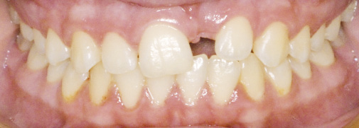
Obr. 2 / Fig. 2.
Výchozí stav intraorálně – zub 21 retinován v nepříznivé poloze. Mezera pro zub 21 je nedostatečná. Zuby 11 a 22 jsou skloněné do mezery, zub 22 je vůči zubu 12 v infrapozici. Je patrný defekt alveolární kosti v oblasti retinovaného zubu / Initial condition intraorally - tooth 21 impacted in an unfavourable position. Space for tooth 21 is not sufficient. Teeth 11 and 22 inclined to the gap, tooth 22 in infraocclusion related to tooth 12. Visible defect of the alveolar bone at the place of the impacted tooth.
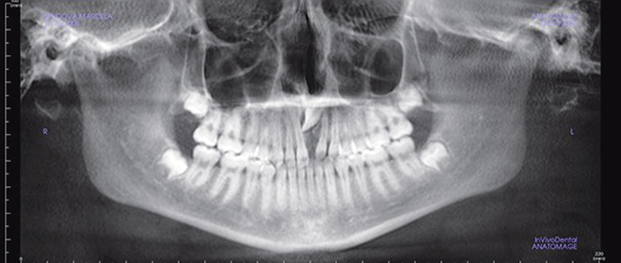
Obr. 3 / Fig. 3
Výchozí OPG – zub 21 uložen v nepříznivé poloze, distorotován o 90°, incizní hranou v kontaktu s kořenem zubu 11. V oblasti retinovaného zubu je patrný defekt alveolární kosti / Initial OPG - tooth 21 situated in an unfavourable position, distorotated by 90°, incisal edge in contact with tooth 11 root. Visible defect of the alveolar bone at the place of the impacted tooth.
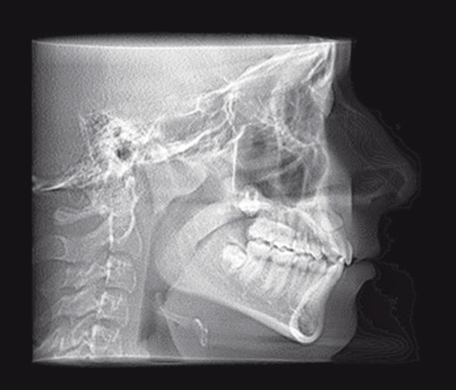
Obr. 4a / Fig. 4a
Výchozí kefalometrický snímek pacientky / Initial cephalogram of the patient
Obr. 4b / Fig,4b
Kefalometrická analýza pacientky před léčbou / Cephalometric analysis of the patient prior to the management
Obr. 5 / Fig. 5
OPG zhotovené po 3 měsících aktivního řazení. Zub 21 bez výrazné změny polohy / OPG taken after 3 months of the active arrangement. Tooth 21 without significant change in its position.
Obr. 6a / Fig. 6a
Situace intraorálně po 7 měsících aktivního řazení, je patrná deformace okluzální roviny a známky otevírání skusu / Condition intraorally after 7 months of the active arrangement; visible deformity of the occlusal plane and signs of bite opening.
Obr. 6b / Fig. 6b
Výřez z CBCT po 7 měsících aktivního řazení. Zub 21 bez výrazné změny polohy, ortodontický attachment pevně fixován k zubu, známky otevírání skusu / CBCT scan after 7 months of the active arrangement. Tooth 21 without significant change in its position, orthodontic attachment fixed firmly on the tooth, signs of bite opening.
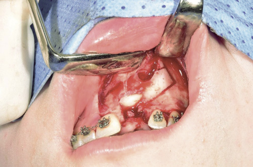
Obr. 7 / Fig. 7
Peroperační fotografie – preparace kostního bločku / Peroperation photography - preparation of the bone
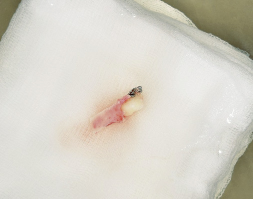
Obr. 8 / Fig. 8
Extrahovaný ankylotický zub 21 s ploténkou vestibulární kompakty. Po dobu preparace lůžka byl zub uchováván ve fyziologickém roztoku / Extracted ankylosed tooth 21 with a disk of vestibular compacta. During the bed preparation the tooth was in saline solution.
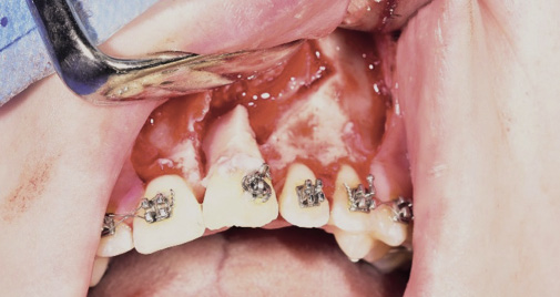
Obr. 9 / Fig. 9
Uložení zubu 21 s kostní ploténkou do upraveného lůžka. Apikálně je patrný defekt odpovídající původnímu uložení ankylotického zubu / Putting of tooth 21 with a bone into the prepared bed. Apically we can see the defect corresponding to the original position of the ankylosed tooth.
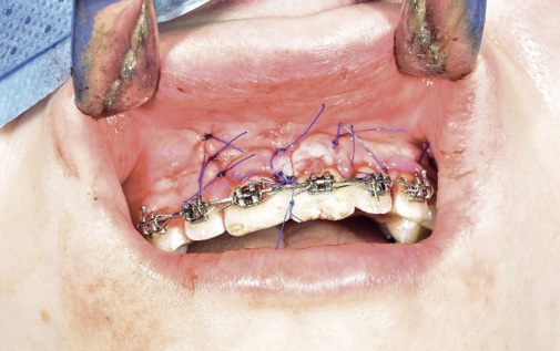
Obr. 10 / Fig. 10
Fixace a sutura – zub byl pomoci flow kompozita a retenční ligatury fixován k sousedním zubům a také pomocí drátěné ligatury k ocelovému oblouku. Pacientce byla doporučena kašovitá strava a šetřící režim / Fixation and suture - the tooth was fixed to surrounding teeth with flow composite and retention ligature, and to the steel arch with wire ligature. The patient was recommended soft food and rest.
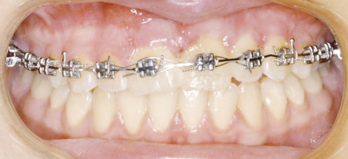
Obr. 11 / Fig. 11
Stav 10 dní po replantaci. Hojení klidné, pacientka bez obtíží / 10 days after replantation. Healing calm, the patient without any problems.
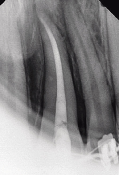
Obr. 12 / Fig. 12
Endodontické ošetření s plněním MTA provedené 3 týdny po zákroku z důvodu dokončeného vývoje kořene replantovaného zubu / Endodontic treatment with MTA filling 3 weeks after the management due to the completed development of the root of the replanted tooth.
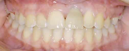
Obr. 13 /Fig. 13
Sejmutí fixního aparátu a recontouring incizálních hran zubů 11-22 fotokompozitními dostavbami z volné ruky. Patrná diskolorace autotransplantovaného zubu / Removal of fixed appliance and recountouring of incisal edges of teeth 11-22 with photocomposite build ups from a free hand. Visible discoloration of the autotransplanted tooth
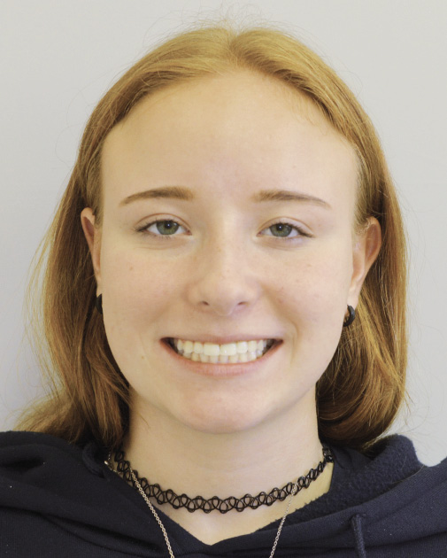
Obr. 14 / Fig. 14
Úsměv pacientky po sejmutí fixního ortodontického aparátu / Smile of the patient after removal of fixed appliance
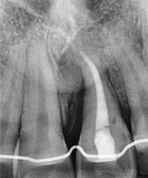
Obr. 15 / Fig. 15
Intraorální rentgenový snímek 1 rok po autotransplantaci, 3 měsíce po sejmutí fixního aparátu. Na snímku lze pozorovat obnovení kostní struktury v oblasti apexu a mezi zuby 21 a 22. Meziálně u zubu 21 kostní defekt přetrvává / X-ray one year after autotransplantation 3 months after removal of fixed appliance. We can see the bone structure in region of the apex and between teeth 21 and 22. The bone defect mesial of tooth 21 remains.

Literatura / References
1. Andreasen JO, Andreasen FM, Andreasen JO. Textbook and color atlas of traumatic injuries to the teeth. 3. ed., 1. print. Copenhagen: Munksgaard [u.a.]; 1994. 771 p.
2. Shafer WG, Hine MK, Levy BM. A textbook of oral pathology. 4. ed., 7. print. Philadelphia: Saunders; 1983. 917 p.
3. Proffit WR. Contemporary orthodontics. 6th edition. Philadelphia, IL: Elsevier; 2018.
4. Isaacson RJ, Strauss RA, Bridges-Poqus A, Peluso AR, Lindauer SJ. Moving an ankylosed central incisor using orthodontics, surgery and distraction osteogenesis. Angle Orthod. 2001 Oct;71(5):411-8. doi.org/10.1043/0003-3219(2001)071<0411:MAACIU>2.0.CO;2
5. Albers DD. Ankylosis of teeth in the developing dentition. Quintessence Int. 1986 May;17(5):303-8.
6. Aoki N, Matsumoto M, Ishii S, Baba J, Ito T, Honda A. Surgical and orthodontic management of an ankylosed mandibular molar with a single-tooth dento-osseous osteotomy: A case report. Journal of Oral and Maxillofacial Surgery, Medicine, and Pathology [Internet]. 2024 Sep [cited 2024 Nov 8];36(5):684-9. Available from: https://linkinghub.elsevier.com/retrieve/pii/S2212555824000036. doi.org/10.1016/j.ajoms.2024.01.002
7. Campbell KM, Casas MJ, Kenny DJ, Chau T. Diagnosis of ankylosis in permanent incisors by expert ratings, Periotest and digital sound wave analysis. Dent Traumatol. 2005 Aug;21(4):206-12. doi.org/10.1111/j.1600-9657.2005.00305.x
8. Sabri R. Treatment of a Class I crowded malocclusion with an ankylosed maxillary central incisor. Am J Orthod Dentofacial Orthop. 2002 Nov;122(5):557-65. doi.org/10.1067/mod.2002.126403
9. Medeiros PJ, Bezerra AR. Treatment of an ankylosed central incisor by single-tooth dento-osseous osteotomy. Am J Orthod Dentofacial Orthop. 1997 Nov;112(5):496-501. doi.org/10.1016/S0889-5406(97)70076-3
10. Epker BN, Paulus PJ. Surgical-orthodontic correction of adult malocclusions: single-tooth dento-osseous osteotomies. Am J Orthod. 1978 Nov;74(5):551-63. doi.org/10.1016/0002-9416(78)90031-3
11. Kofod T, Würtz V, Melsen B. Treatment of an ankylosed central incisor by single tooth dento-osseous osteotomy and a simple distraction device. Am J Orthod Dentofacial Orthop. 2005 Jan;127(1):72-80. doi.org/10.1016/j.ajodo.2003.12.020
12. Lynch SE, editor. Tissue engineering: applications in maxillofacial surgery and periodontics. Chicago: Quintessence Publ. Co; 1999. 297 p. (Quintessence books).
13. Kinzinger GSM, Jänicke S, Riediger D, Diedrich PR. Orthodontic fine adjustment after vertical callus distraction of an ankylosed incisor using the floating bone concept. Am J Orthod Dentofacial Orthop. 2003 Nov;124(5):582-90. doi.org/10.1016/S0889-5406(03)00569-9
14. Malmgren B. Decoronation: how, why, and when? J Calif Dent Assoc. 2000 Nov;28(11):846-54. doi.org/10.1080/19424396.2000.12223133
15. Sapir S, Shapira J. Decoronation for the management of an ankylosed young permanent tooth. Dental Traumatology [Internet]. 2008 Feb [cited 2024 Nov 8];24(1):131-5. Available from: https://onlinelibrary.wiley.com/doi/10.1111/j.1600-9657.2006.00506.x. doi.org/10.1111/j.1600-9657.2006.00506.x
16. Andreasen JO, Paulsen HU, Yu Z, Schwartz O. A long-term study of 370 autotransplanted premolars. Part III. Periodontal healing subsequent to transplantation. Eur J Orthod. 1990;12(1):25-37. doi.org/10.1093/ejo/12.1.25
17. Czochrowska EM, Stenvik A, Zachrisson BU. The esthetic outcome of autotransplanted premolars replacing maxillary incisors. Dent Traumatol. 2002;18(5):237-45. doi.org/10.1034/j.1600-9657.2002.00094.x
MDDr. Kristina Vaidišová
Stomatologická klinika FN Plzeň
Alej Svobody 80, 304 60 Plzeň