Změny parametrů na bočním kefalometrickém snímku po ortognátní operaci u pacientů s rozštěpem
Changes in parameters on lateral cephalograms after orthognathic surgery in patients with cleft lip and palate


MDDr. Ján Staško, Ph.D.1, MUDr. Wanda Urbanová, Ph.D.1,2,3, doc. MUDr. Magdalena Koťová, Ph.D.1,2,3,4,
MDDr. Petra Poláčková, Ph.D.1,2, MUDr. Jana Vašáková, Ph.D.1,2,3, MUDr. Jiří Borovec1,2,3
1 Stomatologická klinika 3. LF UK a Fakultní nemocnice Královské Vinohrady, Praha / Clinic of Stomatology, 3rd Medical Faculty of Charles University and University Hospital Královské Vinohrady, Prague
2 Centrum pro léčbu obličejových rozštěpů FNKV, Praha / Cleft Center, University Hospital Královské Vinohrady, Prague
3 Lékařská fakulta UK v Plzni/ Medical Faculty, Charles University, Pilsen
4 Stomatologická klinika FN a LF UP v Olomouci / Department of Dentistry, University Hospital, Faculty of Medicine and Dentistry, Palacky University, Olomouc
DOI10.61110/50647
Souhrn
Cíl práce: Porovnat změny parametrů měkkých a tvrdých tkání obličeje u pacientů s celkovým jednostranným rozštěpem a pseudoprogenií před a po ortognátní korekci s kontrolní skupinou pacientů s progenním stavem.
Materiál a metodika: Do retrospektivní studie bylo zařazeno 25 pacientů s celkovým jednostranným rozštěpem a pseudoprogenií (9 žen a 16 mužů; studijní skupina – SS) a 13 pacientů se III. skeletální třídou a obráceným skusem bez rozštěpové vady (9 žen a 4 muži; kontrolní skupina – KS), kteří podstoupili ortognátní korekci (Le Fort I „advancement“ s/bez mandibulárním setbackem). Dentoskeletální a měkkotkáňové parametry byly analyzovány na bočních kefalometrických snímcích v časech T1 (před operací), T2 (±14 měsíců po operaci) a T3 (±50 měsíců po operaci). Statistická analýza byla provedena Shapiro-Wilkovými a t-testy.
Výsledky: Mezi časy T2 a T1 jsme u obou skupin pozorovali statisticky významné zvětšení úhlu ANB, WITS, incizálního schůdku, zmenšení úhlu N-A-Po, -1i/APo, u KS navíc zvětšení hloubky skusu mezi T2/T1. Porovnání výsledků T2 a T3 prokázalo nevýznamné změny úhlu ANB a úhlu WITS (–0,38° a +0,31 mm u SS, +0,13° a –1,13 mm u KS). Mezi T3/T2 jsme u SS pozorovali statisticky významné zkrácení Lsʼ/E-linie, Liʼ/E-linie, zmenšení tloušťky horního rtu, goniového úhlu a incizálního schůdku. U KS bylo významné pouze zmenšení tloušťky horního rtu. Při porovnání změn u obou skupin pacientů jsme zjistili u KS signifikantně větší změny hodnoty goniového úhlu a zvětšení hloubky skusu mezi T2/T1, u SS zvětšení vzdálenosti Ls ʼ/E-linie mezi T3/T2.
Závěr: Byl prokázán pozitivní efekt ortognátní korekce na profil tvrdých i měkkých tkání obličeje u obou skupin pacientů. Zjistili jsme statisticky významné rozdíly mezi kontrolní skupinou (KS) a studijní skupinou (SS) ve velikosti změn hodnot goniového úhlu, hloubky skusu a Lsʼ/E-linie před a po operaci. Výsledky jsou více než 14 měsíců po operaci u obou skupin stabilní.
(Ortodoncie 2026, 35, č. 1, s. 27–40)
Abstract
Aim: Comparison of changes in soft and hard tissue facial parameters in patients with unilateral cleft lip and palate and pseudoprognathism prior to and after orthognathic correction compared with a control group of patients with true mandibular prognathism.
Material and method: The retrospective study included 25 patients with unilateral cleft lip and palate and pseudoprognathism (9 females and 16 males; study group = SG), and 13 patients with skeletal class III and reverse overjet without cleft (9 females and 4 males; control group = CG) who underwent orthognathic correction (Le Fort I advancement with/without mandibular setback). Dentoskeletal and soft tissue parameters were analyzed in lateral cephalograms at time points T1 (prior to surgery), T2 (± 14 months after surgery), and T3 (± 50 months after surgery). Statistical analysis was performed with the Shapiro–Wilk test and t-tests.
Results: Between T2 and T1, a statistically significant increase in ANB, Wits, and overjet and a decrease in N-A-Po and −1i/APo were observed in both groups. In CG, an additional statistically significant increase in overbite was observed between T2 and T1. Comparison of results between T2 and T3 confirmed non-significant changes in the ANB angle and WITS (−0.38° and +0.31 mm in SG; +0.13° and −1.13 mm in CG). Between T3 and T2, a statistically significant reduction in Ls’/E-line and decreases in upper lip thickness, the gonial angle, and overjet were recorded in SG. In CG, a statistically significant decrease in upper lip thickness was observed. Comparison of changes between both groups revealed significantly larger changes in the gonial angle and an increase in overbite between T2 and T1 in CG and an increase in Ls’/E-line between T3 and T2 in SG.
Conclusion: A positive effect of orthognathic surgery on the profile of hard and soft facial tissues was demonstrated in both groups of patients. Statistically significant differences between control group (CG) and study group (SG) were found in changes in the gonial angle, overbite, and Ls’/E-line prior to and after surgery. The results were stable more than 14 months after surgery in both groups.
(Ortodoncie 2026, 35, No. 1, p. 27–40)

Klíčová slova: celkový jednostranný rozštěp, ortognátní operace, boční kefalometrický snímek.
Key words: unilateral cleft lip and palate, orthognathic surgery, lateral cephalogram.
Úvod
Rozštěpy horní čelisti představují jedny z nejčastějších vrozených vad obličeje. Výrazně ovlivňují nejen estetický vzhled, ale mají i významný dopad na řeč, žvýkání, dýchání a celkovou psychickou pohodu jedince. Vzhled obličeje hraje zásadní roli při sociálních interakcích, což činí léčbu rozštěpů obličeje klíčovou nejen z medicínského, ale i z psychosociálního hlediska. Limitovaný růst horní čelisti u této skupiny pacientů často vede k výrazné disharmonii obličejových proporcí a následným funkčním problémům.
Disproporce vztahu horní a dolní čelisti lze řešit ovlivněním růstu, ortodontickou kompenzací nebo ortodonticko-chirurgickou terapií [1]. Ortognátní operace je zlatým standardem pro léčbu pacientů s rozštěpem se závažnou skeletální anomálií [2]. Ohledně podílu pacientů s celkovým jednostranným rozštěpem vyžadujících ortodonticko-chirurgickou léčbu se autoři dle dostupných literárních zdrojů rozcházejí, někteří udávají 4 %, jiní až 80 % [2–5]. Studie se shodují, že se po ortognátní operaci u pacientů s rozštěpem zlepšují jak skeletální, tak i měkkotkáňový profil obličeje [6].
Existuje jen málo studií zaměřených na dlouhodobé sledování změn dosažených ortognátní operací u pacientů s rozštěpem. Proto bylo cílem této studie porovnat změny měkkých a tvrdých tkání obličeje u pacientů s celkovým jednostranným rozštěpem s pseudoprogenií před a po ortognátní korekci s kontrolní skupinou pacientů s progenním stavem a zhodnotit stabilitu výsledků v dlouhodobém horizontu.
Materiál a metodika
Byla provedena retrospektivní studie hodnotící změny na bočních kefalometrických snímcích u dvou skupin pacientů, kteří podstoupili ortodonticko-chirurgickou léčbu na Stomatologické klinice Fakultní nemocnice Královské Vinohrady a 3. LF UK v letech 2013-2024. Tato retrospektivní studie byla schválena Etickou komisí FNKV EK-VP/50/2024.
Inkluzní kritéria studijní skupiny (SS): celkový jednostranný rozštěp bez přidruženého dalšího postižení, skeletální růst ukončen, zhotovený boční kefalometrický snímek vhodný pro kefalometrickou analýzu před a s časovým odstupem po operaci. Exkluzní kritéria studijní skupiny: jiný typ rozštěpu, systémové onemocnění, faciální trauma, distrakční osteogeneze, ortognátní operace před ukončením skeletálního růstu. Studijní skupinu tvořilo 25 pacientů s jednostranným celkovým rozštěpem (9 žen a 16 mužů), 11 pacientů mělo pravostranný a 14 pacientů levostranný celkový rozštěp, průměrný věk v době operace byl 21 let. Všichni pacienti ze studijní skupiny podstoupili sérii chirurgických výkonů: cheiloplastiku ve věku 0 - 3 měsíců, palatoplastiku 10-15 měsíců a kostní augmentaci rozštěpu čelisti mezi 8.- 14. rokem věku. U 10 pacientů byla do 14 měsíců po ortognátní operaci provedena plastika nosu.
Inkluzní kritéria kontrolní skupiny (KS): III. skeletální třída, skeletální růst ukončen, zhotovený boční kefalometrický snímek vhodný pro kefalometrickou analýzu před a s časovým odstupem po operaci. Exkluzní kritéria: další operační zákrok na čelistech, faciální trauma. Kontrolní skupinu představovalo 13 pacientů bez rozštěpu (9žen a 4 muži), průměrný věk v době operace byl 23 let.
U pacientů z obou skupin proběhla před ortognátní operací ortodontická příprava fixními ortodontickými aparáty, která spočívala v dekompenzaci čelistní anomálie, u žádného z nich nebyla použita technika „surgery first“. Všichni pacienti podstoupili maxilární advancement a část z nich i mandibulární setback pomocí bilaterální sagitální „split“ osteotomie mandibuly (9 pacientů ve studijní skupině a 9 pacientů v kontrolní skupině). Pacienti obou skupin byli operováni stejným maxilofaciálním chirurgem. Ortognátní operace byly uskutečněny v celkové anestezii, fixace čelistí proběhla s použitím titanových dlah a šroubů. V horní čelisti byla u všech pacientů provedena osteotomie v linii Le Fort I, u části z nich proběhla segmentální osteotomie maxily (10 pacientů studijní skupiny a 3 pacienti kontrolní skupiny). Rigidní mezičelistní fixace a operační „splint“ byly použity pouze v peroperační době. Po operaci byla použita intermaxilární fixace elastickými tahy po dobu 6–8 týdnů následovaná ortodontickým doléčením. U všech pacientů byly zachovány stejné principy předoperační, perioperační a pooperační zdravotní péče.
Zpracování a hodnocení dat
V rámci studie byly hodnoceny boční kefalometrické snímky ve třech časech: před operací po ortodontické přípravě (T1, 25 snímků SS, 13 snímků KS), průměrně 14 měsíců po operaci (T2, 25 snímků SS, 13 snímků KS) a průměrně 50 měsíců po operaci (T3, 13 snímků SS a 8 snímků KS). Minimální rozestup mezi zhotovenými snímky v časech T1 a T2 byl 12 měsíců.
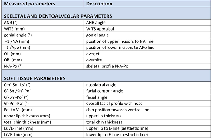
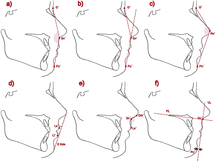
Rentgenové snímky byly zhotoveny a analyzovány v digitální formě. Seznam měřených parametrů je uveden v tabulce 1. Antropometrické body byly konstruovány podle obecných zásad [1]. Hodnocení Po’ (kožního bodu pogonion) k VL (vertical line) proběhlo jako měření vzdálenosti Po’ k linii VL zhotovené jako kolmice k patrové rovině procházející přes kožní bod subnasale (Sn’) (Obr. č. 1). Hodnocení tloušťky horního rtu (upper lip thickness) a brady (total chin thickness) proběhlo podle analýzy měkkých tkání obličeje Tweed-Merrifield [7].
Jeden hodnotitel provedl všechna měření a 10 % z nich bylo po měsíci zopakováno pro zjištění chyby měření.
Statistickou analýzu provedl zkušený biostatistik. Pomocí Shapiro-Wilkova testu normality bylo ověřeno, že většina veličin má normální distribuci. Pro ověření statistické významnosti změn byl použit jednovýběrový t-test. Rozdíly mezi dvěma nezávislými skupinami byly ověřeny pomocí dvouvýběrového t-testu na 5% hladině významnosti. Chyba měření byla kontrolována pomocí Dahlbergovy formule, relativní Dalbergovy chyby a výpočtem koeficientu ICC („Intraclass correlation coefficient“).
Výsledky
Relativní Dahlbergova chyba se pohybovala mezi 0,0 % a 15,4 %. Hodnoty koeficientu ICC byly zjištěny v rozmezí 0,885 až 1,000; což představuje velmi dobrou až perfektní shodu.
Výsledky a výpočet změn kefalometrického měření dentoalveolárních a skeletálních parametrů v časech T2/T1, T3/T1, T3/T2 je uveden v tabulce 2 a 3. V časech T2/T1 i T3/T1 bylo u SS pozorováno statisticky významné zvětšení ANB (pT2/T1 < 0,0001, pT3/T1 = 0,0053), WITS (pT2/T1 < 0,0001, pT3/T1 = 0,0003), incizálního schůdku (pT2/T1 < 0,0001, pT3/T1 = 0,0008) a zmenšení N-A-Po (pT2/T1 < 0,0001, pT3/T1 = 0,0039), –1i/APo (pT2/T1 < 0,0001, pT3/T1 = 0,0003). V časech T3/T2 bylo zjištěno statisticky významné zmenšení goniového úhlu (pT3/T2 = 0,0212) a incizálního schůdku (pT3/T2 = 0,0444).
U KS pacientů bez rozštěpu bylo v časech T2/T1 i T3/T1 pozorováno statisticky významné zvětšení ANB (pT2/T1 = 0,0079, pT3/T1 = 0,0043), WITS (pT2/T1 = 0,0003, pT3/T1 = 0,0050), incizálního schůdku (pT2/T1 = 0,0005, pT3/T1 = 0,0042), hloubky skusu v prvním pozorovaném úseku (pT2/T1 = 0,0132) a zmenšení N-A-Po (pT2/T1 < 0,00, pT3/T1 = 0,0076), –1i/APo (pT2/T1 < 0,0001, pT3/T1 = 0,0003). V časech T3/T2 nebyly statisticky významné změny zjištěny.
Výsledky a výpočet změn kefalometrického měření měkkotkáňových parametrů mezi T2/T1, T3/T1, T3/T2 je uveden v tabulce 2 a 3. U SS bylo mezi T2/T1 a T3/T1 prokázáno statisticky významné zvýšení hodnot G’-Sn’/Sn’-Po’ (pT2/T1 = 0,0001, pT3/T1 = 0,0130) a Ls’/E-linie (pT2/T1 < 0,0001, pT3/T1 = 0,0041) a snížení hodnot G’-Sn’-Po’ (pT2/T1 < 0,0001, pT3/T1 = 0,0047), G’-Pn’-Po’ (pT2/T1 < 0,0001, pT3/T1 = 0,0115), polohy brady (Po’) k VL (pT2/T1 = 0,0002, pT3/T1 = 0,0047) a tloušťky horního rtu (pT2/T1 = 0,0077, pT3/T1 = 0,0204). Mezi T3/T2 došlo ke statisticky významnému zvýšení hodnot G’-Sn’-Po’ (pT3/T2 = 0,0120) a snížení u tloušťky horního rtu (pT3/T2 = 0,0016), Ls’/E-linie (pT3/T2 = 0,0001) a Li’/E-linie (pT3/T2 = 0,0040). U KS v období T2/T1 došlo ke statisticky významnému snížení hodnot Cm’-Sn’-Ls’ a k nárůstu hodnoty Ls’/E-linie (pT2/T1 = 0,0018). V období T3/T1 bylo prokázáno snížení hodnot G’-Sn’-Po’ (pT3/T1 = 0,0261), G’-Pn’-Po’ (pT3/T1 = 0,072), polohy brady (Po’) k VL (pT3/T1 = 0,0410) a nárůst hodnot G’-Sn’/Sn’-Po’ (pT3/T1 = 0,0261), a Ls’/E-linie (pT3/T1 = 0,0211). V čase T3/T2 bylo zaznamenáno statisticky významné zmenšení tloušťky horního rtu (pT3/T2 = 0,0492).
Porovnání změn kefalometrických parametrů mezi skupinami pacientů s rozštěpem a bez rozštěpu, které nastaly v časech T2/T1, T3/T1 a T3/T2 jsou zobrazeny v tabulce 4. Byly zjištěny signifikantní rozdíly ve změně velikosti goniového úhlu (pGo = 0,029) a hloubky skusu (pOB = 0,006) v T2/T1 a v hodnotách Ls’/E-linie v T3/T2 (pLs’/E-line = 0,0041).
Diskuse
U obou skupin pacientů jsme pozorovali příznivý efekt operace na profil měkkých a tvrdých tkání obličeje. V časech T2/T1, stejně i T3/T1, jsme zjistili statisticky významné zvýšení parametrů ANB, WITS, incizálního schůdku a snížení hodnoty –1i/APo, N-A-Po u studijní skupiny pacientů s rozštěpem i kontrolní skupiny, u kontrolní skupiny navíc i zvětšení hloubky skusu mezi T2/T1.
U SS byla hodnota ANB před léčbou průměrně –6,92°, v T2 –1,76° a T3 –2,77°. U KS byly hodnoty ANB –3,38° před operací, v časech T1 a T2 0,15° resp. 0,38°. Oproti tomu Yun et al. naměřili u pacientů s rozštěpem rtu a patra, kteří podstoupili bimaxilární korekci, hodnoty ANB –4,1° před operací vs. 2,5° 6 měsíců po operaci [8]. Tsai et al. u 41 pacientů s celkovým jednostranným rozštěpem metodou 3D CT analýzy zaznamenali zvětšení úhlu ANB z průměrných –4,7° na 3,12° při Le Fort I předsunutí a na 3,37° při bimaxilární korekci [9]. Negativní hodnoty ANB po korekci u pacientů s rozštěpem v čase T2 a T3 v této studii lze vysvětlit významně větší závažností počáteční anomálie a větším operačním posunem čelistí u SS.
Z výsledků této studie vyplývá, že u parametrů na měkkých tkáních došlo mezi T2/T1 i T3/T1 u SS pacientů s rozštěpem ke statisticky významnému zvětšení G’-Sn’/Sn’-Po’, Ls’/E-linie a zmenšení G’-Sn’-Po’, G’-Pn’-Po’, polohy brady k VL a tloušťky horního rtu. Můžeme tedy konstatovat, že došlo ke zlepšení profilu obličeje na měkkých tkáních, což koreluje s dostupnými literárními zdroji.
Hodnoty nazolabiálního úhlu (Cm’-Sn’-Ls’) u SS po operaci byly v T2 průměrně 83,26° a v T3 průměrně 87,76°, přičemž referované hodnoty pro europoidní populaci jsou dle Bergmana et al. 102 ± 8° a podle Anic-Miloševič el al. pro muže 105,4 ± 9,5° a pro ženy 109 ± 7,8° [10,11]. Yun et al. zjistili u pacientů s rozštěpem hodnoty nazolabiálního úhlu 72,7° před operací, oproti 88,7° 6 měsíců po bimaxilární korekci [8]. Heliövaara et al. zaznamenali ve skupině pacientů s celkovým jednostranným rozštěpem signifikantní zvětšení nazolabiálního úhlu (82° vs. 92,9°) v časech imediátně a 6 měsíců po ortognátní operaci [12]. Zjištěné pooperační hodnoty nazolabiálního úhlu u SS z této studie jsou podobného rozsahu jako u výše uvedených výzkumů. U KS jsme zjistili hodnoty nazolabiálního úhlu ve všech časech blízké ideálnímu stavu [13] a vyšší než u studijní skupiny, což přikládáme charakteristickému postižení oblasti nosu a horního rtu u pacientů s celkovým jednostranným rozštěpem.
Esteticky ideální vzdálenost rtu od E-linie je podle Rickettse u europoidní populace pro horní ret 4 mm, pro dolní ret 2 mm [14]. Yun et al. zjistili u pacientů s rozštěpem hodnoty Ls’/E-linie 6,52 mm před vs. 1,81 mm po operaci [8]. Ve zde prezentovaném souboru došlo ke zlepšení postavení horního (z 3,8 mm na 2,63 mm) i dolního rtu vzhledem k E-linii, ideální hodnoty však nebylo dosaženo, především u skupiny pacientů s rozštěpem. Důvodem může být jizevnatá tkáň a z ní rezultující menší elasticita horního rtu u pacientů s rozštěpem, vysoká počáteční diskrepance, vliv korekční operace nazolabiální oblasti u části pacientů s rozštěpem mezi časy T3 a T2, či synergická reakce horního rtu na negativní změnu incizálního schůdku, a tedy i polohy horního řezáku. Tloušťka horního rtu se při maxilárním předsunutí snižuje, to platí pro pacienty s rozštěpovou vadou i bez ní [6]. Byly však popsány velmi odlišné kvantitativní změny ve vztahu k velikosti operačního posunu. Například Yun et al. zaznamenali u pacientů s jednostranným celkovým rozštěpem po bimaxilární korekci nevýznamné zvětšení tloušťky horního rtu o 0,78 mm (z 15,37 mmna 16,15 mm) [8]. Ve zde prezentované studii jsme po operaci zjistili zmenšení tloušťky horního rtu o 1,2 mm v T2/T1 resp. 2,1 mm v T3/T1 u studijní skupiny, u kontrolní skupiny byly změny tloušťky horního rtu kvantitativně menšího rozsahu.
Hodnoty úhlu obličeje byly u SS před operací průměrně 182,7° a po operaci v T2 176,2°; v T3 178,8°. U KS jsme zaznamenali menší změny hodnoty úhlu obličeje (T1 177,7°; T2 173,95°; T3 174,5°), přestože změny mezi T2/T1 byly větší u SS, rozdíl nebyl statisticky významný. Hodnoty úhlu celkového profilu obličeje s nosem u SS byly před operací průměrně 150,5°, po operaci v T2 průměrně 147,1° a v T3 průměrně 147,3°. Heliövaara et al. oproti tomu zaznamenali ve skupině pacientů s celkovým jednostranným rozštěpem mírné zmenšení úhlu celkového profilu obličeje s nosem (139° vs. 136,8°) v časech imediátně a 6 měsíců po ortognátní operaci [12].
Při měření stability výsledků v delším časovém odstupu bylo ve SS z dentálních parametrů v časech T3/T2 zjištěno statisticky významné zmenšení hodnoty incizálního schůdku o 0,77 mm (9,8 %), podobné změny jsme u kontrolní skupiny nepozorovali. Ve studii Patil et al. po Le Fort I s fixací minidlahami u pacientů s celkovým jednostranným rozštěpem se incizální schůdek snížil o 0,9 mm mezi imediátní pooperační dobou a dobou minimálně 1 rok po operaci (průměr 3,5 roku) [15]. V čase T2/T1 došlo k statisticky významným rozdílům ve změně hloubky skusu mezi skupinami, u pacientů s rozštěpem snížení o 0,24 mm a v kontrolní skupině naopak zvýšení o 2 mm. Příčinou těchto rozdílů jsou s největší pravděpodobností odpovídající změny ve velikosti goniového úhlu ve stejném časovém období (průměrně +0,45° u studijní vs. -1° u kontrolní skupiny) a častější bimaxilární korekce u kontrolní skupiny.
Podle mnoha autorů je u pacientů s maxilární hypoplázií a rozštěpem čelisti recidiva pravděpodobnější než u pacientů bez rozštěpu, v odborné literatuře se uvádí 20–40 % sagitální recidivy u této skupiny pacientů [15–20]. Na druhé straně existují studie, které rozdíly ve stabilitě nezjistily [21]. Vzhledem k absenci adekvátních immediátních kefalometrických snímků u souboru pacientů v tomto výzkumu můžeme zhodnotit změny nastolené ortognátní operací, nikoliv míru recidivy. Lze však konstatovat, že měření skeletálních parametrů v časech T3/T2 u SS prokázalo statisticky nevýznamné zmenšení úhlu ANB o –0,38° (7,4% celkového posunu mezi T1 a T2) a zvětšení WITS o 0,31 mm. Jde tedy menší míru změn, než uváfí dlouhodobé monitorování výsledků v odborné literatuře. Statisticky významné bylo zmenšení goniového úhlu o 0,92°. Vzhledem k nízkému podílu bimaxilárních operací u studijní skupiny mohou být tyto změny dány spíše zbytkovým růstem. U KS jsme zjistili statisticky nesignifikantní zvětšení ANB o 0,13° (3,7 % celkového posunu mezi T1 a T2) a zmenšení WITS o 1,13 mm.
Výsledky naší studie podporují závěry autorů, kteří poukazují na minimální skeletální i měkkotkáňové změny po více než 1 roce od operace [5, 23, 24]. Při interpretaci výsledků této studie je však nutné vzít v úvahu určité limitace, jako jsou relativně malá velikost kontrolního souboru, nižší počet pacientů v čase T3, korekční výkony na rtu a nosu u části pacientů studijní skupiny, nehomogenní charakteristiky skupin v některých ohledech, a především retrospektivní sběr dat.
Závěr
Studie prokázala, že ortodonticko-chirurgická léčba je účinnou metodou léčby pseudoprogenie u pacientů s celkovým jednostranným rozštěpem se stabilními výsledky. U většiny sledovaných parametrů na měkkých tkáních a u dentoalveolárních i skeletálních parametrů zlepšení hodnot po operaci přetrvávalo i při sledování v delším časovém odstupu. Byl prokázán pozitivní efekt ortognátní korekce na profil tvrdých i měkkých tkání obličeje u obou skupin pacientů. Zjistili jsme statisticky významné rozdíly mezi kontrolní skupinou a studijní skupinou ve velikosti změn hodnot goniového úhlu, hloubky skusu a Ls’/E-linie před a po operaci. Výsledky jsou více než 14 měsíců po operaci u obou skupin stabilní.
Autoři nemají komerční, vlastnické nebo finanční zájmy na produktech nebo společnostech popsaných v tomto článku.
Introduction
Cleft lip and palate are among the most frequent congenital anomalies of the face. It significantly affects facial esthetics, it also affects speech, mastication, breathing, and overall psychological well-being of an individual. Facial appearance plays a key role in social interactions and, therefore, the treatment of cleft defects is important not only from a medical aspect but also from a psychosocial perspective. Limited maxillary growth in this group of patients often leads to marked facial disproportions and subsequent functional problems.
Disproportion between the upper and lower jaws can be managed by growth modification, orthodontic compensation, or combined orthodontic–surgical therapy [1]. Orthognathic surgery is considered the gold standard for the treatment of patients with cleft lip and palate presenting with severe skeletal discrepancies [2]. According to the available literature, authors are not unanimous regarding the proportion of patients with unilateral cleft requiring orthodontic–surgical treatment, with reported rates ranging from 4% to as high as 80% [2–5]. Nevertheless, studies consistently demonstrate that orthognathic surgery in patients with cleft lip and palate leads to improvement in both skeletal and soft tissue facial profiles [6].
Only a limited number of studies have focused on the long-term follow-up of changes achieved by orthognathic surgery in patients with cleft lip and palate. Therefore, our study aims to compare changes in soft and hard facial tissues in patients with unilateral cleft lip and palate with pseudoprognathism before and after orthognathic correction with a control group of patients with reverse overjet, and to assess the long-term stability of surgical outcomes.
Material and method
The retrospective study assessed changes on lateral cephalograms in two groups of patients who underwent combined orthodontic–surgical treatment at the Clinic of Stomatology, University Hospital Královské Vinohrady, and the 3rd Medical Faculty, Charles University, between 2013 and 2024. The study was approved by the Ethics Committee of the University Hospital Královské Vinohrady (No. EK-VP/50/2024).
Inclusion criteria for the study group (SG) were: unilateral cleft lip and palate without any accompanying anomaly, completed skeletal growth, lateral cephalogram suitable for cephalometric analysis made prior to and after the surgery. Patients with another type of cleft, with systemic disease, facial trauma, distraction osteogenesis, orthognathic surgery performed during growth were excluded. The study group included 25 patients with unilateral cleft lip and palate (9 females and 16 males), 11 patients had a cleft lip and palate on the right and 14 on the left side, and the mean age at the time of surgery was 21 years. All patients in SG underwent a series of surgical interventions: cheiloplasty at 0–3 months of age, palatoplasty at 10–15 months, and alveolar bone grafting between 8 and 14 years of age. 10 patients underwent rhinoplasty within 14 months of the orthognathic surgery.
Inclusion criteria for the control group (CG) were: skeletal class III, completed skeletal growth, lateral cephalogram suitable for cephalometric analysis made prior to and after the surgery. Patients with other jaw surgery and facial trauma were excluded. The control group included 13 non-cleft patients (9 females and 4 males), and the mean age at the time of surgery was 23 years.
Before orthognathic surgery, patients from both groups were treated orthodontically using fixed appliances for anomaly decompensation; the „surgery-first“ approach was not applied in any patient. All patients underwent maxillary advancement, and some also underwent mandibular setback using bilateral sagittal split osteotomy of the mandible (9 patients in the SG and 9 patients in the CG). All surgical procedures were performed by the same maxillofacial surgeon under general anesthesia. Fixation was achieved using titanium plates and screws. In all patients, a Le Fort I maxillary osteotomy was performed; segmental maxillary osteotomy was additionally carried out in some cases (10 patients in the SG and 3 patients in the CG). Rigid intermaxillary fixation and a surgical splint were used only intraoperatively. Postoperatively, intermaxillary fixation with elastic traction was applied for 6–8 weeks, followed by orthodontic treatment. Identical principles of preoperative, perioperative, and postoperative care were applied to all patients.
Processing and evaluation of the data
Lateral cephalograms were evaluated at three time points: prior to surgery after orthodontic pretreatment (T1; 25 cephalograms in the SG and 13 in the CG), approx. 14 months after surgery (T2; 25 cephalograms in the SG and 13 in the CG), and approx. 50 months after surgery (T3; 13 cephalograms in the SG and 8 in the CG). The minimum interval between cephalograms obtained at T1 and T2 was 12 months.
All radiographs were obtained and analyzed in digital format. The list of measured parameters is presented in Table 1. Anthropometric landmarks were identified according to standard definitions [1]. The assessment of the position of Po’ relative to the vertical line (VL) was performed by measuring the distance between the soft tissue pogonion (Po’) and the VL, defined as a perpendicular line to the palatal plane passing through the soft tissue subnasale (Sn’) (Fig. 1). Upper lip thickness and total chin thickness were evaluated according to the Tweed–Merrifield soft tissue facial analysis [7].
All measurements were performed by a single examiner. To assess measurement error, 10% of the measurements were repeated after a one-month interval.
Statistical analysis was performed by an experienced biostatistician. Normality of data distribution was assessed using the Shapiro–Wilk test, which showed that most variables followed a normal distribution. A one-sample t-test was used to determine the statistical significance of changes within groups. Differences between the two independent groups were evaluated using a two-sample t-test at a 5% level of significance. Measurement error was assessed using the Dahlberg formula, relative Dahlberg error, and calculation of the intraclass correlation coefficient (ICC).
Results
The relative Dahlberg error ranged from 0.0% to 15.4%. ICC values ranged from 0.885 to 1.000, indicating very good to excellent agreement.
The results and calculated changes in cephalometric measurements of dentoalveolar and skeletal parameters at T2/T1, T3/T1, and T3/T2 are summarized in Tables 2 and 3. In the SG, statistically significant increases in ANB (pT2/T1 < 0.0001, pT3/T1 = 0.0053), WITS (pT2/T1 < 0.0001, pT3/T1 = 0.0003), and overjet (pT2/T1 < 0.0001, pT3/T1 = 0.0008) were observed at both T2/T1 and T3/T1, along with statistically significant decreases in N-A-Po (pT2/T1 < 0.0001, pT3/T1 = 0.0039) and −1i/APo (pT2/T1 < 0.0001, pT3/T1 = 0.0003). At T3/T2, statistically significant increases in the gonial angle (pT3/T2 = 0.0212) and overjet (pT3/T2 = 0.0444) were observed.
In the CG, statistically significant increases in ANB (pT2/T1 = 0.0079, pT3/T1 = 0.0043), WITS (pT2/T1 = 0.0003, pT3/T1 = 0.0050), overjet (pT2/T1 = 0.0005, pT3/T1 = 0.0042), and overbite at T2/T1 (p = 0.0132) were observed, together with statistically significant decreases in N-A-Po (pT2/T1 < 0.0001, pT3/T1 = 0.0076) and −1i/APo (pT2/T1 < 0.0001, pT3/T1 = 0.0003). No statistically significant changes were observed at T3/T2.
Changes in cephalometric measurements of soft tissue parameters at T2/T1, T3/T1, and T3/T2 are summarized in Tables 2 and 3. In the SG, statistically significant increases in G’-Sn’/Sn’-Po’ (pT2/T1 = 0.0001, pT3/T1 = 0.0130) and Ls’/E-line (pT2/T1 < 0.0001, pT3/T1 = 0.0041), and statistically significant decreases in G’-Sn’-Po’ (pT2/T1 < 0.0001, pT3/T1 = 0.0047), G’-Pn’-Po’ (pT2/T1 < 0.0001, pT3/T1 = 0.0115), chin position (Po’) relative to the vertical line (VL) (pT2/T1 = 0.0002, pT3/T1 = 0.0047), and upper lip thickness (pT2/T1 = 0.0077, pT3/T1 = 0.0204) were observed between T2/T1 and T3/T1. Between T3/T2, a statistically significant increase in G’-Sn’-Po’ (p = 0.0120) and statistically significant decreases in upper lip thickness (p = 0.0016), Ls’/E-line (p = 0.0001), and Li’/E-line (p = 0.0040) were recorded.
In the control group, a statistically significant decrease in Cm’-Sn’-Ls’ and an increase in Ls’/E-line were observed at T2/T1 (p = 0.0018). At T3/T1, statistically significant decreases in G’-Sn’-Po’ (p = 0.0261), G’-Pn’-Po’ (p = 0.072), and chin position (Po’) relative to VL (p = 0.0410), as well as statistically significant increases in G’-Sn’/Sn’-Po’ (p = 0.0261) and Ls’/E-line (p = 0.0211), were observed. At T3/T2, a statistically significant decrease in upper lip thickness was recorded (p = 0.0492).
Comparison of changes in cephalometric parameters between cleft and non-cleft patients at T2/T1, T3/T1, and T3/T2 is presented in Table 4. Statistically significant differences were found in changes in the gonial angle (p = 0.029) and overbite (p = 0.006) at T2/T1, as well as in Ls’/E-line at T3/T2 (p = 0.0041).
Discussion
In both groups of patients, a favorable effect of orthognathic surgery on the profile of hard and soft facial tissues was observed. At both T2/T1 and T3/T1, statistically significant increases in ANB, WITS, and overjet, as well as decreases in −1i/APo and N-A-Po, were recorded in both groups. In addition, a statistically significant increase in overbite at T2/T1 was observed in the control group.
In the SG, the mean ANB value prior to treatment was −6.92°, −1.76° at T2, and −2.72° at T3. In the CG, the corresponding ANB values were −3.38° prior to surgery, 0.15° at T1, and 0.38° at T2. By comparison, Yun et al. reported ANB values of −4.1° before surgery and 2.5° six months after bimaxillary correction in patients with cleft lip and palate [8]. Tsai et al., using three-dimensional CT analysis in 41 patients with unilateral cleft lip and palate, recorded an increase in ANB from a mean of −4.7° to 3.12° after Le Fort I advancement and to 3.37° after bimaxillary correction [9]. The persistence of negative ANB values at T2 and T3 in cleft patients in the present study may be explained by the greater severity of the initial skeletal discrepancy and the larger surgical advancement required in the study group.
The results of this study indicate that, with respect to soft tissue parameters, statistically significant increases in G’-Sn’/Sn’-Po’ and Ls’/E-line and statistically significant decreases in G’-Sn’-Po’, G’-Pn’-Po’, chin position relative to the VL, and upper lip thickness occurred at both T2/T1 and T3/T1 in cleft patients of the study group. It can therefore be concluded that an improvement in the soft tissue facial profile was achieved, which is consistent with the available literature.
In the SG, mean postoperative values of the nasolabial angle (Cm’-Sn’-Ls’) were 83.26° at T2 and 87.76° at T3. Reference values for the Europoid population have been reported as 102 ± 8° by Bergman et al. and as 105.4 ± 9.5° for men and 109 ± 7.8° for women by Anić-Milošević et al. [10,11]. Yun et al. reported nasolabial angle values of 72.7° before surgery and 88.7° six months after bimaxillary correction in cleft patients [8]. Heliövaara et al. observed a significant increase in the nasolabial angle (82° vs. 92.9°) immediately and six months after orthognathic surgery in patients with complete unilateral cleft lip and palate [12]. The postoperative nasolabial angle values observed in the present study are of a similar range to those reported in the above-mentioned studies. In the CG, nasolabial angle values at all time points were close to the ideal range [13] and higher than those in the SG, which may be attributed to the characteristic involvement of the nasal and upper lip region in patients with complete unilateral cleft lip and palate.
According to Ricketts [14], the esthetically ideal distance between the lip and the E-line in the Europoid population is 4 mm for the upper lip and 2 mm for the lower lip. Yun et al. reported Ls’/E-line values of 6.52 mm preoperatively and 1.81 mm postoperatively in cleft patients [8]. In the present sample, improvement in the position of both the upper lip (from 3.8 mm to 2.63 mm) and the lower lip relative to the E-line was observed; however, ideal values were not achieved, particularly in the cleft group.
This may be due to scarred tissue resulting in lower elasticity of the upper lip in cleft patients, high initial discrepancy, impact of surgery of the nasolabial area in some patients performed between T2 and T3, or synergistic response of the upper lip to the reduction in overjet, and thus also upper incisor position. The upper lip thickness is reduced during maxillary advancement, which holds true for both cleft and non-cleft patients [6]. However, different quantitative changes related to the extent of the advancement were described. For example, Yun et al. reported a non-significant increase in upper lip thickness of 0.78 mm (from 15.37 to 16.15 mm) [8]. In our study, we recorded a decrease in upper lip thickness by 1.2 mm at T2/T1 and 2.1 mm at T3/T1 in the SG after surgery. In the CG, changes in upper lip thickness were quantitatively smaller.
In the SG, facial angle values averaged 182.7° before surgery, 176.2° at T2, and 178.8° at T3. In the CG, we observed minor changes in facial angle values (177.7° at T1, 173.95° at T2, and 174.5° at T3). Although the changes between T2/T1 were larger in SG, the difference was not statistically significant. The mean values of the overall facial profile angle including the nose in SG were 150.5° before surgery, 147.1° at T2, and 147.3° at T3. Heliövaara et al., on the other hand, reported a slight decrease in the overall facial profile angle including the nose (139° vs. 136.8°) immediately and 6 months after orthognathic surgery [12].
When measuring the stability of the results over a longer period of time, a statistically significant reduction in overjet of 0.77 mm (9.8%) was observed in the SG at T3/T2. No similar changes were observed in the control group. In the study by Patil et al., following Le Fort I osteotomy with miniplate fixation in patients with unilateral cleft lip and palate, overjet decreased by 0.9 mm between the immediate postoperative period and a minimum of 1 year after surgery (mean follow-up 3.5 years) [15]. At T2/T1, statistically significant differences in overbite changes were observed between the groups, with a reduction of 0.24 mm in cleft patients and an increase of 2 mm in the CG. These differences are most likely due to corresponding changes in the gonial angle over the same time period (mean +0.45° in SG vs. −1° in CG) and the more frequent use of bimaxillary correction in the control group.
According to many authors, patients with maxillary hypoplasia and cleft are more prone to relapse than non-cleft patients, with 20–40% sagittal relapse reported in the literature for this group of patients [15–20]. On the other hand, there are studies that have not found differences in stability [21]. Due to the absence of adequate immediate postoperative cephalometric radiographs in the present patient cohort, it was possible to evaluate changes achieved by orthognathic surgery rather than the actual rate of relapse. Nevertheless, measurements of skeletal parameters at T3/T2 in the study group demonstrated a statistically non-significant reduction in the ANB angle of −0.38° (7.4% of the total change between T1 and T2) and an increase in WITS of 0.31 mm, indicating a smaller magnitude of change than that reported in the literature during long-term follow-up. A statistically significant reduction in the gonial angle of 0.92° was observed. Given the low proportion of bimaxillary procedures in the study group, these changes may be more related to residual growth. In the CG, a statistically non-significant increase in ANB of 0.13° (3.7% of the total change between T1 and T2) and a decrease in WITS of 1.13 mm were observed.
The results of our study support the conclusions of previous authors, who report minimal skeletal and soft tissue changes more than one year after surgery [5, 23, 24]. However, when interpreting the results of this study, certain limitations should be taken into account, including the relatively small size of the CG, the lower number of patients evaluated at T3, corrective procedures of the lip and nose performed in a subset of patients in the SG, partial heterogeneity of the groups in some respects, and, in particular, the retrospective design of the study.
Conclusion
The present study confirmed that combined orthodontic–surgical treatment is an effective method for managing pseudoprognathism in patients with unilateral cleft lip and palate, providing stable results. For most soft tissue parameters, as well as for dentoalveolar and skeletal parameters, postoperative improvements persisted during long-term follow-up. A positive effect of orthognathic correction on the profile of both hard and soft facial tissues was demonstrated in both patient groups. Statistically significant differences between the study group and the control group were identified in the magnitude of changes in the gonial angle, overbite, and Ls’/E-line before and after surgery. The results remained stable for more than 14 months after surgery in both groups.
The authors declare no commercial, proprietary, or financial interests related to the products or companies described in this article.
Tab. 1 / Tab. 1
Seznam kefalometrických parametrů /List of cephalometric analysis parameters.
Obr 1 / Fig. 1
Schematic representation of the technique for measuring distances and angles of cephalometric analysis parameters: a) G‘-Sn’-Po‘ angle; b) G’-Sn’/Sn’-Po’ angle; c) G‘-Pn’-Po‘ angle; d) distance from upper and lower lip to E-line; e) Cm’-Sn’-Ls’ angle; f) distance from Po’ to VL.
Tab. 2 / Tab. 2
Měření skeletálních, dentoalveolárních rozměrů a rozměrů na měkkých tkáních /Measurement of skeletal, dentoalveolar and soft tissue parameters at T1, T2 and T3 in the study and control groups.
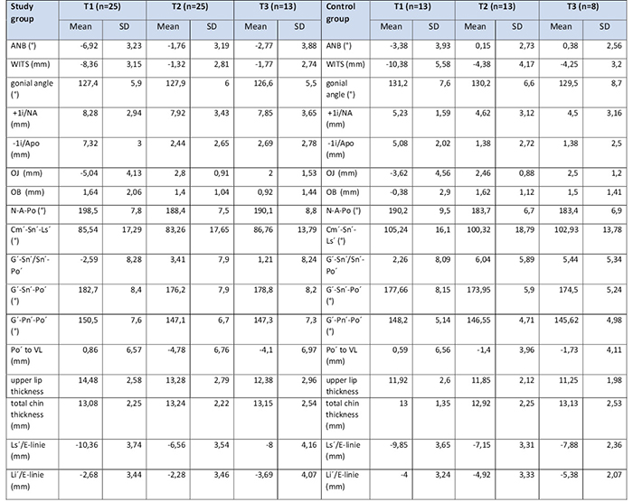
Mean: arithmetic mean; SD: standard deviation.
Tab. 3 / Tab. 3
Změny kefalometrických rozměrů u studijní skupiny/ Calculation of changes in cephalometric measurements of skeletal, dentoalveolar, and soft tissue parameters in the study group.
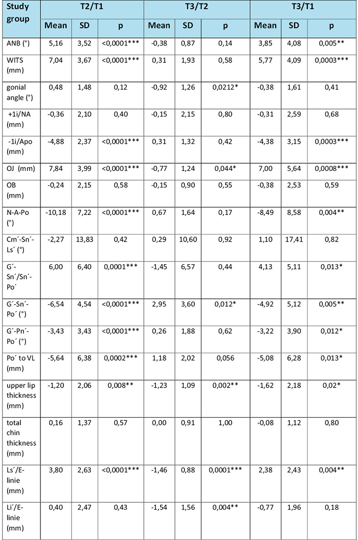
*p < 0,05; **p < 0,01; ***p < 0,001; Mean: arithmetic mean; SD: standard deviation; Min: minimal value; Max: maximal value; p: p-value.
Tab. 4 / Tab. 4
Změny u kontrolní skupiny / Changes in skeletal, dentoalveolar, and soft-tissue cephalometric parameters at T2/T1, T3/T2, and T3/T1 in the control group.
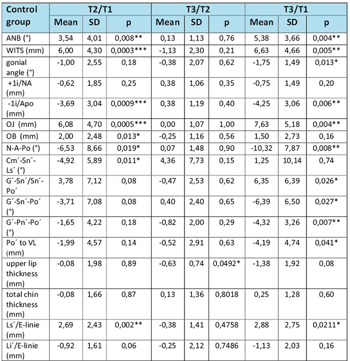
*p < 0,05; **p < 0,01; ***p < 0,001; Mean: arithmetic mean; SD: standard deviation; Min: minimal value; Max: maximal value; p: p value.
Tab. 5 / Tab. 5
Testovaný rozdíl mezi změnami u pacientů s rozštěpy a kontrolní skupinou / Comparison of changes in cephalometric parameters at T2/T1, T3/T1, and T3/T2 between cleft and control groups using the p-value of the two-sample t-test.
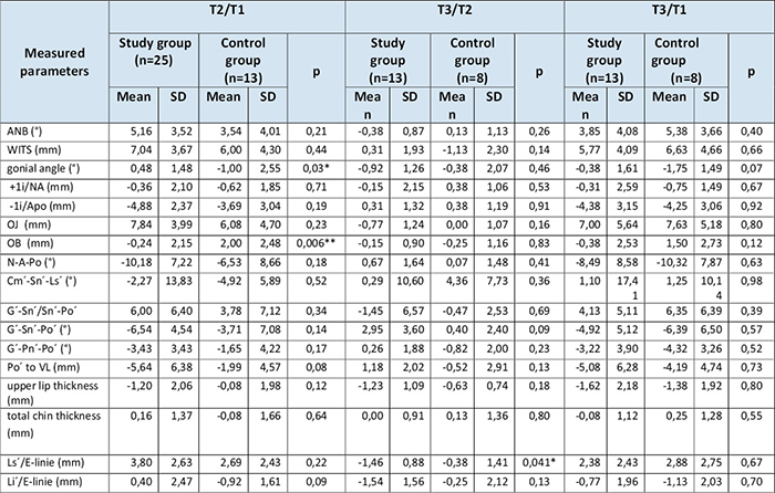
*p < 0,05, **p < 0,01; Mean: arithmetic mean; SD: standard deviation; p: p value.

Literatura / References
1. Proffit W. Contemporary Orthodontics. 6th ed. Philadelphia: Elsevier; 2019.
2. Shetye P, Gibbon T. Cleft and Craniofacial Orthodontics. Hoboken: John Wiley & Sons; 2023. doi.org/10.1002/9781119778387
3. Taib BG, Taib AG, Swift AC, Van Eeden S. Cleft lip and palate: diagnosis and management. Br J Hosp Med. 2015;76:584-91 doi.org/10.12968/hmed.2015.76.10.584
4. Rosenstein S, Dado DV, Kernahan D, Griffith BH, Grasseschi M. The case for early bone grafting in cleft lip and palate: a second report. Plast Reconstr Surg. 1991;87:644-54; discussion 655-6. doi.org/10.1097/00006534-199104000-00006
5. Houston WJ, James DR, Jones E, Kavvadia S. Le Fort I maxillary osteotomies in cleft palate cases. Surgical changes and stability. J Craniomaxillofac Surg. 1989;17:9-15. doi.org/10.1016/S1010-5182(89)80121-0
6. al-Waheidi EM, Harradine NW. Soft tissue profile changes in patients with cleft lip and palate following maxillary osteotomies. Cleft Palate Craniofac J. 1998;35:535-43. doi.org/10.1597/1545-1569_1998_035_0535_stpcip_2.3.co_2
7. Merrifield LL. The profile line as an aid in critically evaluating facial esthetics. Am J Orthod. 1966;52:804-2. doi.org/10.1016/0002-9416(66)90250-8
8. Yun Y, Uhm K, Kim J, Shin D, Choi H, Kim S, et al. Bone and Soft Tissue Changes after Two-Jaw Surgery in Cleft Patients. Arch Plast Surg 2015;42:419-23. doi.org/10.5999/aps.2015.42.4.419
9. Tsai YH, Pai B, Lin HH, Ho CT, Lo LJ. One-Jaw versus Two-Jaw Orthognathic Surgery for Patients with Cleft: A Comparative Study Using 3D Imaging Virtual Surgical Planning. Appl Sci 2022;12:4461. doi.org/10.3390/app12094461
10. Milosevic S, Varga M, Slaj M. Analysis of the soft tissue facial profile by means of angular measurements. Eur J Orthod 2008;30:135-40. doi.org/10.1093/ejo/cjm116
11. Bergman RT. Cephalometric soft tissue facial analysis. Am J Orthod Dentofacial Orthop. 1999;116:373-89. doi.org/10.1016/S0889-5406(99)70222-2
12. Heliövaara A, Ranta R, Hukki J, Rintala A. Skeletal stability of Le Fort I osteotomy in patients with unilateral cleft lip and palate. Scand J Plast Reconstr Surg Hand Surg. 2001;35:43-9. doi.org/10.1080/02844310151032556
13. Fernández-Riveiro P, Smyth E, Suárez-Quintanilla D, Suarez-Cunqueiro M. Angular photogrammetric analysis of the soft tissue facial profile. Eur J Orthod 2003;25:393-9. doi.org/10.1093/ejo/25.4.393
14. Ricketts RM. A foundation for cephalometric communication. Am J Orthod 1960;46:330-57. doi.org/10.1016/0002-9416(60)90047-6
15. Patil S, Basavaraddi SM, Revankar AV, Naik RD, Desai A, Kumar N, et al. Comparative Evaluation of Relapse After Le Fort 1 Osteotomy in Cleft Palate and Non-cleft Individuals: A Longitudinal Study. Cureus. 2024;16:e52298. doi.org/10.7759/cureus.52298
16. Wong FX, Heggie AA, Shand JM, Schneider PM. Skeletal stability of maxillary advancement with and without a mandibular reduction in the cleft lip and palate patient. Int J Oral Maxillofac Surg 2016;45:1501-7. doi.org/10.1016/j.ijom.2016.08.007
17. Saltaji H, Major MP, Alfakir H, Al-Saleh MAQ, Flores-Mir C. Maxillary Advancement With Conventional Orthognathic Surgery in Patients With Cleft Lip and Palate: Is It a Stable Technique? J Oral Maxillofac Surg 2012;70:2859-66. doi.org/10.1016/j.joms.2012.03.009
18. da Silva WS, de Almeida ALPF, Pucciarelli MGR, Neppelenbroek KH, da Silva de Menezes JD, Yaedú RYF, et al. Relapse after Le Fort I surgery in oral cleft patients: a 2-year follow-up using digitized and 3D models. Odontology. 2018;106:445-53. doi.org/10.1007/s10266-018-0351-8
19. Valls-Ontañón A, Fernandes-Ciaccia L, Haas-Junior OL, Hernández-Alfaro F. Relapse-related factors of Le Fort I osteotomy in cleft lip and palate patients: A systematic review and meta-analysis. J Craniomaxillofac Surg 2021;49:879-90. doi.org/10.1016/j.jcms.2021.09.002
20. Figueroa AA, Polley JW, Friede H, Ko EW. Long-term skeletal stability after maxillary advancement with distraction osteogenesis using a rigid external distraction device in cleft maxillary deformities. Plast Reconstr Surg. 2004;114:1382-92; discussion 1393-4. doi.org/10.1097/01.PRS.0000138593.89303.1B
21. Ishihata K, Kibe T, Tezuka M, Seong CH, Maeda-Iino A, Nozoe E, et al. Postoperative stability following orthognathic surgery and final skeletal morphology in class III patients with cleft lip and palate. J Oral Maxillofac Surg Med Pathol 2022;34:704-12. doi.org/10.1016/j.ajoms.2022.04.005
22. Seo J, Yang I-H, Choi JY, Lee J, Baek S-H. Three-Dimensional Facial Soft Tissue Changes After Orthognathic Surgery in Cleft Patients Using Artificial Intelligence-Assisted Landmark Autodigitization. J of Craniofac Surg. 2021;Published Ahead of Print. doi.org/10.1097/SCS.0000000000007712
23. Araujo A, Schendel SA, Wolford LM, Epker BN. Total maxillary advancement with and without bone grafting. J Oral Surg. 1978;36:849-58.
24. Ewing M, Ross RB. Soft tissue response to orthognathic surgery in persons with unilateral cleft lip and palate. Cleft Palate Craniofac J. 1993;30:320-7. doi.org/10.1597/1545-1569_1993_030_0320_strtos_2.3.co_2
MDDr. Ján Staško, PhD.
Stomatologická klinika FNKV Praha
Šrobárova 50, 100 34 Praha 10