Vztah mezi palatinální retencí horního stálého špičáku a anatomií horního stálého postranního řezáku
Relationship between the palatal impacted upper permanent canine and the anatomy of the upper permanent lateral incisor


MDDr. Dana Kloudová, MUDr. Martin Horáček
Ortodontické oddělení Stomatologické kliniky dětí a dospělých 2. LF UK a FN v Motole, Praha / Orthodontic Department of Dental Clinic for Children and Adults 2. LF UK and FN in Motol, Prague
DOI10.61110/50707
Souhrn
Cíl: Cílem studie bylo zjistit, zdali existuje nějaká souvislost mezi palatinální retencí horního stálého špičáku a anatomií horního stálého postranního řezáku
Materiál a metodika: Do výzkumu bylo zařazeno 102 pacientů s jednostrannou a 27 pacientů s oboustrannou palatinální retencí horního stálého špičáku, kterým byly zhotoveny CBCT snímky. Ty byly ve formátu DICOM importovány do zobrazovacího SW InVivo6 (Anatomage, Santa Clara, CA, USA), kde byly horním stálým postranním řezákům měřeny lineární a 3D rozměry. Dále byl hodnocen vývoj kořene a měřen úhel, který svírá dlouhá osa špičáku se střední linií (úhel α
Výsledky: U jednostranné palatinální retence horního stálého špičáku měly postranní řezáky na straně retence významně kratší délku korunky (p = 0,013), bukolingvální rozměr korunky (p = 0,022), šířku na incizi (p = 0,002), délku kořene (p = 0,019), meziodistální rozměr kořene (p = 0,014), bukolingvální rozměr kořene (p = 0,007) i zahnutí kořene (p = 0,026) ve srovnání s kontralaterální skupinou. Ve srovnání s kontrolní skupinou byl statisticky významný rozdíl ještě u rozměrů šířka korunky v krčkové oblasti (MD cervikální) a nejširší meziodistální rozměr na korunce (MD). Postranní řezáky na straně retence špičáku měly statisticky významně menší objem kořene (p = 0,0014) a celého zubu (p = 0,013) ve srovnání s kontralaterální stranou. Ve srovnání s kontrolní skupinou byl statisticky významně menší i objem korunky (p < 0,0001). Na straně retence špičáku se vyskytovalo více čípkovitých řezáků než na kontralaterální straně a v kontrolní skupině. Vývoj kořene postranních řezáků na straně retence špičáku nebyl opožděný, ani nebyla zjištěna souvislost mezi anatomií kořene postranního řezáku a úhlem, který svírá dlouhá osa špičáku se střední linií (úhel α). U oboustranné palatinální retence nebyl zjištěn statisticky významný rozdíl v lineárních a 3D rozměrech postranních řezáků na pravé a levé straně.
Závěr: Byl prokázán statisticky významně vyšší výskyt pozměněné anatomie postranního horního řezáku u palatinálně retinovaného horního stálého špičáku, a to u lineárních i 3D rozměrů. Opožděný vývoj kořene postranních řezáků potvrzen nebyl. Rovněž nebyla potvrzena souvislost mezi anatomií kořene postranního řezáku a úhlem, který svírá dlouhá osa špičáku se střední linií (úhel α). U oboustranné palatinální retence horního špičáku nebyl potvrzen rozdíl v anatomii postranních řezáků na pravé a levé straně.
(Ortodoncie 2026, 35, č. 3, s. 141–159)
Abstract
Aim: The aim of the study was to determine whether there was any link between the palatal impacted upper permanent canine and the anatomy of the upper permanent lateral incisor.
Material and methodology: The study enrolled 102 patients with unilateral and 27 patients with bilateral palatal impacted upper permanent canine to produce CBCT scans. These were imported in DICOM format into the display SW InVivo6 (Anatomage, Santa Clara, CA, USA), where linear and 3D dimensions were measured in permanent lateral incisors. Furthermore, the root development was assessed and the angle between the long axis of the canine with midline (the angle α) was measured.
Results: In unilateral palatal impacted upper permanent canine, lateral incisors on the affected side had a significantly shorter crown length (p = 0.013), buccolingual dimension of the crown (p = 0.022), the width on the incision (p = 0.002), the root length (p = 0.019), the mesiodistal dimension of the root (p = 0.014), the buccolingual dimension of the root (p = 0.007) and the root dilaceration (p = 0.007). Compared to the control group, there was still a statistically significant difference in the width of the crown in the neck region (MD cervical) and the widest mesiodistal dimension on the crown (MD). Lateral incisors on the affected side had statistically significantly less root volume (p = 0.0014) and whole tooth volume (p = 0.013) compared to the contralateral side. The volume of the crown was statistically significantly smaller compared to the control group (p < 0.0001). On the impacted canine side, there were more peg incisors than on the contralateral side and in the control group. The development of lateral incisor root on the impacted canine side was not delayed, nor was a link found between the anatomy of the lateral incisor root and the angle between the long canine axis with and midline (angle α). There was no statistically significant difference in the linear and 3D dimensions of the lateral incisors on the right and left sides of bilaterally impacted canine.
Conclusion: A statistically significantly higher incidence of altered anatomy of the lateral upper incisor was shown in the palatal impacted upper permanent canine, both in linear and 3D dimensions. Delayed development of lateral incisor root has not been confirmed. There was also no confirmation of a link between the anatomy of the root of the lateral incisor and the angle between the long canine axis and midline (angle α). There was no confirmed difference in the anatomy of the lateral incisors on the right and left sides in the bilateral palatal impacted upper canine.
(Ortodoncie 2026, 35, No. 3, p. 141–159)

Klíčová slova: retence horního stálého špičáku, anatomie horního stálého postranního řezáku, CBCT
Keywords: impaction of the upper permanent canine, anatomy of the upper permanent lateral incisor, CBCT
Úvod
Horní stálé špičáky patří mezi nejdůležitější zuby lidské dentice, jsou ovšem relativně často spojeny s retencí, kdy zub neprořeže spontánně do dutiny ústní v období jeho fyziologického prořezávání [1]. Tato dentální anomálie s prevalencí v rozmezí kolem 1,7–4,7 % představuje vždy alespoň do určité míry komplikaci spojenou s prodloužením ortodontické léčby [2], u palatinální retence průměrně o 46,7 týdnů, u vestibulární retence se prodloužení léčby uvádí kratší o několik týdnů [3].
Na etiologii retence horních stálých špičáků existuje několik názorů a v literatuře se dosud nepodařilo dosáhnout shody. Celkové příčiny jsou povšechně relativně vzácné, literatura se omezuje spíše jen na kazuistiky. Uvádí se nejčastěji genetická teorie, dále idiopatické faktory včetně primárního selhání erupce, hypothyreóza, hypopituitarismus, hypoparatyreóza, hypovitaminóza A a D, Downův syndrom, amelogenesis imperfecta, dysostosis cleidocranialis a další vzácná onemocnění, achondroplazie, osteoporóza, abnormální tah svalů a horečnatá onemocnění [4, 5].
Z lokálních faktorů se uvádějí ageneze a anomálie postranního řezáku, opožděný vývoj dentice, transverzální rozměry horní čelisti, prostorové rozměry v zubním oblouku, perzistence či předčasná ztráta dočasného špičáku, nadpočetné zuby, odontomy a další lokální obstrukce, lokální zánětlivé procesy u dočasného špičáku a okolních zubů, cysty, poruchy folikulárního vaku, trauma, dilacerace kořene, ankylóza, abnormální pozice kořene prvního premoláru, abnormální pozice nebo malformace zubního zárodku, dlouhá erupční dráha, zvýšená denzita kosti, úhel mezi přední bází lební a mandibulární linií a rozštěp patra [4, 5, 6].
Z výše uvedených etiologických faktorů se v literatuře nejčastěji uvádí genetická teorie a teorie vedení [6, 7]. U genetické teorie se předpokládá vliv genetických faktorů na retenci horního stálého špičáku, u teorii vedení se předpokládá vliv kořene postranního řezáku, který plní při erupci špičáku funkci vodící lišty. Selhání funkce této vodící lišty (ať už v podobě ageneze či změny tvaru kořene ve smyslu zkrácení, zúžení a zmenšení objemu) vede podle této teorie k retenci horního stálého špičáku.
Cílem této retrospektivní studie bylo zjistit, zdali existuje nějaká statisticky významná závislost mezi palatinální retencí horního stálého špičáku a pozměněnou anatomií horního stálého postranního řezáku. Nulové hypotézy v této studii byly: u jednostranné palatinální retence horního stálého špičáku neexistuje statisticky významný rozdíl v lineárních a 3D rozměrech horního stálého postranního řezáku mezi hlavní, kontralaterální a kontrolní skupinou. Další stanovené nulové hypotézy: mezi hlavní, kontralaterální a kontrolní skupinou neexistuje statisticky významný rozdíl ve výskytu čípkovitých řezáků a vývoje kořene, mezi anatomií kořene horního stálého postranního řezáku a úhlem, který svírá dlouhá osa špičáku se střední linií (úhel α), neexistuje žádná závislost.
Materiál a metodika
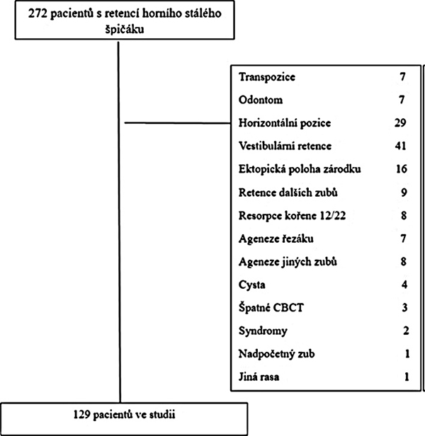
Tato retrospektivní studie byla provedena se souhlasem Etické komise Fakultní nemocnice v Motole a 2. lékařské fakulty Karlovy univerzity v Praze pod číslem EK 870/23. Datový soubor byl tvořen hlavní, kontralaterální a kontrolní skupinou. Pro účely této retrospektivní studie byl soubor pacientů získán z Ortodontického oddělení Stomatologické kliniky dětí a dospělých ve FN Motol, kteří zde byli léčeni v období 2010–2023. Pacienti zařazení do studie splňovali tato kritéria: pacient europoidní rasy, pacienti s jednostrannou, nebo oboustrannou palatinální retencí horního stálého špičáku, plně prořezaný stálý postranní řezák bez výplní, fraktury či jiných patologií, kvalitní CBCT snímek bez artefaktů. Vyřazeni ze studie byli tito pacienti: pacienti s rozštěpy, kraniofaciálními syndromy a jinými vrozenými vadami (onemocněními), pacienti s odontomy v okolí špičáku nebo jinými druhy nádorů, pacienti s nadpočetným zubem v okolí horního špičáku a postranního řezáku, pacienti s folikulárními cystami nebo s jinými typy cyst v okolí horního stálého špičáku, ektopická poloha zárodku horního stálého špičáku, pacienti s horizontální pozicí stálého horního špičáku, pacienti s agenezí horního stálého postranního řezáku, pacienti s agenezí horního stálého špičáku, pacienti jiné než europoidní rasy, pacienti s nekvalitními CBCT snímky, pacienti s dalšími retinovanými zuby, vyjma třetích stálých molárů, pacienti s resorpcí kořene horního stálého postranního řezáku, pacienti s agenezemi dalších stálých zubů, vyjma třetích stálých molárů, pacienti s vestibulární retencí, patologické procesy na korunce či kořenu postranního řezáku a špičáku. Proces vylučování nevhodných pacientů ze souboru zobrazuje obrázek 1.
Z původního počtu 272 pacientů muselo být nakonec vyloučeno 143 pacientů. Velikost studijní skupiny s palatinální retencí horního špičáku tedy činila 129 pacientů (102 pacientů s jednostrannou a 27 pacientů s oboustrannou retencí), z toho 51 mužů (39,53 %; nejvyšší věk 23 let a nejnižší věk 10 let) a 78 žen (60,47 %, nejvyšší věk 20 let a nejnižší 10 let). Průměrný věk celé studijní skupiny byl 13,47 let (SD ±1,79), aritmetický průměr u mužů 13,75 let (SD ± 1,99) a u žen 13,26 (SD ±1,54). Kontrolní skupinu tvořilo 170 pacientů (90 žen, 80 mužů). Všichni pacienti z této skupiny měli fyziologicky prořezané všechny stálé zuby (vyjma třetích stálých molárů) a nevyskytovala se u nich žádná dentální anomálie. CBCT u těchto pacientů bylo zhotoveno pro vyvrácení podezření na fraktury čelistí, nadpočetných zubů apod, které se tímto vyšetřením nepotvrdily. Žádnému pacientovi nebylo indikováno CBCT vyšetření pro potřeby této studie. Věkový průměr byl velice podobný věkovému průměru ve studijní skupině: průměrný věk celé studijní skupiny byl 13,29 let (SD ±1,31), aritmetický průměr u mužů 13,62 let (SD ±1,56) a u žen 13,11 (SD ±1,71).
Všem pacientům, kterým byla diagnostikována palatinální retence horního stálého špičáku na základě klinického vyšetření a OPG, bylo následně pro suspektní resorpci okolních zubů, přítomnost cysty či jiné patologické změny indikováno vyšetření CBCT. Snímky byly provedeny kvalifikovaným rentgenologickým asistentem standardním způsobem na přístroji i-CAT 17-19 (Imaging Science International, Hatfield, PA, USA), s následujícími parametry: FOV 16 cm x 10 cm, napětí 120 kV, proud rentgenky 5 mA, délka snímkování 17 s, tloušťka řezu 0,2 mm, počet nativních snímků 576, velikost izometrického voxelu 0,3 mm. Stejným způsobem bylo zhotoveno i CBCT v kontrolní skupině. Žádné CBCT nebylo zhotoveno pro účely této studie.
Všechny CBCT snímky byly importovány v DICOM formátu do zobrazovacího medicínského softwaru InVivo6 (Anatomage, Santa Clara, CA, USA). Po ořezání zubu od okolních tkání bylo provedeno vlastní měření. K měření vzdáleností bylo použito 2D milimetrové měřítko (přesnost 0,01 mm) a k měření úhlu α 2D úhlové měřidlo (přesnost 0,10). Před samotným měřením v zobrazovacím SW byly nastaveny tři referenční linie: horizontální, sagitální a frontální.
Měřeny byly rozměry lineární a 3D (objem):
Lineární rozměry:
1. Z lineárních rozměrů byly na korunce stálého postranního řezáku měřeny šířka korunky v krčkové oblasti (MD cervikální), šířka incizní hrany (Incize) a nejširší meziodistální rozměr na korunce (MD). Délka korunky byla měřena jako kolmá vzdálenost na vestibulární plošce korunky mezi vrcholem na cementosklovinné hranici a incizí. Bukolingválním rozměrem (BL) se myslí vestibuloorální šířka na cementosklovinné hranici (Obr. 2)
2. Z lineárních rozměrů kořene byly měřeny: délka kořene, která byla definována a měřena jako vzdálenost od cementosklovinné hranice po apex v mm. Poté byla korunka zubu odřezána a kořen natočen tak, aby bylo umožněno měření meziodistálního (MDr) a bukolingválního (BLr) rozměru kořene. Meziodistální rozměr kořene MDr byl definován jako nejširší mesiodistální rozměr kořene (v oblasti cementosklovinné hranice). Bukolingvální rozměr kořene BLr byl definován jako nejširší bukolingvální rozměr kořene (v oblasti cementosklovinné hranice). Obě tyto linie jsou na sebe kolmé. Rozměry byly měřeny rovněž v mm (Obr. 3).
3. Měření délky zahnutí apexu kořene postranního řezáku: Zahnutí kořene bylo definováno jako stav, kdy úhel mezi dlouhou osou zubu a dlouhou osou apikálního zahnutí kořene byl větší než 100 [8]. Délka zahnutí kořene byla měřena jako vzdálenost mezi bodem odklonu od dlouhé osy zubu (kořene) po apex (Obr. 4). Měřeno v mm.
3D rozměry (objem):
4. Celkový objem postranního řezáku, objem korunky a kořene: Zvolený subjekt (zub) byl opět důsledně vyříznut a ořezán o všechny nadbytečné tkáně, aby nedocházelo k nadhodnocení výpočtu objemu. Poté pomocí funkce Volume measurement byl nejdříve změřen celý objem zubu Vt (Obr. 5). Poté byl zub zobrazen zpět do výchozí podoby a funkcí Ořez byl v místě cementosklovinné hranice rozdělen na korunku a kořen. Následně funkcí Volume measurement byl zvlášť vypočítán objem kořene Vt a objem korunky Vc (Obr. 6 a 7).
Další parametry:
5. Přítomnost čípkovitého postranního řezáku: Pro vyhodnocení, zda má řezák čípkovitý tvar, byly porovnány dva rozměry: šířka korunky v krčkové oblasti (MD cervikální) v cementoskolovinné hranici a šířka incize jako vzdálenost dvou nejkrajnějších bodů na incizi (Incize). Postranní řezák byl považován za čípkovitý v případě, že rozměry v krčkové oblasti byly větší než šířka incizní hrany.
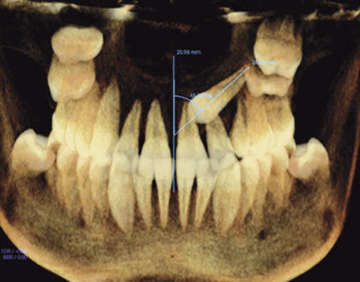
6. Měření úhlu α (tj. úhlu mezi dlouhou osou špičáku a střední linií): Jedná se o úhel mezi střední linií procházející horními středními řezáky, která je kolmá na okluzní linii, a linií reprezentující dlouhou osu retinovaného špičáku. Samotný úhel α je poté měřen pomocí 2D úhlového měřidla (přesnost 0,10).
7. Určení stadia vývoje kořene postranního řezáku: Kořeny postranních řezáků byly hodnoceny podle klasifikace Komínka a Rozkovcové (1984), která sestávala ze sedmi stadií: stadium zubního váčku, stadium počínající mineralizace korunky, stadium pokročilé mineralizace korunky, stadium počáteční tvorby kořene, stadium divergence stěn kořenových kanálků, stadium paralelity kořenových kanálků a stadium konvergence stěn kořenových kanálků.
Všechna měření byla realizována jedním hodnotitelem dvakrát v odstupu tří týdnů. Naměřená data byla zaznamenána do tabulky Excel 2019 (Microsoft, Richmond, USA).
Statistická analýza
Kvantitativní proměnné byly prezentovány pomocí průměrů a směrodatných odchylek (SD), mediánů, minimálních a maximálních hodnot. Shapirovými-Wilkovými testy normality bylo prokázáno normální rozdělení u všech proměnných. Analýza dat byla provedena vzhledem k dostatečně velkému vzorku pomocí parametrických metod. Porovnání 2 závislých vzorků bylo provedeno pomocí párového t-testu. Nezávislé vzorky byly porovnány pomocí analýzy rozptylu (ANOVA) a následnými Dunnettovy post hoc testy. Vzájemný vztah mezi proměnnými byl analyzován pomocí výpočtu Pearsonova korelačního koeficientu a pomocí jednoduché lineární regrese. Hladina významnosti pro všechny testy byla stanovena na hodnotu 0,05. Pokud byla p-hodnota nižší než 0,05, byly považovány rozdíly za statisticky významné, jsou označeny hvězdičkami. Ke statistickému zpracování byl použit statistický software IBM SPSS Statistics for Windows, (Version 23.0. Armonk, NY: IBM Corp). Analýza dat byla provedena ve spolupráci s Mgr. Kateřinou Langovou, Ph.D., Ústav lékařské biofyziky LF UP Olomouc.
Pro výpočet chyby měření bylo provedeno kontrolní měření pro všechny zkoumané proměnné v rozmezí dvou měsíců jedním hodnotitelem. K měření bylo náhodně vybráno třicet pacientů ze studijní, kontralaterální i kontrolní skupiny. Kontrola chyby měření byla provedena pomocí Dahlbergovy formule (D) a relativní Dahlbergovy chyby (RDE) v procentech. Pro zjištění míry absolutní shody mezi prvním a kontrolním měřením byl vypočten koeficient vnitrotřídní korelace (ICC). Relativní Dahlbergova chyba byla spočítána v rozmezí 0,3 % (pro objem zubu) až 5,6 % (pro objem kořene). Hodnoty koeficientu ICC byly zjištěny v rozmezí 0,957 až 0,999 což představuje perfektní shodu.
Výsledky
A. Jednostranná palatinální retence:
Lineární rozměry korunky:
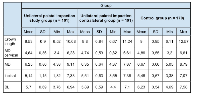
Párovým t-testem bylo prokázáno, že u jednostranně palatinálně retinovaného horního stálého špičáku má horní stálý postranní řezák v hlavní skupině užší korunku než postranní řezák na kontralaterální straně pouze na incizi (p = 0,002). Bukolingvální rozměr korunky v cervikální oblasti je rovněž menší než u postranních řezáků v kontralaterální skupině (p = 0,022) Jiné statisticky významné rozdíly prokázány nebyly. (Tab. 1 a 2). Rovněž bylo prokázáno, že u jednostranně palatinálně retinovaného horního stálého špičáku má horní stálý postranní řezák na postižené straně kratší délku korunky (průměr = 8,53 mm) než postranní řezák na kontralaterální straně (průměr = 8,80 mm, p = 0,013).
Analýza rozptylu prokázala statisticky významné rozdíly v rozměrech korunky mezi porovnávanými skupinami u všech parametrů (p < 0,01). Následně provedené Dunnettovy post-hoc testy prokázaly statisticky významně nižší hodnoty u všech parametrů na postižené straně ve srovnání s kontrolní skupinou. U kontralaterální strany byly statisticky významně nižší hodnoty jen u parametrů MD a BL (Tab. 1 a 2).
Lineární rozměry kořene, zahnutí apexu kořene a vývoj kořene horního stálého postranního řezáku:
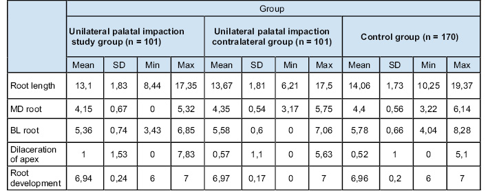
U jednostranně palatinálně retinovaného horního stálého špičáku bylo prokázáno, že horní stálý postranní řezák v hlavní skupině má kratší délku kořene (průměr = 13,10 mm) než postranní řezáky z kontralaterální skupiny (průměr = 13,67 mm, p = 0,019) i z kontrolní skupiny (průměr = 14,1 mm, p < 0,0001). Rozdíl mezi kontralaterální stranou a kontrolní skupinou nebyl statisticky významný (p = 0,147). MDr i BLr rozměry jsou v hlavní skupině významně užší než na kontralaterální straně (p = 0,001 pro MDr, p < 0,0001 pro BLr). U rozměru MDr ANOVA prokázala statisticky významné rozdíly (p = 0,003). Následně provedené Dunnettovy post-hoc testy odhalily statisticky významné rozdíly jen mezi postiženou stranou a kontrolní skupinou (průměr = 4,40 mm, p = 0,001). U rozměru BLr byly naopak zjištěny statisticky významné rozdíly mezi hlavní, kontralaterální i kontrolní skupinou (Tab. 3 a 4).
Výsledky prokázaly, že u jednostranně palatinálně retinovaného horního stálého špičáku mají kořeny horních stálých postranních řezáků na postižené straně významnější zahnutí než kořeny postranních řezáků na kontralaterální straně (p = 0,026) a v kontrolní skupině (p = 0,003). Naopak mezi kontrolní a kontralaterální skupinou nebyl u zahnutí kořene zjištěn žádný statisticky významný rozdíl (p = 0,907) (Tab. 3 a 4). Ve vývoji kořene nebyly zjištěny žádné statisticky významné rozdíly mezi hlavní a kontralaterální skupinou (p = 0,181), kontrolní skupinou (p = 0,485) ani mezi kontrolní a kontralaterální skupinou (p = 0,878) (Tab. 3 a 4).
Objem:
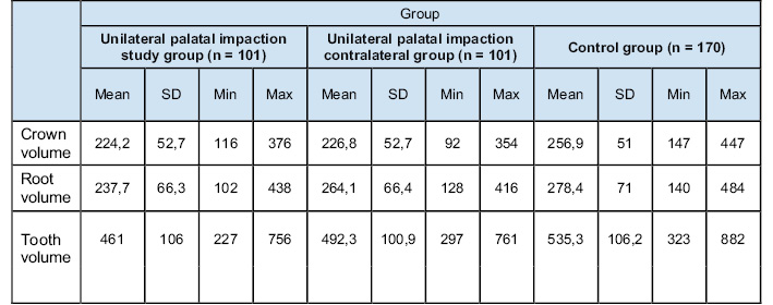
Objem korunky horního stálého postranního řezáku mezi hlavní a kontralaterální skupinou nevykázal žádný statisticky významný rozdíl (p = 0,677), naopak mezi hlavní a kontrolní skupinou (p < 0,0001) i kontralaterální a kontrolní skupinou (p < 0,0001) byly zjištěny statisticky významné rozdíly. Postranní řezáky z hlavní skupiny měly statisticky významně menší objem kořene než řezáky z kontrolní (p < 0,0001) a kontralaterální skupiny (p = 0,014). Rozdíly v objemu kořene mezi kontrolní a kontralaterální skupinou zjištěny nebyly (p = 0,177). Celkový objem zubu byl významně menší u řezáků na straně retence ve srovnání s kontralaterální (p = 0,013) a kontrolní skupinou (p < 0,0001) (Tab. 5 a 6).
Výskyt čípkovitých řezáků:
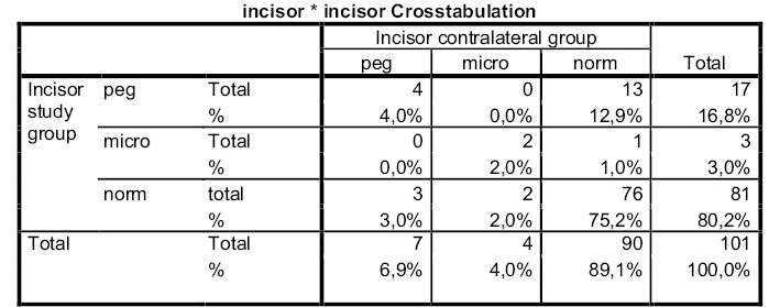
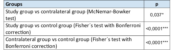
McNemarovým-Bowkerovým testem byl prokázán statisticky významný rozdíl mezi stranami ve výskytu čípkovitých řezáků. Čtyři pacienti mají čípkovité řezáky na obou stranách, 2 pacienti mají mikrodoncii na obou stranách a 76 pacientů má na obou stranách normální řezák. 13 pacientů má na postižené straně čípkovitý řezák a na nepostižené normální tvar (opačná situace nastala pouze u 3 pacientů), jeden pacient má na postižené straně mikrodoncii a na nepostižené má normální tvar (opačná situace je u 2 pacientů) (Tab. 7–8).
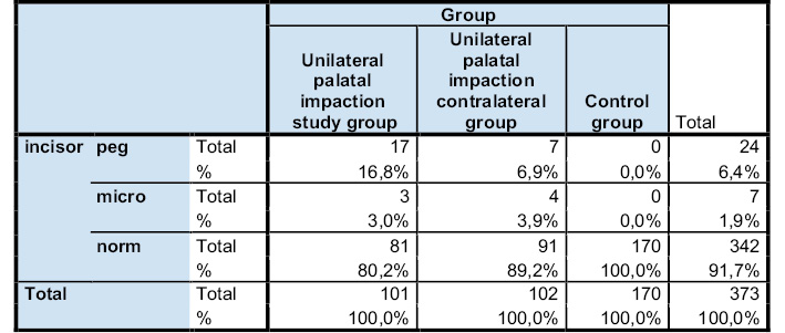
Fisherovým testem bylo prokázáno, že existují rozdíly ve výskytu čípkovitých řezáků (p < 0,0001) u obou skupin ve srovnání s kontrolní skupinou. V kontrolní skupině se vyskytují pouze normální řezáky. V kontralaterální skupině se čípkovité řezáky vyskytují méně často než na postižené straně, ve srovnání s kontrolní skupinou je výskyt čípkovitých řezáků na nepostižené straně statisticky významně vyšší (Tab. 9).
Závislost mezi rozměry kořene postranního řezáku a úhlem mezi střední linií a dlouhou osou retinovaného špičáku:
Mezi rozměry kořene (délka, MDr a BLr) a úhlem, který svírá dlouhá osa špičáku se střední linií (úhel α), neexistuje statisticky významná závislost (Tab. 10).
B. Oboustranná palatinální retence
Rozměry korunky horního stálého postranního řezáku:
Párovým t-testem nebyl prokázán statisticky významný rozdíl mezi pravou a levou stranou v délce korunky horního stálého postranního řezáku (p = 0,121), byly ale prokázány statisticky významně rozdílné hodnoty mezi pravou a levou stranou v MD cervikální (p = 0,044) a v incizi (p = 0,0005) (Tab. 11).
Rozměry kořene horního stálého postranního řezáku:
Párovým t-testem nebyl prokázán statisticky významný rozdíl mezi pravou a levou stranou v MD kořene (p = 0,097), v BL kořene (p = 0,528), v délce kořene (p = 0,179), v zahnutí kořene (p = 0,855), ani ve vývoji kořene (p = 0,161) (Tab. 12).
Diskuse
Podle teorie vedení plní postranní řezák při erupci stálého špičáku funkci jakési vodící lišty, podél které prořezává špičák do dutiny ústní. Pozměněný tvar postranního řezáku, zejména pak tvar kořene, by podle této teorie měl zvyšovat riziko palatinální retence špičáku [6, 9].
Lineární rozměry postranního řezáku u jednostranné palatinální retence
Výsledky této studie se u lineárních rozměrů (měřeno v mm) u jednostranné palatinální retence významně nelišily od předešlých studií, kterých je ve srovnání s objemem relativně mnoho, poněvadž lineární rozměry ve starších studiích byly měřeny na sádrových modelech nebo OPG [10–12]. Becker [13] měřil sice jen na 38 sádrových modelech a pouze rozměry MD a BL, jejich hodnoty ale vyšly podobně jako v této studii (u MD 6,3 vs. 6,5; u BL 5,4 vs. 5,7). Rozdíl ve srovnání s výsledky ve studií Chaushu [14] byl v rozměru MD 0,25 mm a u BL 0,1 mm. Naopak Yan [15] naměřil BL rozměr korunky až o 2 mm delší. Lineární rozměry kořene se nelišily od výsledků většiny studií, řezáky na straně retence měly významně kratší kořen. Gupta [16] naměřil kořen řezáků na straně retence o 0,46 mm kratší ve srovnání s kontrolní skupinou. U nás byl rozdíl s kontralaterální skupinou 0,57 mm a s kontrolní skupinou 0,9 mm. Dubovská [17] ve své studii naměřila rozměry MDr a BLr v průměru o 0,4 mm kratší u řezáků na postižené straně ve srovnání s kontrolní skupinou, v této studii byl v MDr rozměru rozdíl mezi hlavní a kontralaterální skupinou 0,2 mm a mezi hlavní a kontrolní skupinou 0,25 mm. V BLr rozměru měly postranní řezáky na straně retence o 0,22 mm kratší rozměr než řezáky v kontralaterální skupině a o 0,42 mm kratší rozměr než řezáky z kontrolní skupiny.
Objem zubu, korunky a kořene u jednostranné palatinální retence
Pokrok v 3D zobrazovacích technologiích umožňuje měřit nejen lineární či úhlové rozměry zubů, ale umožňuje měřit objem zubů či jiných struktur, jako např. dýchacích cest [18]. Studií měřící objem postranního řezáku je podstatně méně než těch, které měří lineární rozměry. Ve své studii Leonardi [19] měřil z 3D rozměrů pouze objem zubu, jen na 28 pacientech a stejně jako v této studii srovnávali s kontralaterální i kontrolní skupinou. Objem řezáku na straně retence jim stejně jako v této studii vyšel signifikantně menší ve srovnání s kontrolní i kontralaterální skupinou. Ve studii Korala [20], který rovněž měřil jen celý zub, vyšel objem řezáku na straně retence signifikantně menší. Kim [21] měřil jen objem kořene, který vyšel rovněž signifikantně menší. Jen zcela výjimečně se lze setkat s tím, že řezáky na straně palatinální retence měly signifikantně větší objem kořene či zubu ve srovnání s kontralaterální či kontrolní skupinou [22].
Výskyt čípkovitých řezáků, zahnutí a vývoj kořene u jednostranné palatinální retence
Výsledky našeho výzkumu jsou v souladu s většinou dosavadních studií. Černochová [23] zjistila, že ze 165 pacientů s palatinální retencí horního stálého špičáku mělo 9,1 % pacientů čípkovitý řezák, ve studii Beckera [11] 17,2 % pacientů a ve své studii Chaushu [14] objevila 10 čípkovitých řezáků, tj. u 11,36 % pacientů, přičemž 80 % z nich se vyskytovalo u žen.
Vývoj a zakřivení kořene
Výsledky naší studie potvrdily kratší kořen postranních řezáků, ale nikoliv jejich opožděný vývoj. Lövgren [24] naopak ve své studii na 514 pacientech s retinovaným horním stálým špičákem zjistil opožděný vývoj kořene v průměru o 0,2 roku ve srovnání s kontrolní skupinou. Becker [25] ve své studii zase uvádí, že až polovina pacientů s retinovaným špičákem měla opožděný vývoj dentice. U vestibulární retence opožděný vývoj dentice nezaznamenali. U Sajnani [26] byl opožděný vývoj dentice u 30 % pacientů s palatinální retencí. Možné rozdíly výsledků lze zřejmě přičíst metodice výzkumu, zatímco v této studii byla použita metoda dle Komínka a Rozkovcové [27], všechny tyto studie použily Demirjianovu metodu [28–29].
Kořeny řezáků měly rovněž signifikantně delší zakřivení kořene než řezáky z kontralaterální a kontrolní skupiny, asi o 0,5 mm ve srovnání s kontralaterální a kontrolní skupinou. Mezi kontralaterální a kontrolní skupinou statisticky významný rozdíl zjištěn nebyl. Podobné výsledky vyšly v mnoha studiích [8, 30–31]. Jejich autoři shodně uvádí, že se tím potvrzuje teorie vedení, kdy tyto řezáky s abnormálním tvarem kořene neplní plnohodnotně funkci vodící lišty.
Závislost mezi rozměry kořene postranního řezáku a úhlem mezi střední linií a dlouhou osou retinovaného špičáku
Dalším cílem práce bylo rovněž zjistit, zdali existuje nějaká souvislost mezi anatomií kořene postranního řezáku a úhlem, který svírá dlouhá osa špičáku se střední linií (úhel α). Základní myšlenka byla, že pokud by byl kořen nějak pozměněn, např. kratší, prořezávající špičák nenajde vodící lištu, vybočí z erupční dráhy a úhel α se tak zvětší. Kromě délky kořene byly zkoumány ještě rozměry MDr, BLr a objem kořene Vr. Ani u jedné proměnné nebyla zjištěna souvislost s úhlem α. Lze to vysvětlit tím, že úhel α je pravděpodobně ovlivněn jinými faktory, jako je např. anomální poloha zubního zárodku. Srovnání s výsledky jiných studií nejsou dostupná, poněvadž dle našich informací se touto problematikou žádná studie ještě nezabývala.
Oboustranná palatinální retence
Anatomií postranního řezáku u oboustranné retence se zabývalo podstatně méně studií, pravděpodobně kvůli nedostatku velikosti souboru. Ty, které analyzovaly i oboustrannou retenci, si pokládaly stejné hypotézy jako v této studii u jednostranné retence [9, 17]. U oboustranné palatinální retence byl soubor o velikosti 27 pacientů v této studii využit k zodpovězení jiné hypotézy: pokud opravdu platí hypotéza, že palatinální retence je způsobena odlišnou anatomií postranního řezáku, tak by se anatomie postranního řezáku na pravé a levé straně neměla lišit, tedy by nemělo docházet k situaci, že na jedné straně má postranní řezák např. kratší kořen, zatímco na protilehlé straně má kořen normální velikost, byť tam stejně nastala retence špičáku. Mezi pravou a levou stranou by se tedy neměly vyskytovat statisticky významné rozdíly. Výsledky analýzy tuto hypotézu potvrdily: u těchto proměnných, tj. u délky korunky, rozměrech korunky MD a BL, u délky kořene, dále u BLr, MDr a zahnutí kořene nebyl zjištěn statisticky významný rozdíl mezi pravou a levou stranou. Významný rozdíl nebyl naměřen rovněž ani u vývoje kořene či objemu korunky, kořene a celého zubu. Srovnání s jinými studiemi neuvádíme, protože při naší nejlepší znalosti není dostupná žádná studie, která by se zabývala touto výzkumnou otázkou.
Klinická aplikace
Bylo potvrzeno, že palatinální retence horního stálého špičáku má statisticky významnou závislost s pozměněnou anatomií horního stálého postranního řezáku: řezáky měly statisticky významně kratší délku korunky, ve srovnání s kontrolní skupinou byly u palatinální retence naměřeny kratší i další lineární rozměry korunky. Na straně retence byl rovněž potvrzen statisticky významný vyšší výskyt čípkovitých řezáků. Všechny tyto rozměry jsou rozeznatelné v rámci klinického vyšetření, ať už během preventivní prohlídky u praktického zubního lékaře či prvního vyšetření u ortodontisty. Všechny tyto diagnostikované alterované rozměry postranního řezáku by měly vést k indikaci zhotovení OPG přibližně ve věku 9 let, kdy je možné včasné zachycení palatinální retence špičáku. Včasná diagnóza může vést při splnění vhodných indikačních kritérií k včasnému zahájení interceptivní léčby, ať už v podobě extrakcí dočasného špičáku [32], v některých studiích uváděných extrakcí i dočasných prvních molárů [33], rychlé maxilární expanze [34] nebo vytvoření místa v zubním oblouku v oblasti místa prořezání stálého špičáku [35–36].
Rovněž byla potvrzena i statisticky významná souvislost s pozměněnou anatomií kořene: jsou kratší, užší a často mají zakřivení kořene různého stupně. Jsou-li tyto parametry zjištěny na OPG snímcích zhotovených v rámci preventivních prohlídek u PZL, je vhodné pečlivě a pravidelně sledovat erupční dráhu stálého špičáku a v případě splnění indikačních kritérii rovněž přistoupit k zahájení interceptivní léčby [6, 25].
Závěr
– U jednostranné palatinální retence horního stálého špičáku nacházíme pozměněnou anatomii stálého postranního řezáku, a to jak u rozměrů lineárních, tak i u 3D rozměrů. Rovněž je i vyšší výskyt čípkovitých řezáků.
– Postranní řezáky na straně retinovaného špičáku nemají opožděný vývoj.
– Mezi anatomií kořene postranního řezáku a úhlem mezi dlouhou osou špičáku a střední linií nebyla nalezena žádná korelace.
– U oboustranné palatinální retence horního stálého špičáku nebyly nalezeny rozdíly v anatomii kořene postranních řezáků mezi pravou a levou stranou. U korunky byly statisticky významné rozdíly pouze na incizi a MD cervikální.
Autoři nemají komerční, vlastnické nebo finanční zájmy na produktech nebo společnostech popsaných v tomto článku.
Introduction
The upper permanent canines are among the most important teeth of human dentition, however, they are relatively often associated with impaction, where the tooth does not spontaneously erupt into the oral cavity during the period of its physiological eruption [1]. This dental anomaly with prevalence in the range of around 1.7–4.7% is always at least to some extent a complication associated with the extension of orthodontic treatment [2] averaging 46.7 weeks in case of palatal impacted tooth; vestibular impaction treatment is reported to be shorter by several weeks [3].
There are several opinions on the etiology of upper permanent canines impaction, and no consensus has yet been reached in the literature. Overall, the causes are relatively rare, with the literature limited to case reports. Genetic theory is reported most frequently, as well as idiopathic factors including primary eruption failure, hypothyroidism, hypopituitarism, hypoparathyroidism, hypovitaminosis A and D, Down syndrome, amelogenesis imperfecta, dysostosis cleidocranialis and other rare diseases, achondroplasia, osteoporosis, abnormal muscle flexion and febrile diseases [4, 5].
Local factors include agenesis and anomalies of the lateral incisor, delayed dentition development, transverse dimensions of the upper jaw, spatial dimensions in the dental arch, persistence or premature loss of the temporary canine, supernumerary teeth, odontomas and other local obstructions, local inflammatory processes in the temporary canine and surrounding teeth, cysts, follicular sac disorders, trauma, root dillation, ankylosis, abnormal root position of the first premolar, abnormal position or malformation of the tooth germ, long eruption pathway, increased bone density, angle between the anterior base of the cranial and mandibular lines, and cleft palate [4, 5, 6].
Of the aforementioned etiological factors, genetic theory and tooth guiding theory are most often reported in the literature [6, 7]. Genetic theory assumes the influence of genetic factors on the impaction of the upper permanent canine, guidance theory assumes the influence of the root of the lateral incisor, which performs the function of a guide bar during the eruption of the canine. Failure of the function of this guide bar (whether in the form of agenesis or a change in the shape of the root in terms of shortening, narrowing and shrinking the volume) leads, according to this theory, to displaced upper permanent canine.
The objective of this retrospective study was to determine whether there was any statistically significant dependence between the palatal retention of the upper permanent canine and the altered anatomy of the upper permanent lateral incisor. The zero hypotheses in this study were: there is no statistically significant difference in the linear and 3D dimensions of the upper permanent lateral incisor between the major, contralateral and control groups for the unilateral palatal impacted upper permanent canine. Other established null hypotheses: there is no statistically significant difference in the incidence of peg incisors and root development; there is no relationship between the anatomy of the root of the upper permanent lateral incisor and the angle between the canine long axis and the midline (angle α).
Material and methodology
This retrospective study was conducted with the approval of the Ethics Committee of the University Hospital of Motol and the 2nd Medical Faculty of Charles University in Prague under EC No. 870/23. The dataset was made up of main, contralateral and control groups. For the purpose of this retrospective study, a sample of patients were recruited from the Orthodontic Department of The Dental Clinic of Children and Adults at Motol Faculty Hospital who were treated there during the period 2010–2023. Patients enrolled in the study met the following criteria: patients of Europoid race, patients with unilateral or bilateral palatal impacted upper permanent canine, a fully erupted permanent lateral incisor without fillings, fractures or other pathologies, a quality CBCT scan without artifacts. The following patients were excluded from the study: patients with clefts, craniofacial syndromes and other birth defects (diseases), patients with odontomas around the canine or other types of tumors, patients with supernumerary teeth around the upper canine and lateral incisor, patients with follicular cysts or other types of cysts around the upper permanent canine, ectopic germ position of the upper permanent canine, patients with the horizontal position of the permanent upper canine, patients with missing upper permanent lateral incisor, patients of other than Europoid race, patients with poor quality CBCT, patients with other impacted teeth with the exception of third permanent molars, patients with upper permanent lateral incisor root resorption, patients with other missing permanent teeth with the exception of third permanent molars, patients with vestibular impaction, with pathologies of the crown or root of lateral incisor and canine. The process of excluding inappropriate patients from the sample is shown in Figure 1.
Of the initial 272 patients, 143 had to be excluded. Thus, the size of the upper canine palatal impaction study group was 129 patients (102 patients with single-sided and 27 patients with double-sided impaction; 51 men (39.53%; highest age 23 and lowest age 10) and 78 women (60.47%, highest age 20 and lowest 10 years). The mean age of the entire study group was 13.47 years (SD ±1.79), the arithmetic mean for men was 13.75 years (SD ±1.99) and for women was 13.26 (SD ±1.54). The control group consisted of 170 patients (90 women, 80 men). All patients in this group had all their permanent teeth physiologically erupted (except for the third permanent molars) and did not experience any dental anomalies. CBCT in these patients was made to disprove suspected fractures of the jaws, supernumerary teeth, etc., which were not confirmed by this examination. No patients were indicated a CBCT scan for the needs of this study. The age average was very similar to the age average in the study group: the mean age of the entire study group was 13.29 years (SD ±1.31), the arithmetic mean for men was 13.62 years (SD ±1.56), and for women it was 13.11 (SD ±1.71).
All patients diagnosed with palatal impacted upper permanent canine based on clinical examination and OPG were subsequently indicated for CBCT testing due to suspected resorption of surrounding teeth, presence of a cyst or other pathological changes. The scans were taken by a qualified X-ray assistant in the standard manner on the i-CAT 17-19 (Imaging Science International, Hatfield, PA, USA) instrument, with the following parameters: FOV 16 cm x 10 cm, voltage 120 kV, X-ray current 5 mA, length 17 s, cut thickness 0.2 mm, number of native images 576, isometric voxel size 0.3 mm. The CBCT in the control group was also made in the same way. No CBCT was produced for the purpose of this study.
All CBCT images were imported in DICOM format into Inivo6 (Anatomage, Santa Clara, CA, USA) imaging medicine software. After trimming the tooth from surrounding tissues, a measurement was made. A 2D millimeter scale (accuracy 0.01 mm) was used to measure distances and a 2D angular gauge (accuracy 0.1°) was used to measure α. Prior to the actual measurement in the imaging SW, three reference lines were set: horizontal, sagittal and frontal.
Linear and 3D (volume) dimensions were measured:
Linear dimensions:
1. From linear dimensions, the width of the crown in the neck region (MD cervical), the width of the incisal edge (Incision) and the widest mesiodistal dimension on the crown (MD) were measured. The length of the crown was measured as the perpendicular distance on the vestibular plane of the crown between the tip on the cementoenamel junction and the incision. Buccolingual dimension (BL) means vestibulooral width on the cementoenamel junction (Fig. 2).
2. From the linear dimensions of the root, the following measurements were made: the length of the root, which was defined and measured as the distance from the cementoenamel junction to the apex in mm. The crown of the tooth was then cut off and the root rotated to allow for the measurement of the mesiodistal (MDr) and buccolingual (BLr) root dimensions. The mesiodistal dimension of the root MDr was defined as the widest mesiodidal dimension of the root (in the CEJ area). The buccolingual dimension of the root BLr was defined as the widest buccolingual dimension of the root (in the CEJ area). Both of these lines are perpendicular to each other. Dimensions were also measured in mm (Fig. 3).
3. Measurement of lateral incisor root apex dilaceration length: The root dilaceration was defined as the condition when the angle between the long axis of the tooth and the long axis of the apical dilaceration of the root was greater than 100 [8]. The length of the root dilaceration was measured as the distance between the point of deflection from the long axis of the tooth (root) and the apex (Fig. 4). Measured in mm.
3D dimensions (volume):
4. Total lateral incisor volume, crown and root volume: The chosen subject (tooth) was again rigorously cut and trimmed off all the extra tissues to avoid overestimating the volume calculation. Then, using the Volume measurement function, the entire volume of the tooth Vt was first measured (Fig. 5). The tooth was then shown back to its default form and by the Trimming function was divided into a crown and a root at the point of cementoenamel junction. Subsequently using the Volume measurement function we separately calculated the volume of the root Vt and the volume of the crown Vc (Fig. 6 and 7).
Other parameters:
5. Presence of peg lateral incisor: Two dimensions were compared to assess whether the incisor had a cone shape: the width of the crown in the neck region (MDc) in the cementoenamel junction and the width of the incision as the distance of the two most extreme points per incision (Incision). The lateral incisor was considered peg if the dimensions in the neck area were greater than the width of the incisal edge.
6. Measurement of the angle α (i.e. the angle between the long axis of the canine and the middle line): This is the angle between the middle line running through the upper central incisors, which is perpendicular to the occlusive line, and the line representing the long axis of the impacted canine. The angle α itself is then measured using a 2D angular gauge (accuracy 0.1°).
7. Determination of lateral incisor root development stage: The roots of the lateral incisors were evaluated according to the classification by Komínek and Rozkovcová (1984), which consisted of seven stages: the stage of the dental sac, the stage of incipient crown mineralization, the stage of advanced crown mineralization, the stage of incipient root formation, the stage of divergence of root canal walls, the stage of parallelism of root canals, and the stage of convergence of root canal walls.
All measurements were taken by one evaluator twice three weeks apart. The measured data was recorded in the Excel 2019 table (Microsoft, Richmond, USA).
Statistical analysis
Quantitative variables were presented using averages and standard deviations (SDs), medians, minimum and maximum values. Shapiro-Wilk normality tests showed a normal distribution for all variables. Thanks to a sufficiently large sample the analysis of the data was carried out using parameters. A comparison of 2 dependent samples was made using a pair t-test. The independent samples were compared using variance analysis (ANOVA) and subsequent Dunnett post-hoc tests. The interrelationship between the variables was analyzed by calculating the Pearson correlation factor and using a simple linear regression. The significance level for all tests was set at 0.05. If the p-value was less than 0.05, the differences were considered statistically significant, marked with asterisks. IBM statistical software SPSS Statistics for Windows, (Version 23.0) was used for statistical processing (Armonk, NY: IBM Corp). The data analysis was carried out in cooperation with Mgr. Kateřina Langová, Ph.D., Institute of Medical Biophysics, Faculty of Medicine and Dentistry, Palacky University Olomouc.
To calculate the measurement error, a control measurement was made for all the variables examined over a two-month span by a single evaluator. Thirty patients from the study, contralateral and control groups were randomly selected for measurements. The measurement error check was made using Dahlberg formula (D) and relative Dahlberg error (RDE) in percents. An intra-class correlation factor (ICC) was calculated to determine the absolute match rate between the first and the control measurements. The relative Dahlberg error was calculated in the range of 0.3 per cent (for tooth volume) to 5.6 per cent (for root volume). ICC values were found to be in the range of 0.957 to 0.999, which represents a perfect match.
Results
A. Unilateral palatal impaction:
Linear dimensions of the crown:
A pair t-test showed that a single-sided palatal impacted upper permanent canine has a narrower crown than a contralateral incisor on the incision only (p = 0.002). The buccolingual dimension of the crown in the cervical region is also smaller than that of the lateral incisors in the contralateral group (p = 0.022) Other statistically significant differences were not proved. (Tab. 1 and 2). It has also been demonstrated that the single-sided palatal impacted upper permanent canine the upper permanent lateral incisor had a shorter crown length on the affected side (mean = 8.53 mm) than the contralateral lateral incisor (mean = 8.80 mm, p = 0.013).
ANOVA showed statistically significant differences in crown dimensions between the groups compared for all parameters (p < 0.01). Subsequently, Dunnett’s post-hoc tests showed statistically significantly lower values for all parameters on the affected side compared to the control group. For the contralateral side, only the MD and BL parameters were statistically significantly lower (Tab. 1 and 2).
The linear dimensions of the root, the dilaceration of the root apex and the development of the root of the upper permanent lateral incisor:
In case of the unilateral palatal impacted upper permanent canine it was shown that the upper permanent lateral incisor in the study group had a shorter root length (mean = 13.10 mm) than the contralateral group lateral incisors (mean = 13.67 mm, p = 0.019) as well as the control group lateral incisors (mean = 14.1 mm, p < 0.0001). The difference between the contralateral side and the control group was not statistically significant (p = 0.147). Both MDr and BLr dimensions are significantly narrower in the main group than on the contralateral side (p = 0.001 for MDr, p < 0.0001 for BLr). For the MDr dimension, ANOVA showed statistically significant differences (p = 0.003). Subsequently, Dunnett’s post-hoc tests revealed statistically significant differences between the affected side and control group only (mean = 4.40 mm, p = 0.001). In contrast, the BLr dimension showed statistically significant differences between the main, contralateral and control groups (Tab. 3 and 4).
The results showed that in the unilateral palatal impacted upper permanent canine, the roots dilacerations of the upper permanent lateral incisors on the affected side were more significant than those of the contralateral lateral incisors (p = 0.026) and in the control group (p = 0.003). Conversely, there was no statistically significant difference (p = 0.907) between the control and the contralateral group for root dilaceration (Tab. 3 and 4). There were no statistically significant differences in root development between the main and contralateral groups (p = 0.181), the control group (p = 0.485), or the control and contralateral group (p = 0.878) (Tab. 3 and 4).
Volume:
The crown volume of the upper permanent lateral incisor between the major and contralateral groups showed no statistically significant difference (p = 0.677), whereas statistically significant differences were found between the major and control groups (p < 0.0001) as well as the contralateral and control groups (p < 0.0001). Lateral incisors from the main group had statistically significantly less root volume than those from the control (p <0.0001) and contralateral groups (p = 0.014). There were no differences in root volume between the control and contralateral groups (p = 0.177). The total tooth volume was significantly smaller in the incisors on the impaction side compared to the contralateral (p = 0.013) and control group (p < 0.0001) (Tab. 5 and 6).
Peg incisors:
The McNemar-Bowker test showed a statistically significant difference in the incidence of peg incisors between the sides. Four patients had peg incisors on both sides, 2 patients had microdontia on both sides and 76 patients had a normal incisor on both sides. 13 patients had a peg incisor on the affected side and a normal shape on the unaffected side (the opposite situation occurred in only 3 patients), one patient has microdontia on the affected side and a normal shape on the unaffected side (the opposite situation is in 2 patients) (Tab. 7–8).
Fisher’s test showed that there were differences in the incidence of peg incisors (p < 0.0001) between the two groups compared to the control group. Only normal incisors occurred in the control group. In the contralateral group, peg incisors occur less frequently than on the affected side, compared to the control group, the incidence of peg incisors on the non-affected side is statistically significantly higher (Tab. 9).
Correlation between the dimensions of the lateral incisor root and the angle between the midline and the long axis of the impacted canine:
There is no statistically significant correlation between the dimensions of the root (length, MDr and BLr) and the angle between the long axis of the canine and the midline (angle α) (Tab 10).
B. Bilateral palatal impaction
Dimensions of the crown of the upper permanent lateral incisor:
The pair t-test showed no statistically significant difference between the right and left sides of the upper permanent lateral incisor crown length (p = 0.121), but showed statistically significant differences between the right and left sides in the cervical MD (p = 0.044) and in the incision (p = 0.0005) (Tab. 11).
Dimensions of the root of the upper permanent lateral incisor:
The pair t-test showed no statistically significant difference between the right and left sides in the root MD (p = 0.097), in the root BL (p = 0.528), in the root length (p = 0.179), in the root dilaceration (p = 0.855), or in the root development (p = 0.161) (Tab. 12).
Discussion
According to the guidance theory, in the eruption of the permanent canine the lateral incisor acts as a sort of a guide bar, along which the canine erupts into the oral cavity. According to this theory the altered shape of the lateral incisor, especially the shape of the root, should increase the risk of palatal impaction of the canine [6, 9].
Linear dimensions of the lateral incisor in unilateral palatal impaction
The results of this study were not significantly different for linear dimensions (measured in mm) of single-sided palatal impaction from previous studies, of which there are relatively many compared to those dealing with the volume, as linear dimensions in older studies were measured in plaster models or OPG [10–12]. Becker [13] measured only on 38 plaster models and only the dimensions of MD and BL, but their values came out similarly to this study (for MD 6.3 vs. 6.5; u BL 5.4 vs. 5.7). Compared to the results in the study by Chaushu [14] the difference was 0.25 mm in MD and 0.1 mm in BL. In contrast, Yan [15] measured the BL crown dimension up to 2 mm longer. The linear dimensions of the root were not different from those of most studies, with the incisors on the affected side having a significantly shorter root. Gupta [16] measured the root of the incisors on the affected side 0.46 mm shorter compared to the control group. For us, the difference was 0.57 mm with the contralateral group and 0.9 mm with the control group. Dubovská [17] measured the dimensions of MDr and BLr on average 0.4 mm shorter in the incisors on the affected side compared to the control group, in this study the difference between the main and contralateral groups was 0.2 mm in MDr and 0.25 mm between the main and control groups. In BLr dimension, the lateral incisors on the affected side were 0.22 mm shorter than those in the contralateral group and 0.42 mm shorter than those in the control group.
Tooth, crown and root volume in unilateral palatal impaction
Advances in 3D imaging technologies make it possible to measure not only linear or angular dimensions of teeth, but also to measure the volume of teeth or other structures, such as airways [18]. There are significantly less studies measuring lateral incisor volume than those measuring linear dimensions. In his study, Leonardi [19] measured only tooth volume from 3D dimensions, in only 28 patients and, as in this study, compared with both the contralateral and control groups. As in this study, the volume of the incisor on the impaction side came out significantly smaller compared to both the control and contralateral groups. In the Korala [20] study, which also measured only the whole tooth, the volume of the incisor on the impaction side came out significantly smaller. Kim [21] measured only the volume of the root, which also came out significantly smaller. It is only very rare to see that the incisors on the palatal impaction side had significantly greater root or tooth volume compared to the contralateral or control group [22].
Occurrence of peg incisors, dilaceration and root development in unilateral palatal impaction
The results of our study are in line with most studies to date. Černochová [23] found that of the 165 patients with palatal impacted upper permanent canine, 9.1% had a peg incisor; in the study by Becker [11] it was 17.2%, and Chaushu [14] 10 peg incisors, i.e. 11.36% of patients, with 80% of them were found in women.
Root development and dilaceration
The results of our study confirmed the shorter root of lateral incisors, but not their delayed development. Lövgren [24], on the other hand, found delayed root development of an average of 0.2 years compared to the control group in his study of 514 patients with an impacted upper permanent canine. In turn, Becker [25] reports in his study that up to half of patients with an impacted canine had delayed dental development. For vestibular retention, they did not experience delayed dentition development. In Sajnani [26], 30% of patients with palatal impaction retention experienced delayed dental development. The possible differences in results are probably attributable to the research methodology, whereas in this study Komínek and Rozkovcová [27] method was used, all of these studies used Demirjian’s method [28–29].
Incisor roots also had significantly longer root dilaceration than incisors from the contralateral and control groups, about 0.5 mm compared to the contralateral and control groups. There was no statistically significant difference between the contralateral and control groups. Similar results came out in many studies [8, 30–31]. Their authors consistently state that this confirms the theory of guiding, whereby these incisors with an abnormal shape of the root do not fully perform the function of the guide bar.
Correlation between the dimensions of the root of the lateral incisor and the angle between the midline and the long axis of the impacted canine
Another goal of the work was also to determine whether there was any connection between the root anatomy of the lateral incisor and the angle between the canine long axis and the midline (angle α). The basic idea was that if the root had been altered in any way, e.g. a shorter, erupting canine would not find a guide bar, it would deflect of the eruption path and thus the angle α would increase. In addition to the root length, the dimensions of MDr, BLr and the volume of root Vr were examined. Neither variable was found to be related to the angle α. This can be explained by the fact that the angle α is likely to be influenced by other factors, such as the anomalous position of the tooth germ. Comparisons with the results of other studies are not available because, according to our information, no study has yet looked at this issue.
Bilateral palatal impaction
Significantly fewer studies looked at the anatomy of the lateral incisor in the bilateral impaction, probably due to a lack of sample size. Those that analyzed bilateral impaction worked with the same hypotheses as in this study for unilateral impaction [9, 17]. In bilateral palatal impaction, the 27-patient sample in this study was used to answer another hypothesis: if the hypothesis that palatal impaction is caused by a different anatomy of the lateral incisor is valid, then the anatomy of the lateral incisor on the right and left should not differ, so there should not be a situation where on one side the incisor has, for example, a shorter root, while on the opposite side the root has a normal size, although there is impacted canine anyway. Thus, there should be no statistically significant differences between the right and left sides. The results of the analysis confirmed this hypothesis: for these variables, i.e. crown length, crown dimensions MD and BL, root length, further for BLr, MDr and root dilaceration, there was no statistically significant difference between the right and left sides. A significant difference was also not measured in the development of the root or volume of the crown, root and whole tooth. We do not make comparisons with other studies because, to our best knowledge, no study is available to address this research question.
Clinical application
It has been confirmed that the palatal impaction of the upper permanent canine has a statistically significant correlation with the altered anatomy of the upper permanent lateral incisor: the incisors had significantly shorter crown compared to the control group, as well as other linear dimensions of the crown. A statistically significantly higher incidence of peg incisors was also confirmed on the impaction side. All of these dimensions are discernible in a clinical examination, whether during a dental practitioner’s preventive examination or a first examination in an orthodontist’s. All of these diagnosed alternating dimensions of the lateral incisor should lead to the indication of OPG production at approximately 9 years of age, when early diagnosis of the palatal impaction of the canine is possible. When the appropriate criteria are met, early diagnosis may lead to the early start of interceptive treatment, whether in the form of temporary canine extractions [32], in some studies are reported also extractions of temporary first molars [33], rapid maxillary expansion [34], or securing of a space in the dental arch for the permanent canine eruption [35–36].
A statistically significant correlation with altered root anatomy has also been confirmed: they are shorter, narrower and often have root dilaceration of varying degrees. If these parameters are detected on OPG scans made as part of the preventive examination scans, it is advisable to closely and regularly monitor the eruption pathway of the permanent canine and, if the criteria are met, also proceed to the initiation of the interceptive treatment [6, 25].
Conclusion
– In the unilateral palatal impaction of the upper permanent canine, we find the altered anatomy of the permanent lateral incisor, both in linear and 3D dimensions. There is also a higher incidence of peg incisors.
– Lateral incisors on the impacted canine side do not develop late.
– No correlation was found between the root anatomy of the lateral incisor and the angle between the long axis of the canine and the midline.
– There were no differences in the anatomy of the root of the lateral incisors between the right and left sides in the bilateral palatal impaction of the upper permanent canine. For crown, the statistically significant differences were found only for incision and MD cervical.
Authors do not have commercial, proprietary or financial interests in the products or companies described in this article.
Obr. 1/ Fig. 1
Vyloučení nevhodných pacientů ze studie / Exclusion of ineligible patients from study
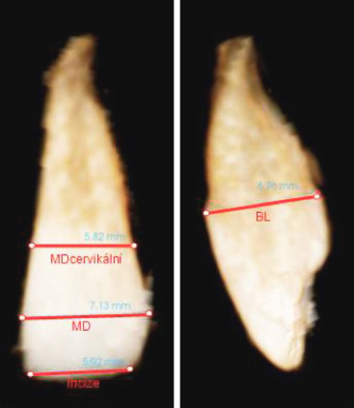
Obr. 2/ Fig. 2
Měření rozměrů korunky v meziodistálním (MD) a bukolingválním rozměru (BL) / Measurement of crown dimensions in the mesiodistal (MD) and buccolingual (BL) directions
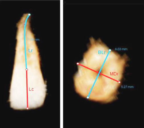
Obr. 3/ Fig. 3
Měření délky korunky a kořene a dalších rozměrů kořene / Measurement of the crown and root lengths and other root dimensions
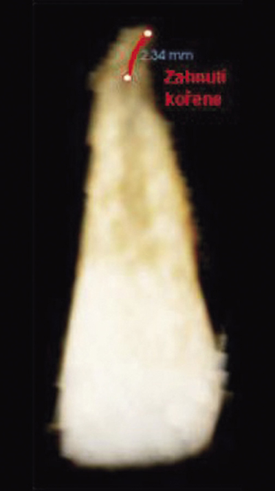
Obr. 4 / Fig. 4
Délka zahnutí kořene / Length of root dilaceration
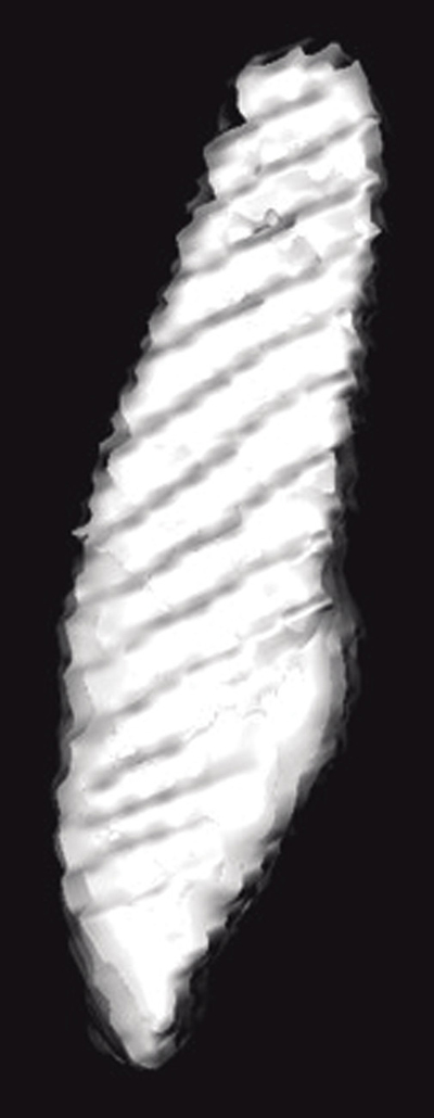
Obr. 5 / Fig. 5
Vizualizace naměřeného objemu vystřiženého postranního řezáku / Visualisa tion of measured volume of cut-out lateral incisor
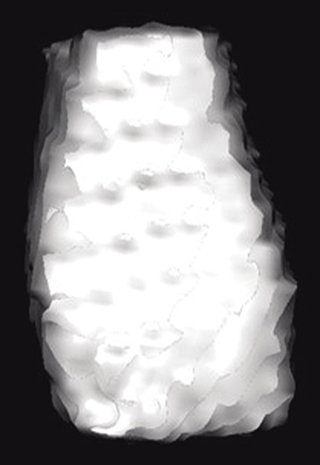
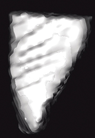
Obr. 6 a, b / Fig. 6 a, b
Vizualizace naměřeného objemu korunky postranního řezáku / Visualisation of measured volume of crown of lateral incisor
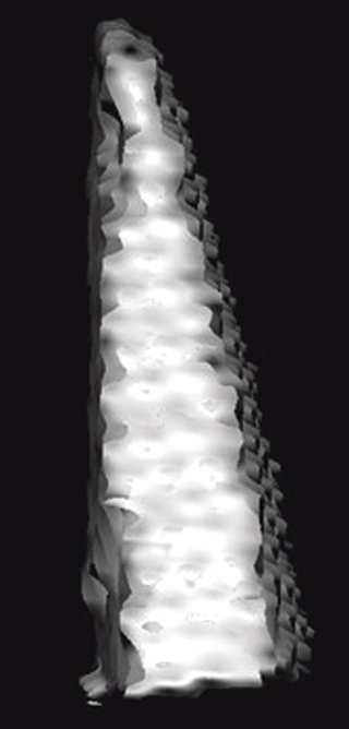

Obr. 7 a, b / Fig. 7 a, b
Vizualizace naměřeného objemu vystřiženého kořene zubu / Visualisation of measured volume of cut-out root of lateral incisor
Obr. 8/ Fig. 8
Úhel α mezi dlouhou osou špičáku a střední linií / Angle α between long axis of canine and midline
Tab. 1 / Tab. 1
Rozměry korunky horní stálého postranního řezáku u jednostranné retence horního stálého špičáku / Crown dimensions of maxillary permanent lateral incisor by unilateral impaction of maxillary permanent canine
Tab. 2 / Tab. 2
Rozměry korunky horního stálého postranního řezáku u jednostranné retence horního stálého špičáku / Crown dimensions of maxillary permanent lateral incisor by unilateral impaction of maxillary permanent canine

Tab. 3 / Tab. 3
Rozměry kořene horního stálého postranního řezáku u jednostranné retence horního stálého špičáku / Root dimensions of maxillary permanent lateral incisor by unilateral impaction of maxillary permanent canine
Tab. 4 / Tab. 4
Rozměry kořene horního stálého postranního řezáku u jednostranné retence horního stálého špičáku / Root dimensions of maxillary permanent lateral incisor by unilateral impaction of maxillary permanent canine

Tab. 5 / Tab. 5
Objem horního stálého postranního řezáku / Volume of maxillary permanent lateral incisor
Tab. 6 / Tab. 6
Rozměry kořene / paired t-test and Dunnett’s tests – root dimensions/p>

Tab. 7 / Tab. 7
Porovnání výskytu čípkovitých řezáků na postižené (v řádcích) a nepostižené (ve sloupcích) straně / Comparison in incidence of peg lateral incisors on affected side (in lines) and unaffected side (in columns)
Tab. 8 / Tab. 8
Porovnání skupin ve výskytu čípkovitého řezáku / Comparison in groups in incidence of peg incisor
Tab. 9 / Tab 9
McNemarův-Bowker test a Fisherovy testy / p-values of McNemar-Bowker test and Fisher’s tests
Tab. 10 / Tab. 10
Korelační analýza mezi rozměry kořene postranního řezáku a úhlem, který svírá dlouhá osa špičáku se střední linií (úhel α) / p-values of correlation analysis among root dimensions of lateral incisor and angle between long axis of canine and midline

Tab. 11 / Tab. 11
Rozměry korunky horního stálého postranního řezáku u oboustranné retence horního stálého špičáku / Crown dimensions of maxillary lateral permanent incisor by bilateral impaction of maxillary permanent canine
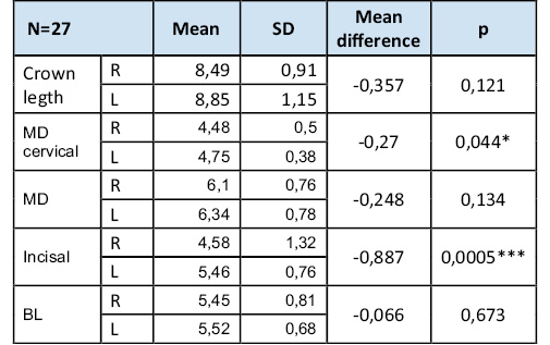
R - pravá strana, right side. L – levá strana, left side
Tab. 12 / Tab. 12
Rozměry kořene horního stálého postranního řezáku u oboustranné retence horního stálého špičáku / Root dimensions of maxillary lateral permanent incisor by bilateral impaction of maxillary permanent canine
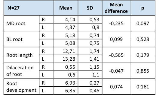
R - pravá strana, right side, L – levá strana, left side

Literatura / References
1. Kamínek M. et. al. Ortodoncie. Galén, Praha, 2014, Galén; 2014.
2. Lövgren ML, Dahl O, Uribe P, Ransjö M, Westerlund A. Prevalence of impacted maxillary canines – an epidemiological study in a region with systematically implemented interceptive treatment. Eur J Orthod. 2019; 41(5): 454-459. doi: 10.1093/ejo/cjz056
3. oh PKT, Puöemotov A, Nguyen H, Pinto N, Olive R. Treatment duration by morphology and location of impacted maxillary canines: A cone-beam computed tomography investigation. Am J Orthod Dentofacial Orthop. 2024;166(2):160-170. doi: 10.1016/j.ajodo.2024.04.010
4. Litsas G, Acar A. A review of early displaced maxillary canines: etiology, diagnosis and interceptive treatment. OpenDent J. 2011; 5: 39-47. doi: 10.2174/1874210601105010039
5. Jacobs SG. The impacted maxillary canine. Further observations on aethiology, radiographic localization, prevention/interception of impaction, and when to suspect impaction. Aus Dent J.1996;41(5):310-316. doi: 10.1111/j.1834-7819.1996.tb03139.x
6. Becker A, Chaushu S. Etiology of maxillary canine impaction: A review. Amer. J. Orthodont. dentofacial Orthop. 2015; 148(4): 557-567. doi: 10.1016/j.ajodo.2015.06.013
7. Peck S, Peck L, Kataja M. The pallataly displaced canine as a dental anomaly of genetic origin. Angle Orthodont. 1994;64(4):249-256. doi: 10.1043/0003-3219(1994)064<0249:WNID>2.0.CO;2
8. Cao D, Shao B, Izadikhah I, Xie L, Wu B, Li H, Yan B. Root dilaceration in maxillary impacted canines and adjacent teeth: A retrospective analysis of the difference between buccal and palatal impaction. Am J Orthod Dentofac Orthop Off Publ Am Assoc Orthod Its Const Soc Am Board Orthod. 2021;159(2):167–74. doi: 10.1016/j.ajodo.2019.12.019
9. Becker A, Sharabi S, Chuashu S. Maxillary tooth size variation in dentitions with palatal canine displacement. Eur J Orthodont. 2002(3);24:313-318. doi: 10.1093/ejo/24.3.313
10. Brin I, Becker A, Shalhav M. Position of the maxillary permanent canine in relation to anomalous or missing lateral incisors: a population study. Eur. J. Orthodont. 1986; 8(1): 12-16. doi: 10.1093/ejo/8.1.12
11. Becker A, Smith P, Behar R. The incidence of anomalous lateral incisors in relation to palatally-displaced cuspids. Angle Orthodont. 1981; 51(1): 24-29. doi: 10.1043/0003-3219(1981)051<0024:TIOAML>2.0.CO;2
12. Anic-Milosevic, Varga S, Mestrovic S, Lapter-Varga and Slaj M. Dental and occlusal feaures in patients with palatally displaced canines. Eur. J. Orthod. 2009; 31(4): 367-373. doi: 10.1093/ejo/cjp014
13. Becker A, Gillis I, Shpack N. The etiology of palatal displacement of maxillary canines. Clin. Orthodont. Res. 1999;2(2):62-66. doi: 10.1111/ocr.1999.2.2.62
14. Chaushu S, Sharabi S, Becker A. Dental morphologic characteristics of normal versus delayed developing dentitions with palatally displaced canines. Amer. J. Orthodont. dentofacial. Otrhop. 2002;121(4):339-46. doi: 10.1067/mod.2002.121559
15. Yan B, Sun Z, Fields H, Wang L, Luo L. Etiologic factors for buccal and palatal maxillary canine impaction: A perspective based on cone-beam computed tomography analyses. Amer. J. Orthodont. dentofacial Orthop. 2013; 143(4):527-534. doi: 10.1016/j.ajodo.2012.11.021
16. Gupta A, Chandna A, Taragi H, Bhattacharya P and Bhandari R. Relationship of the impacted maxillary canine with the morphology of adjacent incisor – a retrospective study. International Journal of advanced research. 2024;12(07):716-718. doi: 10.21474/IJAR01/19105
17. Dubovská I. el. al. Lateral incisor morphology in patients with impacted canine: A CT Study, IOSR-JDMS 2015;14:1-5. doi: 10.6084/M9.FIGSHARE.1488613.V1
18. Tang H, Zhou Q, Li H, Zheng F, Cui X, Jiang J. Exploring long-term changes and influencing factors of the upper airway in patiens with a skeletal Class II relationship after mandibular advancement with maxillary setback surgery: A comprehensive 2-year follow-up nvestigation. Amer. J. Orthodont.dentofacial. Orthop. 2024; 165(5):520-532e. doi: 10.1016/j.ajodo.2023.11.010
19. Leonardi R, Muraglie S, Crimi S, Pirroni M, Musumeci G and Perrotta R. Morphology of palatally displaced canines and adjacent teeth, a 3-D evaluation from cone-beam computed tomographic images. BMC Oral Health. 2018; 18(1):156. doi: 10.1186/s12903-018-0617-0
20. Koral S, Özçirpici AA, Tunçer NI. Association Between Impacted Maxillary Canines and Adjacent Lateral Incisors: A Retrospective Study with COne Beam Computed Tomography. Turkish Journal of Orthodontics. 2021;34(4):207-213. doi: 10.5152/TurkJOrthod.2021.20148
21. Kim Y, Hyun HK, Jang KT. Morphological relationship analysis of impacted maxillary canines and the adjacent teeth on 3-dimensional reconstructed CT images. Angle Orthod. 2017;87(4):591-597. doi: 10.2319/0003-3219-87.5.791
22. Shahabi M, Zarch HH, Shadman Z, Ahrari F. Root volume measurements of maxillary canines and lateral incisors in patients with unilateral maxillary canine impaction. Dental Press J Orthod. 2024;29(4):e242416. doi: 10.1590/2177-6709.29.4.e242416.oar
23. Černochová P. Poruchy prořezávání horního stálého špičáku. Habilitační práce v oboru ortodoncie. Brno,2010.
24. Lövgren ML, Ransjö, Uribe P, Westerlund A. Dental age in children with impacted maxillary canines. Acta Odontologica Scandinavica. 2021;79(4): 289-295. doi: 10.1080/00016357.2020.1843189
25. Becker A, Chaushu S. Dental age in maxillary canine ectopia. Am J Orthod Dentofacial Orthop. 2000; 117(6):657-662.
26. Sajnani A, King N. Dental age of children and adolescents with impacted maxillary canines. Journal of Orofacial Orthopedics. 2012;73:359-364. doi: 10.1007/s00056-012-0106-z
27. Komínek, Jaroslav; Rozkovcová, Eva. Metoda určování zubního věku a její význam pro praxi. Pokroky ve stomatologii, 1984, 2: 175-208.
28. Demirjian A, Goldstein H. New systems for dental maturity based on seven and four teeth. Ann Hum Biol.1976;3(5):411-421. doi: 10.1080/03014467600001671
29. Demirjian A, Goldstein H, Tanner JM. A new system of dental age assessment. Hum Biol. 1973; 45(2):211-227.
30. Hettiarachchi PVKS, Olive RJ, Monsour P. Morphology of palatally impacted canines: A case-controlled cone-beam volumetric tomography study. Am J Orthod Dentofac Orthop. 2017; 151(2): 357-362. doi: 10.1016/j.ajodo.2016.06.044
31. Dekel E, Nucci L, Weill T, Flores-Mir C, Becker A, Perillo L, Chaushu S. Impaction of maxillary canines and its effect on the position of adjacent teeth and canine development: A cone-beam computed tomography study. Am J Orthod Dentofac Orthop. 2021; 159(2):135-147. doi: 10.1016/j.ajodo.2020.10.015
32. Ericson S, Kurol J. CT diagnosis of ectopically erupting maxillary canines a case report. Eur J Orthod. 1988;10(2):115-21. doi: 10.1093/ejo/10.2.115
33. Alessandri Bonetti G, Zanarini M, Incerti Parenti S, Marini I, Gatto MR. Preventive treatment of ectopically erupting maxillary permanent canines by extraction of deciduous canines and first molars: A randomized clinical trial. Am J Orthod Dentofacial Orthop. 2011;139(3):316-23. doi: 10.1016/j.ajodo.2009.03.051
34. Baccetti T, Mucedero M, Leonardi M, Cozza P. Interceptive treatment of palatal impaction of maxillary canines with rapid maxillary expansion: a randomized clinical trial. Am J Orthod Dentofacial Orthop. 2009;136(5):657-61. doi: 10.1016/j.ajodo.2008.03.019
35. Baccetti T, Leonardi M, Armi P. A randomized clinical study of two interceptive approaches to palatally displaced canines. Eur.J.Orthodont. 2008;30(4): 381-385
36. Leonardi M, Armi P, Franchi L, Baccetti T. Two interceptive approaches to palatally displaced canines: a prospective longitudinal study. Angle Orthod. 2004;74(5):581-6. doi: 10.1043/0003-3219(2004)074<0581:TIATPD>2.0.CO;2
MDDr. Dana Kloudová
Ortodontické oddělení
Stomatologická klinika dětí a dospělých
2. lékařská fakulta UK a FN Motol
V Úvalu 84, 130 06 Praha 5