Invaginace gingivy u ortodontické extrakční terapie
Gingival Invagination in Orthodontic Extraction Therapy




MDDr. Matúš Stráňava1, doc. MUDr. Miloš Špidlen, Ph.D.1, Mgr. Kateřina Langová, Ph.D.2, doc. MUDr. Ivo Marek, Ph.D.1,3
1 Ortodontické oddělení Kliniky zubního lékařství lékařské fakulty Univerzity Palackého v Olomouci a Fakultní nemocnice Olomouc / Department of Orthodontics, Clinic of Dentistry, Faculty of Medicine and Dentistry, Palacký University Olomouc and University Hospital Olomouc
2 Ústav lékařské biofyziky lékařské fakulty Univerzity Palackého v Olomouci / Institute of Medical Biophysics, Faculty of Medicine and Dentistry, Palacký University Olomouc
3 Stomatologické centrum, s. r. o., Břeclav / Centre of Stomatology, Břeclav
DOI10.61110/50723
Souhrn
Cíl práce: Cílem prezentované prospektivní studie bylo zjistit, jaké je prevalence invaginace gingivy, a zda existuje souvislost mezi přítomností, případně rozsahem invaginace a recidivou extrakčních mezer ve fázi retence. Dále bylo zjišťováno, jestli má na recidivu extrakčních mezer vliv zvolený retenční protokol.
Soubor a metoda: Do studie bylo zařazeno 71 pacientů s 219 extrahovanými zuby. Všichni pacienti absolvovali ortodontickou extrakční terapii fixním aparátem a dostavili se po dobu trvání studie na sejmutí fixního aparátu nebo kontrolu ve fázi retence. U pacientů byla sondována hloubka invaginace gingivy, měřena šířka zbytkové mezery, a data byla statisticky zpracována pomocí Mann-Whitney U-testu, Kruskal-Wallis testu, Spearmanova korelačního koeficientu a testu ANOVA pro syntetizované chyby.
Výsledky: Invaginace byla nalezena po sejmutí fixního aparátu vestibulárně v 78 % kvadrantů s extrahovaným zubem a v 45 % orálně. Výskyt invaginace gingivy byl častější v dolní čelisti. Byla nalezena korelace mezi hloubkou sondáže a recidivou extrakční mezery. Nebyla nalezena souvislost mezi retenčním protokolem a přítomností recidivy v extrakční mezeře.
Závěr: Invaginace gingivy je častý jev vznikající při ortodontické léčbě s extrakcemi stálých zubů a je nutné s jeho výskytem počítat, stejně tak se zvýšeným rizikem recidivy extrakčních mezer.
(Ortodoncie 2026, 35, č. 3, s. 161–177)
Abstract
Aim: The aim of this prospective study was to determine the prevalence of gingival invagination and whether there is a correlation between the presence or extent of invagination and the recurrence of extraction spaces during the retention phase. Furthermore, the study investigated whether the chosen retention protocol influences the recurrence of extraction spaces.
Material and Method: The study included 71 patients with 219 extracted teeth. All patients underwent orthodontic extraction therapy with fixed appliances and attended follow-up appointments during the study period for removal of the fixed appliance or check-ups during the retention phase. The depth of gingival invagination was probed in patients, the width of the residual space was measured, and the data were statistically analyzed using the Mann-Whitney U-test, the Kruskal-Wallis test, Spearman’s correlation coefficient, and the ANOVA test for synthesized errors.
Results: After removal of the fixed appliance, gingival invagination was found vestibularly in 78% of quadrants with an extracted tooth and orally in 45%. The incidence of gingival invagination was more common in the mandible. A correlation was found between probing depth and recurrence of the extraction site. No association was found between the retention protocol and the presence of recurrence in the extraction site.
Conclusion: Gingival invagination is a common phenomenon occurring during orthodontic treatment involving the extraction of permanent teeth, and its occurrence must be anticipated, as must the increased risk of extraction space recurrence.
(Ortodoncie 2026, 35, No. 3, p. 161–177)

Klíčová slova: invaginace gingivy, extrakční terapie, recidiva, retence
Keywords: gingival invagination, extraction therapy, recurrence, retention
Úvod
Invaginace gingivy je častý nález po ortodontickém uzávěru extrakční mezery fixním aparátem [1, 2]. Jedná se o meziodistální lineární invaginace interproximální gingivální tkáně, přítomné po ortodontickém uzávěru extrakční mezery, případně diastematu nebo tremat [3]. Nomenklatura v odborné literatuře není jednotná, je možné se setkat i s označením rozštěp gingivy, případně duplikatura gingivy [1, 3].
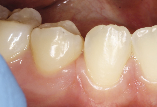
Klinický obraz invaginace může být od povrchového záhybu na gingivě (Obr. 1) až po vestibulo-orálně procházející lézi dělící papilu na meziální a distální část [4]. Vstup invaginace je možné rozeznat aspekcí a její hloubku sondáží. Pro diagnostiku není jednoznačně určena hloubka sondáže, některé studie uvádějí 1 milimetr [5], jiné 2 milimetry [4]. Sondovat invaginaci je možné horizontálně (vestibulárně a orálně) a vertikálně.
Histologické preparáty z invaginace gingivy a jejího okolí ukazují na hyperkeratinizaci epitelu s hlubokou proliferací na okrajích invaginace [6, 7]. Bohaté cévní zásobení přítomné na preparátech poukazuje na zvýšený metabolismus. Taktéž je přítomné zvýšené množství glykosaminoglykanů značící vysokou elasticitu tkáně, která může být odpovědná za recidivu mezer [3, 8].
Mechanismus vzniku invaginace není v literatuře úplně objasněný, ale nejčastěji jsou v odborném tisku uváděny dvě teorie. První, „mechanická“ teorie tvrdí, že rozhodujícím faktorem je nedostatečná, případně žádná remodelace gingivy při pohybu zubu danou oblastí. Měkké tkáně se tak stlačí a vytvoří masu gingivy, která je příčinou vzniku invaginace. Druhá teorie říká, že invaginace vzniká na podkladu atrofie alveolárního výběžku po extrakci zubu. Jsou také diskutovány další faktory s možným vlivem na vznik invaginace, jako například časový rámec od extrakce po začátek pohybu zubů, hustota kosti, velikost alveolárního výběžku, biotyp gingivy, technika extrakce, velikost mezery, proliferace pojivové tkáně a epitelu, změna topografie a morfologie kostní tkáně, narušený vývoj volné gingivy a transseptálních periodontálních vláken, mikrofraktury, mechanika ortodontické terapie, kouření, a polymorfizmus interleukinu-1 [3, 9–11].
Dlouhodobé následky invaginace gingivy na orální zdraví jsou téměř neznámé [9]. Přítomnost invaginace může negativně ovlivňovat možnost pacientů provádět důkladně ústní hygienu v dané oblasti [5]. Nahromadění měkké tkáně v zóně tlaku může mít kromě vzniku invaginace za následek recidivu extrakčních mezer po ortodontické terapii [10]. Kromě přebytku tkání v zóně tlaku se na recidivě ale pravděpodobně podílí i tkáňové napětí v zóně tahu [12, 13]
Soubor a metoda
Kohortová prospektivní studie probíhala v době od 1. 10. 2024 do 31. 8. 2025 na ortodontickém oddělení Fakultní nemocnice Olomouc a Univerzity Palackého v Olomouci. Výzkum byl realizován na základě kladného stanoviska etické komise FNOL a LF UP (jednací číslo 108/24).
Do experimentu byli konsekutivně zařazováni pacienti a děleni do dvou souborů, jejichž výsledky byly vyhodnocovány nezávisle na sobě.
Do Souboru 1 byli zařazování pacienti, kteří navštívili ortodontické oddělení v době trvání studie z důvodu ukončení aktivní fáze ortodontické terapie – sejmutí fixního aparátu. Soubor 1 zahrnoval celkem 42 pacientů s 124 extrahovanými zuby.
Do Souboru 2 byli zařazování pacienti, kteří navštívili ortodontické oddělení v době trvání studie z důvodu kontroly ve fázi retence a to v rozmezí jednoho měsíce až devíti let od ukončení aktivní fáze terapie. Soubor 2 zahrnoval 29 pacientů s 95 extrahovanými zuby. Tento soubor byl použit pro posouzení vývoje invaginace gingivy v delším časovém horizontu.
Pro oba soubory platila shodná kritéria pro zařazení:
1. Pacient byl po extrakci alespoň jednoho plně prořezaného stálého zubu ohraničeného meziálně i distálně sousedními stálými zuby, s následným ortodontickým uzávěrem mezery. Extrakce retinovaných nebo semiretinovaných zubů nebyly do studie zařazeny.
2. Poskytnutí písemného informovaného souhlasu pacienta s provedením klinického měření a zařazením do studie.
3. Žádný chirurgický zákrok v oblasti uzavírané mezery.
4. Kompletní ortodontická dokumentace před začátkem terapie.
5. Absence protetické práce nebo výplně na aproximálních plochách zubů sousedících s mezerou.
6. Pacient bez vrozené vývojové vady orofaciální oblasti, bez závažných úrazů a bez onemocnění parodontu.
Všichni pacienti absolvovali ortodontickou terapii pod vedením lékařů ortodontického oddělení Kliniky zubního lékařství LF UP Olomouc pomocí fixních aparátů, s použitím zámků s preskripcí Roth (Ortho Organizers, Carlsbad, USA).
Klinická měření zahrnovala detekci případné invaginace gingivy v oblasti uzávěru extrakční mezery a její sondáž vestibulárně i orálně. Sondáž byla prováděna v horizontálním směru, v nejhlubším místě invaginace pomocí parodontologické sondy kalibrované na 1 milimetr (LM Dental, Parainen, Finsko), (Obr. 2). Všechna měření byla prováděna hlavním autorem studie.
Pro posouzení, zda je invaginace přítomná, nebo ne, byla použita metodika dle Robertsona et al. [5], kde Robertson definuje invaginaci jako rýhu v gingivě hlubší než 1 mm.
Pokud nebyly nalezeny známky invaginace – tedy pokud rýha v gingivě nebyla hlubší než 1 mm, byla přiřazena hodnota 0 mm. Pokud byla nalezena úplná invaginace (sonda procházela skrz papilu z vestibulární strany na orální), byla přiřazena hodnota 9 mm jak vestibulární, tak orální invaginaci.
Dále byla zjišťována přítomnost a případně velikost mezery pomocí měrek na stripping kalibrovaných na jednu desetinu milimetru (Ortho Arch, Schaumburg, USA), (Obr. 3). Jako šířka mezery byla zaznamenána největší velikost měrky, která vešla mezi aproximální plochy bez použití nadměrné síly. V případě, že šířka mezery překračovala 0,5 mm, bylo použito více měrek najednou a jejich hodnoty byly sečteny.
Vzhledem k tomu, že se často u jednoho pacienta neshoduje výskyt invaginace ve všech kvadrantech, byla každá extrakční mezera hodnocena samostatně jako jeden subjekt.
Zjištěné údaje byly zaznamenány do tabulky v programu Excel (Microsoft, Redmond, USA), kde byla také provedena základní analýza dat.
U pacientů ze Souboru 1 bylo měření provedeno ve třech časových bodech: v den sejmutí fixního aparátu (T0), při první kontrole ve fázi retence 1 měsíc od sejmutí fixního aparátu (T1), a při druhé kontrole retence 4 měsíce od sejmutí fixního aparátu (T2).
Sledovanými parametry byly: doba, která uplynula od odeslání pacienta k extrakci po zahájení aktivní terapie, doba aktivní terapie, věk pacienta při zahájení a ukončení aktivní terapie, pohlaví, biotyp gingivy, zda se jedná o horní nebo dolní zubní oblouk, levou nebo pravou stranu, volba snímací retence, a vybrané diagnostické údaje před zahájením terapie: hloubka skusu (mm), incizální schůdek (mm), hodnota úhlů NS-ML (⁰), ANB (⁰), a hodnota WITS (mm).
Pacienti byli do skupin dle biotypu gingivy rozděleni pomocí testu parodontální sondou na prostředním řezáku – když sonda po zavedení do gingiválního sulku prosvítá, jedná se o slabý biotyp, když ne, jedná se o silný biotyp gingivy.
U pacientů ze Souboru 2 bylo hodnocení provedeno pouze jednou, v období retence v rozmezí jednoho měsíce až devíti let od ukončení aktivní fáze terapie. Byla zaznamenána doba od ukončení aktivní terapie.
Statistické zpracování
V Souboru 1 byla sledována korelace hloubky invaginace gingivy s věkem pacientů, délkou terapie, a vybranými skeletálními a dentálními znaky, dále korelace mezi hloubkou invaginace a velikostí reziduální mezery.
V Souboru 2 byla sledována korelace hloubky invaginace gingivy s velikostí reziduální mezery, dále mezi hloubkou sondáže a dobou od sejmutí fixního aparátu.
V souboru 1 byl navíc sledován rozdíl ve velikosti reziduální mezery v časech T1 a T0, T2 a T0 v jednotlivých kvadrantech rozdělených do 4 skupin dle retenčního protokolu. Retenční protokoly byly následující:
1. Deskový retenční aparát doporučen používat po dobu 8 hodin denně (D8).
2. Deskový retenční aparát doporučen používat po dobu alespoň 16 hodin denně první měsíc po sejmutí fixního aparátu, pak 8 hodin denně (D16).
3. Fóliový retenční aparát doporučen používat po dobu 8 hodin denně (F8).
4. Fóliový retenční aparát doporučen používat po dobu alespoň 16 hodin denně první měsíc po sejmutí fixního aparátu, pak 8 hodin denně (F16).
Čtyři kvadranty s fixní retencí byly ze souboru vyřazeny z důvodu malého vzorku.
Statistická analýza dat byla prováděna v programu Statistica (Tibco, Palo Alto, USA).
Vzhledem k nenormální distribuci dat byly použity neparametrické testy. U porovnání dvou skupin byl použit Mann-Whitney U test, u vícero skupin Kruskal – Walis test, a na posouzení korelace Spearmanův korelační koeficient.
Pro zohlednění cluster efektu byla dále provedena analýza rozptylu pro syntetizované chyby s mezerou jako závislou proměnnou, invaginací gingivy vestibulárně a orálně jako nezávislými proměnnými, a ID číslem pacienta jako náhodným faktorem. Pro zachování neparametrické povahy testu byly absolutní hodnoty nahrazeny pořadím hodnot.
Jako hladina statistické významnosti byla stanovena hodnota p = 0,05.
Chyba měření
U 7 pacientů (22 extrahovaných zubů) bylo provedeno měření hloubky orální i vestibulární invaginace v čase T0 dvakrát, ráno při sejmutí fixního aparátu a podruhé odpoledne při předání snímací retence.
Chyba měření byla kontrolována pomocí Dahlbergovy formule a relativní Dahlbergovy chyby. Míra absolutní shody mezi prvním a kontrolním měřením byla ověřena výpočtem koeficientu ICC (Intraclass correlation coefficient). Výskyt systematické chyby byl ověřen pomocí párového t-testu.
Relativní Dahlbergova chyba byla 14,9 % pro vestibulární invaginaci a 15,6 % pro orální invaginaci.
Hodnoty koeficientu ICC byly zjištěny 0,989 (pro vestibulární invaginaci) a 0,993 (pro orální invaginaci), což představuje perfektní shodu. Párovým t-testem nebyla zjištěna systematická chyba.
Výsledky
Soubor 1
1. Ze 124 kvadrantů s extrahovaným zubem byla při sejmutí fixního aparátu v 78 % přítomná invaginace vestibulárně a v 45 % orálně. Zastoupení jednotlivých hodnot sondáže je znázorněno na histogramu (Obr. 4).
2. Invaginace gingivy byla statisticky významně častější a závažnější v dolní čelisti než v horní čelisti. Nebyl zjištěn statisticky významný vliv pohlaví nebo biotypu gingivy (Tab. 2).
3. Nebyla zjištěna souvislost mezi věkem pacienta při nasazení či sejmutí fixního aparátu, ani délkou doby, která uplynula mezi odesláním pacienta na extrakci zubu a nasazením fixního aparátu a invaginací gingivy. Nebyl nalezen vztah mezi hodnotou incizálního schůdku, hloubkou skusu, úhlu báze lební s mandibulární linií, úhlu ANB, a hodnoty Wits s invaginací gingivy (Tab. 3).
4. Byla nalezena statisticky významná pozitivní korelace mezi sondáží invaginace vestibulárně i orálně a velikostí mezery. Korelace v čase T0 byla slabá. Nejsilnější korelace byla mezi sondáží orální invaginace v časech T1 a T2 a velikostí mezery v časech T1 a T2 (Tab. 4).
5. V čase T0, T1 i T2 byla zjištěna signifikantní asociace mezi vestibulární invaginací a velikostí mezery i mezi orální invaginací a velikostí mezery. Model zahrnoval náhodný efekt pacienta, který byl taktéž signifikantní, což indikuje přítomnost variability mezi pacienty (Tab. 5–7).
6. Nebyl nalezen statisticky významný rozdíl v recidivě extrakčních mezer v krátkodobém horizontu u pacientů s různými retenčními protokoly (Tab. 8).
Soubor 2
1. Byla nalezena statisticky významná slabá pozitivní korelace mezi sondáží invaginace vestibulárně i orálně a velikostí mezery (Tab. 9).
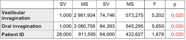
2. Byla nalezena signifikantní asociace mezi vestibulární invaginací a velikostí mezery i mezi orální invaginací a velikostí mezery. Model zahrnoval náhodní efekt pacienta, který byl taktéž signifikantní (Tab. 10).
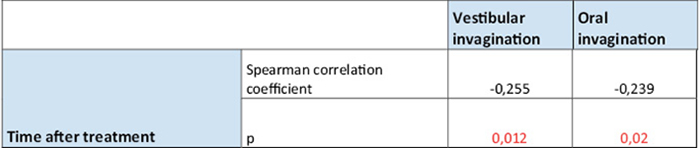
3. Při posouzení změn v dlouhodobém horizontu 1 měsíce až 9,3 roku byla nalezena velmi slabá negativní monotónní statisticky významná korelace mezi dobou, která uplynula od sejmutí fixního aparátu a hloubkou invaginace orálně i vestibulárně. (Tab. 11).
Diskuse
Invaginace gingivy se vyskytla při sejmutí fixního aparátu v 78 % případů vestibulárně a 45 % případů orálně. Její prevalence se v literatuře udává mezi 30–100 % [1, 3–5, 10, 11, 14, 15]. Velikost rozptylu je způsobena tím, že někteří autoři považují za rozhodující hloubku sondáže, od které považují invaginaci za přítomnou, jeden milimetr [5], kdežto jiní dva milimetry [4]. Navíc jsou studie často postavené na rozdílné metodice a hodnotí přítomnost invaginace v jiných časových bodech (například ještě před sejmutím fixního aparátu [3, 10], nebo naopak několik měsíců až roků v retenci [5, 14]).
Nebyla nalezena souvislost mezi hloubkou sondáže invaginace a pohlavím pacienta, což je v souladu s výsledky Bertlové et al. [16].
Naproti tomu Al-Jundi et al. [15] popisuje vyšší výskyt invaginace u mužů.
Hlubší sondáž invaginace gingivy byla nalezena v dolní čelisti než v horní čelisti.
Circunsová et al. [4] také popisuje, že je větší tendence k tvorbě invaginace v dolní čelisti. Naznačuje i vyšší výskyt invaginací vestibulárně než orálně, tento rozdíl však nebyl statisticky významný, statisticky významný nebyl ani vliv sklonu špičáku.
Naproti tomu Bertlová et al. [16] nalezla větší výskyt invaginace v horní čelisti.
Reichert et al. [14] zjistil, že je větší tendence k tvorbě invaginace gingivy v dolní čelisti než v horní. V dolní čelisti se v jeho souboru vyskytovala úplná invaginace až v 25 % případů. Kromě toho také popisuje hlubší sondáž invaginace vestibulárně než orálně.
Araújo et al. [17] ve studii na psech zjistil, že po extrakci zubu probíhá po dobu prvních 8 měsíců hojení větší ztráta vertikálního rozměru alveolární kosti vestibulárně než lingválně. To může vysvětlovat větší výskyt invaginace vestibulárně v naší studii. Reichert et al. [14] tento rozdíl připisuje i anatomii. Vzhledem ke tvaru zubního oblouku musí při uzávěru mezery překonat vestibulární povrch zubu větší délku než orální, a tedy prochází přes větší množství měkkých tkání.
Větší výskyt invaginace gingivy v dolní čelisti může vysvětlovat pomalejší pohyb zubů v dolní čelisti [1] nebo jiná anatomie kosti a dynamika hojení tkání [14].
V předkládané studii nebyla nalezena souvislost mezi biotypem gingivy a hloubkou sondáže invaginace gingivy, což je v souladu s výsledky Circunsové et al. [4].
Naproti tomu Bertlová et al. [16] a Stappert et al. [10]. tvrdí, že je větší výskyt a závažnost invaginace gingivy u pacientů se slabým biotypem gingivy. Biotyp ale určovali pomocí jiné metody, ještě před extrakcí, a to transgingivální sondáží jehlou při bázi meziální a distální papily u extrahovaného zubu, na rozdíl od předkládané studie, kdy byli pacienti do skupin dle biotypu gingivy rozděleni pomocí testu parodontální sondou na prostředním řezáku – když sonda po zavedení do gingiválního sulku prosvítá, jedná se o slabý biotyp [18–20].
Nepotvrdila se korelace sondáže invaginace s věkem pacientů, ani s délkou trvání ortodontické terapie, ani s délkou doby od extrakce zubu po zahájení terapie.
Bertlová et al. [16] naproti tomu popisuje, že u pacientů, u kterých uběhlo 8 týdnů od extrakce zubu do zahájení retrakce špičáků, byl větší výskyt invaginace gingivy než u pacientů, kde byl zub extrahován týden před zahájením pohybu. Větší výskyt invaginace byl také pozorován u pacientů s rychlejším pohybem zubů a u pacientů s dehiscencí bukální kompakty po extrakci.
Stappert et al. [10] tvrdí, že větší riziko vzniku invaginace je u pacientů s pomalým pohybem zubů do extrakční mezery. Dále zjistil, že je větší závažnost invaginace u dospělých pacientů než u adolescentů.
Reichert et al. ve své následné studii nenašel rozdíl ve výskytu invaginace gingivy mezi skupinou pacientů, kde se zahájil ortodontický uzávěr mezery 2–4 týdny od extrakce, a druhou skupinou, kde se čekalo na ortodontický pohyb více než 12 týdnů [21]. Naopak v jiné studii popisuje u skupiny pacientů s invaginací statisticky významně delší čas od extrakce po zahájení uzávěru mezery oproti skupině bez invaginace [14].
Daraqel et al. [22] v metaanalýze popisuje rychlejší pohyb zubů do extrakční mezery u pacientů, kde se distalizace zahájila brzy po extrakci, neuvádí ale statisticky významný rozdíl ve výskytu invaginace.
Jedním z limitů předkládané studie je, že je čas od extrakce zubu po uzávěr mezery pouze odhadován. Čas, který uplynul od odeslání pacienta k extrakci po provedení extrakce na jiném pracovišti může být různý, také doba nivelizace, po které se začne s distalizací zubů. I celková doba uzavírání mezery nemusí být nutně závislá na době aktivní terapie. Na rozdíl od toho studie Bertlové et al. [16] pracuje s přesnějším časem uzávěru mezery, protože extrakce byly provedeny až po nivelizaci.
V předkládané studii se nepotvrdila korelace invaginace gingivy s některým ze sledovaných skeletálních nebo dentálních znaků.
Naproti tomu Han et al. [23] zjistil větší výskyt invaginace gingivy u normodivergentních a hyperdivergentních pacientů. Dále Bertlová et al. [16] zkoumala, zda má růstová tendence vliv na výskyt invaginace, ale analýza nebyla provedena z důvodu malého vzorku.
Celkově se ale nezdá, že by existoval spolehlivý skeletální nebo dentální parametr, který by dokázal předpovědět riziko vzniku invaginace gingivy.
Při posouzení změn v dlouhodobém horizontu 1 měsíce až 9,3 roku byla nalezena velmi slabá negativní monotónní korelace mezi dobou, která uplynula od sejmutí fixního aparátu a hloubkou invaginace. Byla potvrzena jako statisticky signifikantní, tedy ne náhodná.
Jinými slovy je zde slabý náznak tendence, že čím je delší doba od sejmutí, tím je menší invaginace, ale nelze to považovat za pravidlo.
Circunsová et al. [4] také našla tendenci k nižšímu výskytu invaginace u pacientů, u kterých uplynula delší doba od ukončení aktivní fáze ortodontické terapie.
Kurol et al. [7] tvrdí, že zbytnění papil ustupuje spolu s tím, jak se zmírňuje hyperplazie gingivy způsobená přítomností fixního aparátu po ukončení terapie. To může vysvětlovat nižší hodnoty sondáže invaginací delší dobu po sejmutí fixního aparátu, dle Kurola ale na histologických preparátech zůstává proliferace epitelu charakteristická pro invaginaci gingivy zjevná ještě po dlouhou dobu.
Stappert et al. [24] tvrdí, že je souvislost mezi výskytem a rozsahem invaginace a recidivou extrakčních mezer. Recidiva byla u 43 % extrakčních míst s invaginací oproti 37 % u extrakčních míst bez invaginace a stoupala při zvyšující se ploše měkkých tkání v prostoru invaginace.
Byla nalezena pozitivní korelace mezi hloubkou sondáže invaginace a přítomností reziduální mezery, přičemž hodnota sondáže orálně se jeví pro výskyt mezer významnější než vestibulárně.
To je v souladu s výsledky Reicherta et al. [14], který v souboru pacientů 3 měsíce v retenci popisuje přítomnost zbytkové mezery u 30 % případů s invaginací gingivy. Ve skupině bez invaginace gingivy se nenašla žádná zbytková mezera.
Naproti tomu Circunsová et al. [4] nenašla vztah mezi výskytem a závažností invaginace a recidivou extrakčních mezer.
Dle Wehrbeina et al. [25] je také potřeba u pacientů s invaginací gingivy počítat s větší časovou náročností uzávěru extrakční mezery.
Často udávaná příčina recidivy extrakčních mezer je stlačení transseptálních periodontálních vláken v místě uzavřené extrakční mezery [26]. Erikson et al. [12] přirovnává tyto tkáně k jizvě.
V naší studii se nepotvrdilo, že by na recidivu mezery v krátkodobém horizontu měl vliv výběr retenčního snímacího aparátu, ani doba, po kterou má denně pacient snímací aparát používat.
Limitem studie je, že není objektivně potvrzeno, jaká byla skutečná spolupráce pacientů. To by bylo možné pouze při použití snímačů, například teploty [27].
Cíl retence ale není pouze zabránit recidivě extrakčních mezer.
Achenbrennerová [28] tvrdí, že deskový a vakuově tvarovaný foliový retenční aparát mají podobnou klinickou účinnost, i když fóliový aparát lépe brání recidivě stěsnání ve frontálním úseku. Lepší spolupráce i pohodlí pacientů bylo v případě foliového aparátu, ten má ale ve srovnání s deskou vyšší míru poškození.
Jäderbergová et al. [29] porovnávala 2 skupiny pacientů 6 měsíců po sejmutí fixního aparátu: jedna skupina používala vakuově tvarovaný fóliový retenční aparát 3 měsíce v délce celého dne, pak jenom na noc, kdy druhá skupina nosila aparát celý den pouze jeden týden. Nebyl nalezen statisticky významný rozdíl Littleova indexu, hloubky skusu, ani incizálního schůdku mezi skupinami.
Jiné studie porovnávají používání Hawleyho deskového retenčního aparátu pouze na noc po dobu jednoho roku a používaní celý den po dobu 6 měsíců a pak 6 měsíců pouze na noc. Nebyl zjištěn žádný statisticky významný rozdíl Littleova indexu [30]. Doporučuje se však, že doba používání dekového aparátu by neměla klesnout pod 8 hodin denně [31].
Gill et al. [32] tvrdí, že není statisticky významný rozdíl v efektivitě používaní foliového retenčního aparátu pouze na noc nebo během dne i noci z hlediska Littleova indexu, incizálního schůdku, nebo hloubky skusu 6 měsíců po sejmutí fixního aparátu.
Při porovnání foliového a deskového aparátu někteří autoři nenašli statisticky významný rozdíl při sledování mezišpičákové vzdálenosti, mezimolárové vzdálenosti, Littleova indexu, incizálního schůdku nebo hloubky skusu [33–35]. Dle jiných studií je foliový retenční aparát efektivnější v boji se stěsnáním frontálního úseku [36, 37]. Další výhoda fóliového retenčního aparátu je v jeho lepší toleranci pacienty [38]. Naproti tomu je deskový aparát odolnější a umožňuje settling [38, 39].
Prohlášení o financování
Tento výzkum nebyl financován z grantových projektů, veřejných ani komerčních zdrojů ani ze zdrojů neziskových organizací.
Závěr
1. Invaginace gingivy je častý nežádoucí efekt extrakční ortodontické terapie.
2. Při posouzení změn v dlouhodobém horizontu 1 měsíce až 9,3 roku byla nalezena velmi slabá negativní monotónní korelace mezi dobou, která uplynula od sejmutí fixního aparátu a hloubkou invaginace. Byla potvrzena jako statisticky signifikantní, tedy ne náhodná. Je zde slabý náznak tendence, že čím je delší doba od sejmutí, tím je menší invaginace, ale nelze to považovat za pravidlo.
3. Výskyt invaginace gingivy je častější v dolní čelisti, neexistuje však žádný parametr, který by dokázal spolehlivě předpovědět, u kterého pacienta se invaginace gingivy vyskytne.
4. Při sejmutí fixního aparátu byla nalezena velmi slabá pozitivní korelace mezi sondáží invaginace gingivy a velikosti zbytkové mezery. To naznačuje, že mezera je většinou při ukončení aktivní léčby úplně uzavřena a invaginace v tom nehraje významnější roli. Naproti tomu v krátkodobé retenci (1 až 3 měsíce) byla nalezena silnější pozitivní korelace. Nejsilnější pozitivní korelace byla přítomná mezi invaginací orálně a šířkou mezery v čase T2.
5. Nebyl zjištěn vliv použitého retenčního protokolu na recidivu extrakčních mezer. Jedné se však o krátkodobé pozorování a nebyla ověřována spolupráce pacientů při dodržování retenčního protokolu.
Autoři nemají komerční, vlastnické nebo finanční zájmy na produktech nebo společnostech popsaných v tomto článku
Introduction
Gingival invagination is a common finding following orthodontic closure of an extraction space with fixed appliances [1, 2]. It refers to interdistal linear invaginations of the interproximal gingival tissue, present following orthodontic closure of an extraction space, or possibly a diastema or trema [3]. The nomenclature in the professional literature is not uniform; terms such as gingival cleft or gingival duplication may also be encountered [1, 3].
The clinical presentation of invagination can range from a superficial fold on the gingiva (Fig. 1) to a vestibulo-orally extending lesion dividing the papilla into mesial and distal parts [4]. The entrance to the invagination can be identified by visual inspection, and its depth by probing. There is no clear consensus on the diagnostic depth of probing; some studies report 1 millimeter [5], others 2 millimeters [4]. The invagination can be probed horizontally (vestibularly and orally) and vertically.
Histological specimens from the gingival invagination and its surrounding area show hyperkeratinization of the epithelium with deep proliferation at the edges of the invagination [6, 7]. The rich vascular supply present in the specimens indicates increased metabolism. There is also an increased amount of glycosaminoglycans, indicating high tissue elasticity, which may be responsible for the recurrence of spaces [3, 8].
The mechanism underlying the development of invagination has not been fully elucidated in the literature, but two theories are most frequently cited in the professional press. The first, „mechanical“ theory posits that the decisive factor is insufficient, or even absent, gingival remodeling as the tooth moves through the affected area. The soft tissues are thus compressed, forming a gingival mass that causes the invagination. The second theory states that invagination arises due to atrophy of the alveolar ridge following tooth extraction. Other factors with a potential influence on the development of invagination are also discussed, such as the time frame from extraction to the start of tooth movement, bone density, the size of the alveolar ridge, gingival biotype, extraction technique, the size of the gap, proliferation of connective tissue and epithelium, changes in bone tissue topography and morphology, impaired development of free gingiva and transseptal periodontal fibers, microfractures, orthodontic treatment mechanics, smoking, and interleukin-1 polymorphism [3, 9–11].
The long-term effects of gingival invagination on oral health are largely unknown [9]. The presence of invagination may negatively affect patients’ ability to perform thorough oral hygiene in the affected area [5]. In addition to causing invagination, the accumulation of soft tissue in the compression zone may result in the recurrence of extraction spaces following orthodontic treatment [10]. However, in addition to excess tissue in the compression zone, tissue tension in the tension zone likely also contributes to recurrence [12, 13].
Material and Methods
This prospective cohort study was conducted from October 1, 2024, to August 31, 2025, at the Orthodontic Department of the Olomouc University Hospital and Palacký University in Olomouc. The research was conducted based on the approval of the Ethics Committee of the University Hospital Olomouc and Palacký University (case number 108/24).
Patients were consecutively enrolled in the study and divided into two Samples, the results of which were evaluated independently of one another.
Sample 1 included patients who visited the orthodontic department during the study period to complete the active phase of orthodontic treatment–removal of fixed appliances. Sample 1 comprised a total of 42 patients with 124 extracted teeth.
Sample 2 included patients who visited the orthodontic department during the study period for a follow-up visit during the retention phase, ranging from one month to nine years after the completion of the active phase of treatment. Sample 2 included 29 patients with 95 extracted teeth. This cohort was used to assess the development of gingival invagination over a longer time horizon.
The same inclusion criteria applied to both samples:
1. The patient had undergone extraction of at least one fully erupted permanent tooth flanked medially and distally by adjacent permanent teeth, followed by orthodontic closure of the space. Extractions of impacted or semi-impacted teeth were not included in the study.
2. The patient provided written informed consent for clinical measurements and inclusion in the study.
3. No surgical intervention in the area of the space to be closed.
4. Complete orthodontic documentation prior to the start of treatment.
5. Absence of prosthetic work or fillings on the occlusal surfaces of teeth adjacent to the space.
6. Patient without congenital developmental defects of the orofacial region, without serious trauma, and without periodontal disease.
All patients underwent orthodontic treatment under the supervision of orthodontists from the Orthodontic Department of the Faculty of Dentistry, Palacký University Olomouc, using fixed appliances with Roth-prescribed brackets (Ortho Organizers, Carlsbad, USA).
Clinical measurements included the detection of any gingival invagination in the area of the closure of the extraction site and its probing both vestibularly and orally. Probing was performed horizontally at the deepest point of the invagination using a periodontal probe calibrated to 1 millimeter (LM Dental, Parainen, Finland) (Fig. 2). All measurements were performed by the lead author of the study.
To assess whether invagination was present or not, the methodology described by Robertson et al. [5] was used, in which Robertson defines invagination as a groove in the gingiva deeper than 1 mm.
If no signs of invagination were found—that is, if the groove in the gingiva was no deeper than 1 mm—a value of 0 mm was assigned. If complete invagination was found (the probe passed through the papilla from the vestibular side to the oral side), a value of 9 mm was assigned to both the vestibular and oral invaginations.
Furthermore, the presence and, if applicable, the size of the space were determined using stripping gauges calibrated to one-tenth of a millimeter (Ortho Arch, Schaumburg, USA) (Fig. 3). The width of the gap was recorded as the largest gauge size that fit between the approximal surfaces without the use of excessive force. If the space width exceeded 0.5 mm, multiple gauges were used simultaneously, and their values were summed.
Since the occurrence of invagination often varies across quadrants in a single patient, each extraction space was evaluated separately as a single entity.
The collected data were entered into a spreadsheet in Excel (Microsoft, Redmond, USA), where basic data an Sample 1, measurements were taken at three time points: on the day the fixed appliance was removed (T0), at the first retention check-up 1 month after removal of the fixed appliance (T1), and at the second retention check-up 4 months after removal of the fixed appliance (T2).
The parameters monitored were: the time elapsed from the patient’s referral for extraction to the start of active therapy, the duration of active therapy, the patient’s age at the start and end of active therapy, gender, gingival biotype, whether the upper or lower dental arch was involved, the left or right side, choice of removable retention, and selected diagnostic data prior to the start of therapy: overbite (mm), overjet (mm), NS-ML angle (⁰), ANB angle (⁰), and WITS value (mm).
Patients were divided into samples based on gingival biotype using a periodontal probe test on the central incisor—if the probe is visible after insertion into the gingival sulcus, it is a thin biotype; if not, it is a thick gingival biotype.
For patients in Sample 2, the evaluation was performed only once, during the retention period ranging from one month to nine years after the end of the active phase of therapy. The time elapsed since the end of active therapy was recorded.
Statistical Analysis
In Sample 1, the correlation between gingival invagination depth and patient age, duration of therapy, and selected skeletal and dental characteristics was examined, as well as the correlation between invagination depth and residual space size.
In Sample 2, the correlation between the depth of gingival invagination and the size of the residual space was examined, as well as the correlation between probing depth and the time elapsed since removal of the fixed appliance.
In Sample 1, the difference in residual space size at time points T1 and T0, T2 and T0 was also examined in individual quadrants divided into 4 groups according to the retention protocol. The retention protocols were as follows:
1. It is recommended to wear the plate retainer for 8 hours a day (D8).
2. It is recommended to wear the plate retainer for at least 16 hours a day during the first month after the fixed appliance is removed, then 8 hours a day (D16).
3. It is recommended to wear the foil retainer for 8 hours a day (F8).
4. It is recommended to wear the foil retainer for at least 16 hours a day during the first month after removal of the fixed appliance, then 8 hours a day (F16).
The four quadrants with fixed retention were excluded from the dataset due to the small sample size.
Statistical analysis of the data was performed using the Statistica software (Tibco, Palo Alto, USA).
Due to the non-normal distribution of the data, nonparametric tests were used. The Mann-Whitney U test was used to compare two groups, the Kruskal–Wallis test for multiple groups, and Spearman’s correlation coefficient to assess correlation.
To account for the cluster effect, an analysis of variance was further performed with synthesized errors with spaces as the dependent variable, vestibular and oral gingival invagination as independent variables, and the patient’s ID number as a random factor. To preserve the nonparametric nature of the test, absolute values were replaced with rank orders.
A p-value of 0.05 was set as the level of statistical significance.
Measurement Error
In 7 patients (22 extracted teeth), measurements of the depth of both the oral and vestibular invagination were taken twice at time T0: once in the morning when the fixed appliance was removed, and again in the afternoon when the removable retainer was handed over.
Measurement error was assessed using Dahlberg’s formula and the relative Dahlberg error. The degree of absolute agreement between the first and control measurements was verified by calculating the ICC (Intraclass Correlation Coefficient). The presence of systematic error was verified using a paired t-test.
The relative Dahlberg error was 14.9% for vestibular invagination and 15.6% for oral invagination.
The ICC values were found to be 0.989 (for vestibular invagination) and 0.993 (for oral invagination), representing perfect agreement. No systematic error was detected by the paired t-test.
Results
Sample 1
1. Of the 124 quadrants with an extracted tooth, vestibular invagination was present in 78% and oral invagination in 45% when the fixed appliance was removed. The distribution of individual probing depths is shown in the histogram (Fig. 4).
2. Gingival invagination was statistically significantly more frequent and severe in the mandible than in the maxilla. No statistically significant influence of gender or gingival biotype was found (Table 2).
3. No association was found between the patient’s age at the time of fixed appliance placement or removal, nor the length of time elapsed between the patient’s referral for tooth extraction and fixed appliance placement, and gingival invagination. No relationship was found between the overjet value, overbite, the angle of the cranial base to the mandibular line, the ANB angle, and the WITS value with gingival invagination (Table 3).
4. A statistically significant positive correlation was found between the probing depth of vestibular and oral invagination and the size of the space. The correlation at time T0 was weak. The strongest correlation was between the probing depth of oral invagination at times T1 and T2 and the size of the space at times T1 and T2 (Table 4).
5. At time points T0, T1, and T2, a significant association was found between vestibular invagination and space size, as well as between oral invagination and space size. The model included a random patient effect, which was also significant, indicating the presence of variability among patients (Tables 5–7).
6. No statistically significant difference was found in the short-term recurrence of extraction spaces among patients with different retention protocols (Table 8).
Sample 2
1. A statistically significant, weak positive correlation was found between the measurement of vestibular and oral invagination and the size of the space (Table 9).
2. A significant association was found between vestibular invagination and space size, as well as between oral invagination and gap size. The model included a random patient effect, which was also significant (Table 10).
3. When assessing changes over the long term (1 month to 9.3 years), a very weak negative monotonic statistically significant correlation was found between the time elapsed since removal of the fixed appliance and the depth of invagination both orally and vestibularly. (Table 11).
Discussion
Gingival invagination occurred upon removal of the fixed appliance in 78% of cases on the vestibular side and in 45% of cases on the oral side. Its prevalence is reported in the literature to range from 30% to 100% [1, 3–5, 10, 11, 14, 15]. The wide range is due to the fact that some authors consider a probing depth of one millimeter to be the threshold at which invagination is present [5], while others use two millimeters [4]. Furthermore, studies are often based on different methodologies and assess the presence of invagination at different time points (for example, before the removal of the fixed appliance [3, 10], or conversely, several months to years into retention [5, 14]).
No correlation was found between the probing depth of gingival invagination and the patient’s gender, which is consistent with the results of Bertl et al. [16].
In contrast, Al-Jundi et al. [15] describe a higher incidence of invagination in men.
Deeper gingival invagination probing was found in the mandible than in the maxilla.
Circuns et al. [4] also report a greater tendency for invagination to form in the mandible. They also suggest a higher incidence of invagination in the vestibular region than in the oral region; however, this difference was not statistically significant, nor was the influence of canine inclination.
In contrast, Bertl et al. [16] found a higher incidence of invagination in the upper jaw.
Reichert et al. [14] found that there is a greater tendency for gingival invagination to occur in the lower jaw than in the upper jaw. In their study, complete invagination occurred in up to 25% of cases in the lower jaw. In addition, he also describes deeper probing of the invagination on the vestibular side than on the oral side.
Araújo et al. [17] found in a study on dogs that, during the first 8 months of healing following tooth extraction, there is greater loss of vertical dimension of the alveolar bone on the vestibular side than on the lingual side. This may explain the higher incidence of invagination on the vestibular side in our study. Reichert et al. [14] also attribute this difference to anatomy. Due to the shape of the dental arch, when closing the gap, the vestibular surface of the tooth must traverse a greater length than the oral surface and thus passes through a greater amount of soft tissue.
The higher incidence of gingival invagination in the mandible may be explained by slower tooth movement in the mandible [1] or by different bone anatomy and tissue healing dynamics [14].
In the present study, no association was found between gingival biotype and the probing depth of gingival invagination, which is consistent with the results of Circuns et al. [4].
In contrast, Bertl et al. [16] and Stappert et al. [10] claim that the incidence and severity of gingival invagination are higher in patients with a weak gingival biotype. However, they determined the biotype using a different method, prior to extraction, namely transgingival probing with a needle at the base of the mesial and distal papillae of the extracted tooth, unlike in the present study, where patients were divided into groups according to gingival biotype using a periodontal probe test on the central incisor – if the probe is visible after insertion into the gingival sulcus, it indicates a weak biotype [18–20].
No correlation was found between gingival invagination probing and patient age, the duration of orthodontic treatment, or the time elapsed from tooth extraction to the start of treatment.
Bertl et al. [16], on the other hand, report that patients in whom 8 weeks elapsed between tooth extraction and the start of canine retraction had a higher incidence of gingival invagination than patients in whom the tooth was extracted one week before the start of movement. A higher incidence of invagination was also observed in patients with faster tooth movement and in patients with buccal compact dehiscentia following extraction.
Stappert et al. [10] state that there is a higher risk of invagination in patients with slow tooth movement into the extraction site. They also found that the severity of invagination is greater in adult patients than in adolescents.
In their follow-up study, Reichert et al. found no difference in the incidence of gingival invagination between a group of patients in whom orthodontic closure of the space was initiated 2–4 weeks after extraction and a second group in which orthodontic movement was delayed for more than 12 weeks [21]. Conversely, another study reports a statistically significantly longer time from extraction to the start of space closure in the group of patients with gingival invagination compared to the group without invagination [14].
Daraqel et al. [22] describe in a meta-analysis faster tooth movement into the extraction space in patients where distalization began soon after extraction, but they do not report a statistically significant difference in the incidence of invagination.
One of the limitations of the present study is that the time from tooth extraction to space closure is only estimated. The time elapsed from the patient’s referral for extraction to the actual extraction at another facility may vary, as may the duration of leveling before tooth distalization begins. Furthermore, the total duration of space closure may not necessarily depend on the duration of active therapy. In contrast, the study by Bertl et al. [16] uses a more precise time for space closure, as extractions were performed only after leveling.
In the present study, no correlation was found between gingival invagination and any of the observed skeletal or dental features.
In contrast, Han et al. [23] found a higher incidence of gingival invagination in normodivergent and hyperdivergent patients. Furthermore, Bertl et al. [16] investigated whether growth tendency influences the incidence of invagination, but the analysis was not performed due to the small sample size.
Overall, however, there does not appear to be a reliable skeletal or dental parameter capable of predicting the risk of gingival invagination.
When assessing changes over a long-term period ranging from 1 month to 9.3 years, a very weak negative monotonic correlation was found between the time elapsed since the removal of the fixed appliance and the depth of retraction. This correlation was confirmed as statistically significant, i.e., not random.
In other words, there is a weak indication of a trend that the longer the time since removal, the less severe the invagination, but this cannot be considered a rule.
Circuns et al. [4] also found a tendency toward a lower incidence of invagination in patients for whom a longer period had elapsed since the end of the active phase of orthodontic therapy.
Kurol et al. [7] state that papillary hypertrophy subsides as gingival hyperplasia caused by the presence of fixed appliances resolves following the completion of treatment. This may explain the lower probing depths of invaginations a longer time after removal of the fixed appliance; however, according to Kurol, the epithelial proliferation characteristic of gingival invagination remains evident on histological specimens for a long time.
Stappert et al. [24] state that there is a correlation between the occurrence and extent of invagination and the recurrence of extraction sites. Recurrence occurred in 43% of extraction sites with invagination compared to 37% in extraction sites without invagination, and it increased with the expanding area of soft tissue within the invagination site.
A positive correlation was found between the probing depth of the invagination and the presence of a residual space, with the oral probing depth appearing to be more significant for the occurrence of gaps than the vestibular probing depth.
This is consistent with the findings of Reichert et al. [14], who, in a cohort of patients 3 months into retention, reported the presence of residual spaces in 30% of cases with gingival invagination. No residual gaps were found in the group without gingival invagination.
In contrast, Circuns et al. [4] found no relationship between the occurrence and severity of invagination and the recurrence of extraction spaces.
According to Wehrbein et al. [25], it is also necessary to anticipate that closing the extraction space will take longer in patients with gingival invagination.
A frequently cited cause of extraction socket recurrence is compression of the transseptal periodontal fibers at the site of the closed extraction socket [26]. Erikson et al. [12] compare these tissues to scar tissue.
In our study, it was not confirmed that the choice of the removable retention appliance or the duration of daily use by the patient had an effect on the recurrence of the gap in the short term.
A limitation of the study is that the actual level of patient compliance was not objectively confirmed. This would only be possible with the use of sensors, such as temperature sensors [27].
However, the goal of retention is not merely to prevent the recurrence of extraction spaces.
Achenbrennerová [28] states that plate-type and vacuum-formed foil retainers have similar clinical efficacy, although the foil retainer is more effective at preventing relapse of crowding in the anterior region. Patient compliance and comfort were better with the foil retainer; however, it has a higher rate of damage compared to the plate-type retainer.
Jäderberg et al. [29] compared two groups of patients 6 months after removal of fixed appliances: one group used a vacuum-formed foil retainer for 3 months full-time, then only at night, while the other group wore the retainer full-time for only one week. No statistically significant difference was found in the Little Index, overbite, or overjet between the groups.
Other studies compare the use of a Hawley plate retainer worn only at night for one year with use throughout the day for 6 months followed by 6 months of nighttime-only wear. No statistically significant difference in the Little Index was found [30]. However, it is recommended that the duration of retainer wear should not fall below 8 hours per day [31].
Gill et al. [32] state that there is no statistically significant difference in the effectiveness of using a foil retainer only at night or both day and night in terms of the Little Index, overjet, or overbite 6 months after removal of the fixed appliance.
When comparing foil and plate apparatus, some authors did not find a statistically significant difference in tracking between canine distances, between molar distances, Little index, overjet or overbite [33–35]. According to other studies, the foil retainer is more effective in fighting the crowding of the frontal segment [36, 37]. Another advantage of the foil retainer is in its better patient tolerance [38]. In contrast, the plate retainer is more durable and allows settling [38, 39].
Funding statement
This research was not funded by grant projects, public or commercial sources, or nonprofit organizations.
Conclusion
1. Gingiva invagination is a common adverse effect of extractional orthodontic therapy.
2. When assessing changes in the long term of 1 month to 9.3 years, a very weak negative monotonous correlation was found between the time elapsed since the removal of the fixed appliance and the depth of invagination. It was confirmed as statistically significant, i.e. not random. There is a slight suggestion that the longer the time since removal, the less invagination, but this cannot be considered a rule.
3. The incidence of gingiva invagination is more common in the lower jaw, but there is no parameter that can reliably predict which patient will experience gingiva invagination.
4. When the fixed apparatus was removed, a very weak positive correlation was found between the gingiva invagination probe and the size of the residual space. This suggests that the gap is mostly closed when active treatment is completed, and invagination does not play a major role in this. By contrast, a stronger positive correlation was found in short-term retention (1 to 3 months). The strongest positive correlation was present between oral invagination and T2 time space width.
5. There was no effect of the retention protocol used on the recurrence of extraction spaces. However, the observation was short-term and patient cooperation in compliance with the retention protocol was not verified.
Authors do not have commercial, proprietary or financial interests in the products or companies described in this article.
Obr. 1 / Fig. 1
Invaginace gingivy v místě extrahovaného zubu 44. / Gingival invagination at the site of the extracted tooth 44.
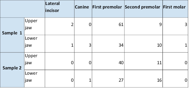
Tab. 1 / Tab. 1
Zastoupení jednotlivých extrahovaných zubů v souborech. / Distribution of individual extracted teeth in the samples.
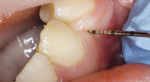
Obr. 2 / Fig. 2
Sondáž invaginace gingivy 3 mm v místě extrahovaného zubu 34. / Probing of gingival invagination 3 mm at the site of the extracted tooth 34.
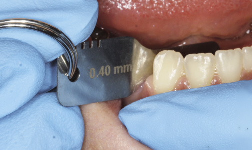
Obr. 3 / Fig. 3
Mezera 0,4 mm v místě extrahovaného zubu 44. / Space reopening of 0.4 mm at the site of the extracted tooth 44.
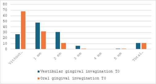
Obr. 4 / Fig. 4
Histogram sondáže invaginace gingivy, Soubor 1. Na ose Y četnost hloubky sondáže v jednotlivých poextrakčních lokalitách. / Histogram of gingival invagination probing, Sample 1. The Y-axis shows the frequency of probing depth in individual post-extraction sites.
Tab. 2 / Tab. 2
Porovnání hloubky invaginací mezi horní a dolní čelistí, mezi pohlavími a mezi silným a slabým biotypem gingivy, Soubor 1. / Comparison of invagination depth between the upper and lower jaw, between genders, and between thick and thin gingival biotypes, Sample 1.
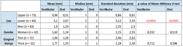
Tab. 3 / Tab. 3
Korelace věku pacienta, délky trvání terapie, skeletálních a dentálních znaků s hloubkou sondáže invaginace vestibulárně a orálně, Soubor 1. / Correlation of patient age, duration of therapy, skeletal and dental characteristics with vestibular and oral invagination, Sample 1.
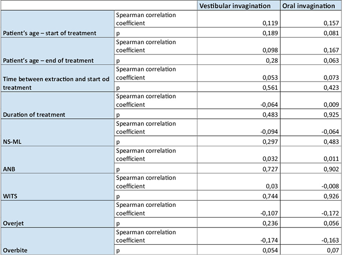
Tab. 4 / Tab. 4
Korelace sondáže invaginace gingivy s velikostí mezery v časech T0, T1, T2, Soubor 1. / Correlation of gingival invagination probing with space size at times T0, T1, T2, Sample 1.

Tab. 5 / Tab. 5
Závislost velikosti mezery na sondáži invaginace gingivy vestibulárně a orálně se zohledněním cluster efektu, Soubor 1, T0. / Dependence of space size on probing of vestibular and oral gingival invagination, taking into account the cluster effect, Sample 1, T0.

SV – stupně volnosti efektu / degrees of freedom (effect), MS – střední čtverec efektu / mean square (effect), SV – stupně volnosti chyby / degrees of freedom (error), MS – střední čtverec chyby / mean square (error), F – testová statistika / test statistic
Tab. 6 / Tab. 6
Závislost velikosti mezery na sondáži invaginace gingivy vestibulárně a orálně se zohledněním cluster efektu, Soubor 1, T1. / Dependence of space size on probing of vestibular and oral gingival invagination, taking into account the cluster effect, Sample 1, T1.

SV – stupně volnosti efektu / degrees of freedom (effect), MS – střední čtverec efektu / mean square (effect), SV – stupně volnosti chyby / degrees of freedom (error), MS – střední čtverec chyby / mean square (error), F – testová statistika / test statistic
Tab. 7 / Tab. 7
Závislost velikosti mezery na sondáži invaginace gingivy vestibulárně a orálně se zohledněním cluster efektu, Soubor 1, T2. / Dependence of space size on probing of vestibular and oral gingival invagination, taking into account the cluster effect, Sample 1, T2.

SV – stupně volnosti efektu / degrees of freedom (effect), MS – střední čtverec efektu / mean square (effect), SV – stupně volnosti chyby / degrees of freedom (error), MS – střední čtverec chyby / mean square (error), F – testová statistika / test statistic
Tab. 8 / Tab. 8
Otevírání mezery v časech T1 a T2 oproti času T0 v jednotlivých skupinách dle použitého retenčního protokolu, Soubor 1. / Space reopening at times T1 and T2 compared to time T0 in individual groups according to the retention protocol used, Sample 1.
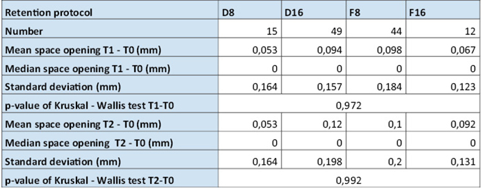
Tab. 9 / Tab. 9
Korelace sondáže invaginace gingivy s velikostí mezery, Soubor 2. / Correlation of gingival invagination probing with space size, Sample 2.

Tab. 10 / Tab. 10
Závislost velikosti mezery na sondáži invaginace gingivy vestibulárně a orálně se zohledněním cluster efektu, Soubor 2. / Dependence of space size on probing of vestibular and oral gingival invagination, taking into account the cluster effect, Sample 2.
SV – stupně volnosti efektu / degrees of freedom (effect), MS – střední čtverec efektu / mean square (effect), SV – stupně volnosti chyby / degrees of freedom (error), MS – střední čtverec chyby / mean square (error), F – testová statistika / test statistic
Tab. 11 / Tab. 11
Korelace sondáže invaginace gingivy s dobou od sejmutí fixního aparátu, Soubor 2. / Correlation of gingival invagination probing with time since removal of the fixed appliance, Sample 2.

Literatura / References
1. Gölz L, Reichert C, Jäger A. Gingival invagination — a systematic review. Fortschr Kieferorthop. 2011;72(6):409–20. doi: 10.1007/s00056-011-0046-z
2. Reichert C, Kutschera E, Nienkemper M, Scharf S, Mengel M, Fimmers R, et al. Influence of time after extraction on the development of gingival invagination: study protocol for a multicenter pilot randomized controlled clinical trial. Trials. 2013;14:108. doi: 10.1186/1745-6215-14-108
3. Bertl K. Gingivainvaginationen bei kieferorthopädischem Lückenschluss. Wissen Kompakt. 2015;9(3):139–46. doi: 10.1007/s11838-015-0009-0
4. Circuns ALR, Tulloch JFC. Gingival invagination in extraction sites of orthodontic patients: Their incidence, effects on periodontal health, and orthodontic treatment. Am J Orthod. 1983;83(6):469–76. doi: 10.1016/0002-9416(83)90245-2
5. Robertson PB, Schultz LD, Levy BM. Occurrence and Distribution of Interdental Gingival Clefts following Orthodontic Movement into Bicuspid Extraction Sites. J Periodontol. 1977;48(4):232–5. doi: 10.1902/jop.1977.48.4.232
6. Wehrbein H, Fuhrmann R, Andreas A, Diedrich P. Die Bedeutung von Gingivaduplikaturen beim orthodontischen Lückenschluß. Fortschr Kieferorthop. 1993;54(6):231–6. doi: 10.1007/BF02179832
7. Kurol J, Rönnerman A, Heyden G. Long-term gingival conditions after orthodontic closure of extraction sites. Histological and histochemical studies. Eur J Orthod. 1982;4(2):87–92. doi: 10.1093/ejo/4.2.87
8. Rönnerman A, Thilander B, Heyden G. Gingival tissue reactions to orthodontic closure of extraction sites: Histologic and histochemical studies. Am J Orthod. 1980;77(6):620–5. doi: 10.1016/0002-9416(80)90153-0
9. Reichert C, Wenghoefer M, Kutschera E, Götz W, Jäger A. Alveolenerhalt mit einem nanopartikulärem Hydroxylapatit verringert den Ausprägungsgrad von Gingivaduplikaturen – eine prospektive klinische Studie. Fortschr Kieferorthop. 2014;75(1):7–15. doi: 10.1007/s00056-013-0175-7
10. Stappert D, Geiman R, Zadi ZH, Reynolds MA. Gingival clefts revisited: Evaluation of the characteristics that make one more susceptible to gingival clefts. Am J Orthod Dentofacial Orthop. 2018;154(5):677–82. doi: 10.1016/j.ajodo.2018.01.018
11. Gölz L, Reichert C, Dirk C, Jäger A. Retrospective investigation of gingival invaginations : Part II: microbiological findings and genetic risk profile. Fortschr Kieferorthop. 2012;73(5):387–96. doi: 10.1007/s00056-012-0098-8
12. Erikson BE, Kaplan H, Aisenberg MS. Orthodontics and transeptal fibers: A histological interpretation of repair phenomena following the removal of first premolars with retraction of the anterior segment. Am J Orthod Oral Surg. 1945;31(1):1–20. doi: 10.1016/S0096-6347(45)90056-1
13. Redlich M, Shoshan S, Palmon A. Gingival response to orthodontic force. Am J Orthod Dentofacial Orthop. 1999;116(2):152–8. doi: 10.1016/s0889-5406(99)70212-x
14. Reichert C, Gölz L, Dirk C, Jäger A. Retrospective investigation of gingival invaginations : Part I: Clinical findings and presentation of a coding system. Fortschr Kieferorthop. 2012;73(4):307–16. doi: 10.1007/s00056-012-0082-3
15. Al-Jundi A, Sabbagh BA, Baskaradoss JK. Evaluation of Periodontal Changes Adjacent to Extraction Sites during Upper Canine Retraction. J Contemp Dent Pract. 2017;18(2):117–25. doi: 10.5005/jp-journals-10024-2001
16. Bertl K, Neuner H, Meran A, Bertl MH, Reich I, Nemec M, et al. Does the time-point of orthodontic space closure initiation after tooth extraction affect the incidence of gingival cleft development? A randomized controlled clinical trial. J Periodontol. 2020;91(5):572–81. doi: 10.1002/JPER.19-0376
17. Araújo MG, Lindhe J. Dimensional ridge alterations following tooth extraction. An experimental study in the dog. J Clin Periodontol. 2005;32(2):212–8. doi: 10.1111/j.1600-051X.2005.00642.x
18. Fischer KR, Künzlberger A, Donos N, Fickl S, Friedmann A. Gingival biotype revisited—novel classification and assessment tool. Clin Oral Investig. 2018;22(1):443–8. doi: 0.1007/s00784-017-2131-1
19. Sharma VK, Singh D, Srivastava R, Chaturvedi TP, Khairnar M, Singh AK. Assessment of gingival biotype in different facial patterns: A cross-sectional study. Natl J Maxillofac Surg. 2023;14(1):63–7. doi: 10.4103/njms.njms_490_21
20. Malhotra R, Grover V, Bhardwaj A, Mohindra K. Analysis of the gingival biotype based on the measurement of the dentopapillary complex. J Indian Soc Periodontol. 2014;18(1):43–7. doi: 10.4103/0972-124X.128199
21. Reichert C, Kutschera E, Plötz C, Scharf S, Gölz L, Fimmers R, et al. Incidence and severity of gingival invaginations associated with early versus late initiation of orthodontic space closure after tooth extraction. Fortschr Kieferorthop. 2017;78(5):415–25. doi: 10.1007/s00056-017-0098-9
22. Daraqel B, Mheissen S, Li J, Khan H, Allan S, Zheng L. The effect of early versus delayed space closure on the rate of orthodontic tooth movement: a systematic review and meta-analysis. Eur J Orthod. 2023;45(6):680–9. doi: 10.1093/ejo/cjad015
23. Han M, Li SH, Yao Y, Zhao Y, You LP, Zheng Q, et al. Risk factors for gingival invagination: A retrospective study. J Clin Periodontol. 2024;51(9):1199–209. doi: 10.1111/jcpe.14005
24. Stappert D, Bakhsh A, Scott C. Prospective Clinical Investigation of Orthodontic Relapse from Gingival Clefts. Dent Oral Biol Craniofacial Res. 2020 Mar 31;1–7. doi: 10.31487/j.DOBCR.2020.01.05
25. Wehrbein H, Fuhrmann R, Andreas A, Diedrich P. Die Bedeutung von Gingivaduplikaturen beim orthodontischen Lückenschluß. Fortschr Kieferorthop. 1993;54(6):231–6. doi: 10.1007/BF02179832
26. Edwards JG. The prevention of relapse in extraction cases. Am J Orthod. 1971;60(2):128–44. doi: 10.1016/0002-9416(71)90029-7
27. Vagdouti G, Karvouni E, Bitsanis E, Koletsi D. Objective evaluation of compliance after orthodontic treatment using Hawley or vacuum-formed retainers: A 2-center randomized controlled trial over a 3-month period. Am J Orthod Dentofacial Orthop. 2019;156(6):717-726.e2. doi: 10.1016/j.ajodo.2019.07.008
28. Achenbrennerová S. Srovnání efektivity retenční desky a foliového retaineru. Odborná práce ke specializační atestaci z ortodoncie. Plzeň; 2013.
29. Jäderberg S, Feldmann I, Engström C. Removable thermoplastic appliances as orthodontic retainers—a prospective study of different wear regimens. Eur J Orthod. 2012;34(4):475–9. doi: 10.1093/ejo/cjr040
30. Shawesh M, Bhatti B, Usmani T, Mandall N. Hawley retainers full- or part-time? A randomized clinical trial. Eur J Orthod. 2010;32(2):165–70. doi: 10.1093/ejo/cjp082
31. Atik E, Taner T, Aksu M. Is wear time of Hawley retainers measured with microsensors related to mandibular arch stability? Fortschr Kieferorthop. 2025;86(1):49–57. doi: 10.1007/s00056-023-00495-x
32. Gill DS, Naini FB, Jones A, Tredwin CJ. Part-time versus full-time retainer wear following fixed appliance therapy: a randomized prospective controlled trial. World J Orthod. 2007;8(3):300–6. doi: 10.1093/xxx
33. Kaya Y, Tunca M, Keskin S. Comparison of Two Retention Appliances with Respect to Clinical Effectiveness. Turk J Orthod. 2019;32(2):72–8. doi: 10.5152/TurkJOrthod.2019.18045
34. Ashari A, Xian L, Mohamed AMFS, Wahab RMA, Kit YC, Tata MD, et al. One-year comparative assessment of retention of arch width increases between modified vacuum-formed and Hawley retainers: A multicenter randomized clinical trial. Angle Orthod. 2021;92(2):197. doi: 10.2319/050921-363.1
35. Barlin S, Smith R, Reed R, Sandy J, Ireland AJ. A retrospective randomized double-blind comparison study of the effectiveness of Hawley vs vacuum-formed retainers. Angle Orthod. 2011;81(3):404–9. doi: 10.2319/072610-437.1
36. Rowland H, Hichens L, Williams A, Hills D, Killingback N, Ewings P, et al. The effectiveness of Hawley and vacuum-formed retainers: A single-center randomized controlled trial. Am J Orthod Dentofacial Orthop. 2007;132(6):730–7. doi: 10.1016/j.ajodo.2006.06.019
37. Ramazanzadeh B, Ahrari F, Hosseini ZS. The retention characteristics of Hawley and vacuum-formed retainers with different retention protocols. J Clin Exp Dent. 2018;10(3):e224–31. doi: 10.4317/jced.54511
38. Saleh M, Hajeer MY, Muessig D. Acceptability comparison between Hawley retainers and vacuum-formed retainers in orthodontic adult patients: a single-centre, randomized controlled trial. Eur J Orthod. 2017;39(4):453–61. doi: 10.1093/ejo/cjx024
39. Kara B, Yilmaz B. Occlusal contact area changes with different retention protocols: 1-year follow-up. Am J Orthod Dentofacial Orthop. 2020;157(4):533–41. doi: 10.1016/j.ajodo.2019.05.020
MDDr. Matúš Stráňava
Klinika zubního lékařství FNOL a UPOL
Palackého 12, 772 00 Olomouc