Vliv růstové rotace mandibuly na stabilitu bilaterální sagitální split osteotomie s předsunutím mandibuly
The effect of mandibular growth rotation on the stability of bilateral sagittal split osteotomy with mandibular advancement




MDDr. Tereza Šulcová¹, MUDr. Hana Böhmová¹, Ing. Patrice Marek, Ph.D², MDDr. Jiří Hálek¹
¹ Ortodontické oddělení Stomatologické kliniky LF UK a FN Plzeň / Department of Orthodontics, Dental Clinic, Faculty of Medicine, Charles University and University Hospital, Plzeň
² Katedra matematiky, Fakulta aplikovaných věd, Západočeská univerzita v Plzni / Department of Mathematics, Faculty of Applied Sciences, University of West Bohemia in Plzeň
DOI10.61110/50741
Souhrn
Cíl práce Hlavním cílem studie bylo zjistit, zda po bilaterální sagitální split osteotomii s předsunutím mandibuly dochází ke skeletální či dentální recidivě a zda na stabilitu má vliv růstová rotace dolní čelisti.
Materiál a metodika: Zkoumaný soubor tvořilo 34 pacientů – 20 žen a 14 mužů, kteří podstoupili ortodonticko-chirurgickou léčbu. Typ operace byl u všech pacientů stejný – bilaterální sagitální split osteotomie s předsunutím mandibuly. U všech pacientů byl proveden pouze tento operační výkon. Ortognátní operace se u všech pacientů uskutečnila ve Fakultní nemocnici Plzeň – Lochotín na oddělení Ústní, čelistní a obličejové chirurgie. Soubor byl dle hodnoty SN/ML rozdělen na 3 skupiny – skupina A (SN/ML < 27°), skupina N (SN/ML = 27–39°) a skupina P (SN/ML > 39°). Měření probíhalo na kefalometrických snímcích zhotovených před zahájením terapie (T1), před ortognátní operací (T2), 6 týdnů po ortognátní operaci (T3), 6 měsíců po ortognátní operaci (T4) a 2 roky po ortognátní operaci (T5).
Výsledky: V období T3–T4 byl zaznamenán nejvýraznější posun mandibuly dorzálně ve skupině P. V této skupině se úhel ANB zvětšil průměrně o 0,61°, SNB se zmenšil průměrně o –0,87°, bod B se na ose x posunul dorzálně průměrně o –1,48 mm a bod Me průměrně o –1,46 mm. Ve skupině A byl zaznamenán naopak mírný posun mandibuly ventrálně, kdy se úhel ANB zmenšil průměrně o –0,12°, SNB se zvětšil průměrně o 0,17°, bod B se na ose x posunul ventrálně průměrně o 0,90 mm a bod Me průměrně o 1,04 mm. V období T4–T5 byl zaznamenán posun bodu B na ose x dorzálně průměrně o –0,39 mm a bodu Me průměrně o –0,46 mm a protruze dolních řezáků průměrně o 0,58 mm. Mezi skupinami nebyl zaznamenán rozdíl.
Závěr: Výsledky studie naznačují, že růstová rotace má vliv na stabilitu ortodonticko-chirurgické léčby. U posteriorotovaných pacientů byla zaznamenána výraznější skeletální recidiva v sagitálním i vertikálním směru. K nejvýraznější skeletální recidivě v sagitálním i vertikálním směru došlo v období do 6 měsíců po ortognátní operaci.
(Ortodoncie 2026, 35, č. 3, s. 179–190)
Abstract
Aim: The main aim of the study was to determine whether skeletal or dental relapse occurs following bilateral sagittal split osteotomy with mandibular advancement, and whether growth-related rotation of the mandible influences stability.
Material and method: The study cohort comprised 34 patients – 20 women and 14 men – who underwent orthodontic-surgical treatment. The type of surgery was the same for all patients – bilateral sagittal split osteotomy with mandibular advancement. This was the only surgical procedure performed on all patients. The orthognathic surgery was performed on all patients at the University Hospital Plzeň – Lochotín, Department of Oral and Maxillofacial Surgery. The cohort was divided into 3 groups according to the SN/ML value – group A (SN/ML < 27°), group N (SN/ML = 27–39°) and group P (SN/ML > 39°). Measurements were taken from cephalometric radiographs taken before the start of treatment (T1), before orthognathic surgery (T2), 6 weeks after orthognathic surgery (T3), 6 months after orthognathic surgery (T4) and 2 years after orthognathic surgery (T5).
Results: The most pronounced dorsal displacement of the mandible was recorded in group P during the T3–T4 period. In this group, the ANB angle increased by an average of 0.61°, the SNB angle decreased by an average of –0.87°, point B shifted dorsally on the x-axis by an average of –1.48 mm, and point Me by an average of –1.46 mm. In group A, by contrast, a slight ventral displacement of the mandible was recorded, with the ANB angle decreasing by an average of –0.12°, the SNB angle increasing by an average of 0.17°, point B shifting ventrally on the x-axis by an average of 0.90 mm, and point Me by an average of 1.04 mm. During the T4–T5 period, a shift of point B on the x-axis dorsally by an average of –0.39 mm and of point Me by an average of –0.46 mm was recorded, along with an average protrusion of the lower incisors of 0.58 mm. No difference was observed between the groups.
Conclusion: The results of the study suggest that growth rotation influences the stability of orthodontic-surgical treatment. More pronounced skeletal relapse was observed in both the sagittal and vertical planes in posteriorly rotated patients. The most pronounced skeletal relapse in both the sagittal and vertical planes occurred within 6 months of orthognathic surgery.
(Ortodoncie 2026, 35, No. 3, p. 179–190)

Klíčová slova: stabilita, ortognátní operace, BSSO, růstová rotace, ortodonticko-chirurgická terapie
Keywords: stability, orthognathic surgery, BSSO, growth rotation, orthodontic-surgical therapy
Úvod
Volba terapie II. skeletální třídy závisí na více faktorech, zejména na závažnosti ortodontické anomálie, hlavní motivaci a spolupráci pacienta [1–4]. Ortodonticko-chirurgická terapie je vhodná u pacientů s výraznější skeletální vadou, u kterých by snaha o její kompenzaci mohla vést spíše k poškození pacienta [3, 4]. Další indikací pro ortodonticko-chirurgickou léčbu je motivace pacienta ke změně profilu. Ortodontická terapie v porovnání s ortodonticko-chirurgickou ovlivňuje měkké tkáně obličeje a výsledný profil pouze minimálně [2, 5].
Výsledek ortodonticko-chirurgické terapie a její stabilita závisí na plánu léčby, jejím průběhu, předoperačním předléčení, operační technice, konečné artikulaci, retenci, a v neposlední řadě i na přirozených změnách, které probíhají u každého jedince po ukončení terapie [6]. Skeletální recidivu ovlivňuje zejména neuromuskulární adaptace, adaptace temporomandibulárního kloubu, růst a volba operační techniky. Bilaterální sagitální split osteotomie (BSSO) s předsunutím mandibuly je obecně považována za stabilní operaci. Někteří autoři ale uvádějí, že tomu tak je pouze za určitých podmínek [7]. U pacientů s posteriorotací totiž během tohoto výkonu často dochází k rotaci předního segmentu, aby se docílilo zkrácení spodní obličejové třetiny a ve frontálním úseku chrupu byl zajištěn překus. Během CCW rotace dochází zároveň k natažení suprahyoidních svalů, je to tedy nestabilní posun [8–12]. Obecně mají totiž nejlepší předpoklad stabilního výsledku ty operace, během kterých dojde k uvolnění tahu měkkých tkání, hůře jsou na tom ty operace, u kterých dojde naopak k jejich natažení [2, 13]. Z tohoto důvodu se někteří autoři u těchto pacientů přiklánějí k impakci maxily s autorotací mandibuly nebo bimaxilární operaci. BSSO s předsunutím mandibuly indikují pouze u pacientů s průměrnou či výraznou anteriorotací [7].
Materiál
Zkoumaný soubor tvořilo 34 pacientů – 20 žen a 14 mužů, kteří podstoupili ortodonticko-chirurgickou léčbu. Do souboru byli zařazeni pacienti s I. a II. skeletální třídou dle ANB, u kterých nebylo možné dosáhnout stabilního, funkčního a estetického výsledku pouze ortodontickou léčbou.
Kritéria zařazení do souboru
1. Pacient má ukončenou ortodonticko-chirurgickou terapii.
2. Pacient je I. nebo II. skeletální třídy.
3. Vztah zubních oblouků je v II. třídě podle Anglea, 1. nebo 2. oddělení.
4. Operační výkon byl pouze na mandibule (advancement mandibuly), bez genioplastiky.
5. Ortognátní operace byla provedena v dospělosti.
6. Po ortognátní operaci byly zhotoveny rentgenologické snímky v časech dle stanoveného protokolu.
7. Pacient byl minimálně 2 roky po ortognátní operaci.
8. Celkově zdravý pacient.
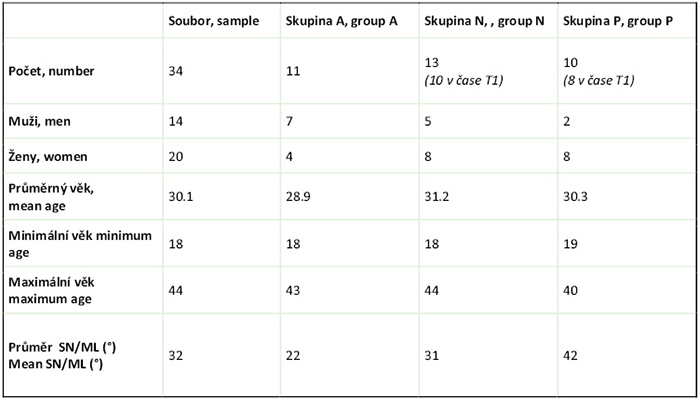
Soubor byl následně rozdělen na 3 skupiny dle hodnoty úhlu mandibulární linie SN/ML. Základní charakteristiky celého souboru a jednotlivých skupin jsou popsány v tabulce (tab. 1).
1. Skupina A (výrazná anteriorotace) – do této skupiny byli zařazeni pacienti s hodnotou SN/ML menší než 27° (průměrná hodnota SN/ML = 22°) a sestávala z 11 pacientů.
2. Skupina N (průměrná anteriorotace) – do této skupiny byli zařazeni pacienti s hodnotou SN/ML v rozpětí 27–39° (průměrná hodnota SN/ML = 31°) a sestávala ze 13 pacientů (v čase T1 z 10 pacientů).
3. Skupina P (růst blízký posteriorotaci) – do této skupiny byli zařazeni pacienti s hodnotou SN/ML větší než 39° (průměrná hodnota SN/ML = 42°) a sestávala z 10 pacientů (v čase T1 z 8 pacientů).
Metodika
Ortodontická příprava a ortodontické doléčení probíhalo v soukromých ortodontických praxích ortodontistů, nebo na ortodontickém oddělení Stomatologické kliniky ve FN Plzeň. Ortognátní operace byla u všech pacientů uskutečněna ve Fakultní nemocnici Plzeň na oddělení Ústní, čelistní a obličejové chirurgie mezi lety 2012 –2022. Operační technika byla u všech pacientů stejná, stanovený typ operace u všech pacientů byla BSSO s předsunutím mandibuly, modifikace Hunsuck/Epker. Jednotlivé segmenty dolní čelisti byly fixovány minidlahami. Na každou stranu se standardně dávala jedna dlaha. Využívaný byl systém MatrixMANDIBLE™, u pacientů operovaných v pozdější době systém MatrixORTHOGNATHIC™, vždy se dvěma či třemi šroubky na každé straně osteotomie. Klinické a rentgenologické pooperační kontroly pacientů se odehrály na ortodontickém oddělení Stomatologické kliniky FN Plzeň – Lochotín.
Všechny kefalometrické snímky byly vyhodnoceny jednou osobou v programu OnyxCeph3™ (Image Instruments, Chemnitz, Německo) v maximálním možném zvětšení. Pro stanovení chyby měření bylo měření některých parametrů u 10 náhodně vybraných pacientů zopakováno. Hodnocené kefalometrické snímky byly zhotovoveny před zahájením terapie (T1), před ortognátní operací (T2), 6 týdnů po ortognátní operaci (T3), 6 měsíců po ortognátní operaci (T4) a 2 roky po ortognátní operaci (T5). U všech pacientů ze souboru byly vyhodnoceny snímky v časech T2–T5. Snímky pořízené v čase T1 byly dostupné pouze u 29 pacientů ze souboru.
Parametry byly zvoleny tak, aby se z nich dala odvodit skeletální a dentální stabilita v sagitálním a vertikálním směru. Pro měření některých parametrů byla využívána horizontální (osa x) a vertikální (osa y) referenční linie. Osa x je odvozena od linie SN, svírá s ní úhel 7° a prochází bodem S. Je tedy přibližně rovnoběžná s Frankfurtskou horizontálou. Úhel 7° byl zvolen z toho důvodu, že většinou odpovídá horizontále při přirozeném postavení hlavy a na kefalometrickém snímku je tato linie lépe reprodukovatelná než Frankfurtská horizontála. Osa y je kolmá na osu x a prochází bodem S (obr. 1). Pomocí referenčních linií je pak možné zaznamenat přesný posun stanovených bodů v horizontálním i vertikálním směru. Kefalometrické snímky byly vyhodnoceny i standardním způsobem a k určení stability byly využity i některé lineární a úhlové parametry vyjmenované níže.
Skeletální parametry
SNB – úhel popisuje anterioposteriorní postavení mandibuly ve vztahu k přední bazi lební
ANB – úhel anterioposteriorní postavení čelistí vůči sobě
SNPog – úhel popisuje anterioposteriorní postavení mandibuly ve vztahu k přední bazi lební ve stupních
Go-Me – délka těla dolní čelisti
ΔBx – sagitální změna bodu B promítnutá na osu x
ΔMex – sagitální změna bodu Me promítnutá na osu x
SN/ML – úhel vyjadřující sklon mandibuly k přední bazi lební
NL/ML – úhel vyjadřující sklon mandibuly k palatinální rovině
N-Me – přední výška obličeje
ArGoMe – goniový úhel
ΔBy – vertikální změna bodu B promítnutá na osu y
ΔMey – vertikální změna bodu Me promítnutá na osu y
Dentální parametry
IS – incizální schůdek, vzdálenost incizních hran horních a dolních řezáků paralelní s okluzní rovinou
+1/SN – úhel osy horního centrálního řezáku k přední bazi lební
-1/ML – úhel osy dolního centrálního řezáku k mandibulární rovině
HS – hloubka skusu, vzdálenost incizních hran horních a dolních řezáků kolmá na okluzní rovinu.
Nejprve byla testována změna jednotlivých parametrů mezi po sobě jdoucími časy. Následně bylo zjišťováno, zda se skupiny A, N a P ve změnách těchto parametrů mezi sebou liší. Pro testování celého souboru v po sobě jdoucích časech byl použit párový t-test, pokud nebyla zamítnuta normalita dat. V opačném případě byl použit Wilcoxonův párový test. Normalita byla vyhodnocena pomocí Shapirova-Wilkova testu. Pro testování odlišnosti skupin byl v případě nezamítnutí normality ve skupinách a nezamítnutí rovnosti rozptylů ve skupinách použit test ANOVA s post-hoc analýzou (Tukeyův test). V opačném případě byl použit Kruskalův-Wallisův test s post-hoc analýzou (Dunnův test s Bonferroniho korekcí). Minimální hladina významnosti u všech testovaných hypotéz byla 5 %.
Výsledky
V celém souboru v průběhu ortodontické přípravy (v čase T1–T2) došlo k mírné CW rotaci mandibuly, a kaudálního posunu bodů B a Me. Došlo k zvětšení incizálního schůdku průměrně o 1,08 mm a retruzi dolních řezáků průměrně o –5,46°. Ve sklonu horních řezáků nebyly zaznamenány významné změny. Mezi skupinami nebyly zaznamenány statisticky významné změny.
V době ortognátní operace (v čase T2–T3) došlo očekávaně k významným změnám v parametrech popisujících polohu mandibuly, IS a HS. Změny byly významné jak ve vertikálním, tak v horizontálním směru. Nejvýraznější operační posun mandibuly byl dle posunu bodu Me na ose x zaznamenán ve skupině P (průměrně o 6,0 mm) a ve skupině A byl posun nejmenší (průměrně o 3,56 mm). Ve vertikálním směru bylo v našem souboru zaznamenáno největší zvětšení vertikálního rozměru naopak ve skupině A, SN/ML se v této skupině zvětšil průměrně o 4,0°, NL/ML o 3,79° a goniový úhel o 6,16°. Ve skupině P bylo zaznamenáno mírné zmenšení hodnoty SN/ML (průměrně o –0,72°), což by potvrzovalo counter-clockwise (CCW) rotaci předního segmentu mandibuly.
V období od 6 týdnů do 6 měsíců po ortognátní operaci (v čase T3–T4) došlo v celém souboru ke statisticky významnému zmenšení hodnoty u parametrů SNB průměrně o –0,35° a Go-Me průměrně o –0,62 mm. Nejvýraznější posun mandibuly dorzálně byl zaznamenán ve skupině P. V této skupině se úhel ANB zvětšil průměrně o 0,61°, SNB se zmenšil průměrně o –0,87°, bod B se na ose x posunul dorzálně průměrně o –1,48 mm a bod Me průměrně o –1,46 mm. Ve skupině A byl zaznamenán naopak mírný posun mandibuly ventrálně, kdy se úhel ANB zmenšil průměrně o –0,12°, SNB se zvětšil průměrně o 0,17°, bod B se na ose x posunul ventrálně průměrně o 0,9 mm a bod Me průměrně o 1,04 mm. Tyto výsledky odpovídají mírné změně ve smyslu původní růstové rotace mandibuly.
U skeletálních vertikálních parametrů v celém souboru v prvních 6 měsících po operaci nebyla zaznamenána statisticky významná změna u žádného parametru. V parametru ΔBy ale byla zaznamenána rozdílnost mezi skupinami, kdy ve skupině P došlo k posunu bodu B kaudálně průměrně o 0,7 mm a ve skupině A naopak k jeho posunu kraniálně průměrně o –0,57 mm.
Dentálně bylo v celém souboru v období do 6 měsíců od operace zaznamenáno zvětšení incizálního schůdku průměrně o 0,36 mm,
V období od 6 měsíců do 2 let po ortognátní operaci (v čase T4–T5) byl v celém souboru na ose x zaznamenán dorzální posun bodu B průměrně o –0,39 mm a bodu Me průměrně o –0,46 mm. Ve vertikálním směru nebyly zaznamenány žádné statisticky ani klinicky významné změny. V celém souboru byla zaznamenána protruze dolních řezáků průměrně o 0,58 mm.
Shrnutí statisticky významných výsledků pro vybrané skeletální parametry ΔBx, ΔMex, ΔBy a ΔMey jsou uvedeny v tabulkách Tab. 2–4.
Diskuse
V průběhu ortodontické předoperační přípravy došlo v našem souboru ke zvětšení incizálního schůdku a retruzi dolních řezáků. V postavení horních řezáků nebyly v celkovém souboru zaznamenány statisticky významné změny. Výsledky naší studie jsou v souladu s tím, co uvádí Proffit. Horní řezáky většinou zůstávají přibližně ve stejném postavení, případně se protrudují. Dolní řezáky se retrudují. Tím se zvětšuje incizální schůdek a vzniká tak prostor pro předsun mandibuly [1, 5].
U všech skeletálních vertikálních parametrů došlo v průběhu ortodontické přípravy ke statisticky významnému zvětšení hodnot. Tyto změny pravděpodobně souvisejí s dentálními změnami, tedy s cíleným zvyšováním skusu [14].
Nejvýraznější operační posun mandibuly byl dle posunu bodu Me na ose x zaznamenán ve skupině P a ve skupině A byl posun nejmenší. Podobné rozložení souboru zaznamenali i Mobarak a Eggensperger. Ve studii Mobaraka byl průměrný posun u posteriorotovaných pacientů 7,6 mm a u anteriorotovaných pacientů 4,4 mm [9]. Eggensperger uvádí průměrný posun u posteriorotovaných 6,3 mm a u anteriorotovaných 4,5 mm [10]. Ve skupině A a N byla zaznamenána CW rotace mandibuly. Ve skupině P byla dle SN/ML zaznamenána CCW rotace mandibuly.Tyto rozdíly mezi skupinami mohou být způsobeny rozdílnými nároky na estetiku obličeje v těchto dvou skupinách [1, 15–17].
V období do 6 měsíců po operaci došlo k dorzálnímu posunu mandibuly a ke zvětšení IS. Dle horizontálnímu posunu bodů B a Me došlo k dorzálnímu posunu mandibuly zejména ve skupině P. Ve skupině A byl zaznamenán naopak mírný posun mandibuly ventrálně. Dle vertikálního posunu bodu B byla ve skupinách A a N zaznamenána CCW rotace mandibuly, ve skupině P byla zaznamenána CW rotace mandibuly. Tyto výsledky odpovídají mírné změně ve smyslu původní růstové rotace mandibuly. Naše výsledky jsou částečně v souladu se studií Mobaraka, který v sagitálním směru zaznamenal největší recidivu taktéž u posteriorotovaných pacientů, kdy průměrná recidiva v této skupině byla 2,7 mm. U anteriorotovaných zaznamenal recidivu průměrně o 1,2 mm. Zároveň ale uvádí, že u posteriorotovaných docházelo v jeho souboru ke kontinuální recidivě po celé zkoumané období (až do 3 let po operaci) a u anteriorotovaných došlo k největší recidivě v období do 2 měsíců po operaci [9]. V tomto se naše výsledky liší, v našem souboru byl ve skupině A zaznamenán naopak posun mandibuly ventrálně. Posun mandibuly ventrálně byl u některých pacientů zaznamenán i ve studii Mihalika, který jej odůvodňoval pokračujícím růstem mandibuly [18]. Vzhledem k tomu, že v našem souboru došlo k posunům už v krátkodobém horizontu do 6 měsíců po operaci, spíše než k růstu bychom se jako k příčině přiklonili k názoru, že jde o tah svalů [7].
U skeletálních vertikálních parametrů v celém souboru v prvních 6 měsících po operaci nebyla zaznamenána statisticky významná změna u žádného parametru, ale byla zaznamenána rozdílnost mezi skupinami, kdy ve skupině P došlo k posunu bodu B kaudálně a ve skupině A naopak k jeho posunu kraniálně. Posun bodu B na ose y může potvrzovat probíhající recidivu v obou skupinách. To, že v celém souboru byla celková změna vertikálních parametrů statisticky nevýznamná, může být ovlivněno tím, že ve skupině P došlo spíše k nárůstu a ve skupině A naopak k poklesu hodnot. V celém souboru byla tedy celková změna minimální. Zaznamenané změny mohou souviset s probíhající dentální recidivou v laterálních segmentech, což se ale v naší studii cíleně nezjišťovalo. Tuto souvislost tudíž nemůžeme s jistotou potvrdit ani vyvrátit, ale může být námětem pro další výzkum. Mobarak ve své studii zaznamenal největší míru recidivy ve vertikálním směru u anteriorotovaných pacientů, průměrně o –1,1 mm. U posteriorotovaných byla recidiva menší, průměrně 0,6 mm [9].
Zvětšení incizálního schůdku souviselo nejspíše se sagitální skeletální recidivou vady. Podobná pozorování učinil Paunonen, který taktéž zaznamenal souvislost mezi skeletální sagitální recidivou a nárůstem incizálního schůdku [19]. Sklon horních a dolních řezáků zůstával v našem souboru bez statisticky významných změn. Je ale nutné zmínit, že v tomto období měla většina pacientů stále nasazený fixní ortodontický aparát a probíhalo ortodontické pooperační doléčení, tudíž mohou být tyto hodnoty ovlivněny a zkresleny fixním aparátem a nemusí nutně potvrzovat stabilitu.
V období od 6 měsíců do 2 let od operace došlo v celém souboru dle posunu bodů B a Me k dorzálnímu posunu mandibuly.
Mezi skupinami nebyly v žádném skeletálním parametru zaznamenány významné rozdíly, což je v rozporu se studií Mobaraka, který udává, že u posteriorotovaných pacientů docházelo k největší míře recidivy mezi 1. a 3. rokem po operaci (průměrně o 1,0 mm v bodě Pog) a u anteriorotovaných pacientů v tomto časovém období k recidivě nedocházelo [9]. K jinému výsledku dospěl i Eggensperger, který v období mezi 6 až 14 měsíci po operaci zaznamenal větší skeletální sagitální recidivu u posteriorotovaných pacientů (průměrně o 2,3 mm v bodě Pog) a u anteriorotovaných nezaznamenal žádné významné změny. Ve vertikálním směru odlišnosti v recidivě mezi skupinami ve své studii nepopisuje [10].
V období od 6 měsíců do 2 let od operace byla v celém souboru zaznamenána protruze dolních řezáků, která mohla být způsobena jejich kompenzační reakcí na probíhající skeletální sagitální recidivu, což popisují i jiní autoři [7, 18].
Závěr
Během ortognátní operace (bilaterální sagitální split osteotomie s předsunutím mandibuly bez genioplastiky) byly v naší studii zaznamenány rozdíly mezi anteriorotovanými a posteriorotovanými pacienty. U anteriorotovaných pacientů došlo operačním zákrokem ke clockwise rotaci, u posteriorotovaných pacientů spíše ke counter-clockwise rotaci předního segmentu mandibuly.
V období od 6 týdnů do 6 měsíců byl u posteriorotovaných pacientů v horizontálním směru zaznamenán dorzální posun mandibuly, u anteriorotovaných ventrální posun. Ve vertikálním směru byla u posteriorotovaných zaznamenána clockwise rotace, u anteriorotovaných counter-clockwise rotace.
V období od 6 měsíců do 2 let po ortognátní operaci došlo v celém souboru k mírné recidivě v horizontálním směru a protruzi dolních řezáků. Ve vertikálním směru ani mezi skupinami nebyly zaznamenány žádné statisticky významné rozdíly.
Výsledky studie naznačují, že růstová rotace má vliv na stabilitu ortodonticko-chirurgické léčby. U posteriorotovaných pacientů byla zaznamenána výraznější skeletální recidiva v sagitálním i vertikálním směru.
Autoři nemají komerční, vlastnické nebo finanční zájmy na produktech nebo společnostech popsaných v tomto článku.
Introduction
The choice of treatment for Class II skeletal malocclusion depends on several factors, particularly the severity of the orthodontic anomaly, the patient’s primary motivation and cooperation [1–4]. Orthodontic-surgical therapy is suitable for patients with a more pronounced skeletal anomaly, in whom attempts to compensate for it could lead to harm to the patient [3, 4]. Another indication for orthodontic-surgical treatment is the patient’s motivation to change their profile. Compared to orthodontic-surgical treatment, orthodontic therapy has only a minimal effect on the soft tissues of the face and the resulting profile [2, 5].
The outcome of orthodontic-surgical therapy and its stability depend on the treatment plan, its course, preoperative preparation, surgical technique, final occlusion, retention, and, last but not least, on the natural changes that occur in every individual after the completion of therapy [6]. Skeletal relapse is influenced primarily by neuromuscular adaptation, adaptation of the temporomandibular joint, growth, and the choice of surgical technique. Bilateral sagittal split osteotomy (BSSO) with mandibular advancement is generally considered a stable procedure. However, some authors state that this is only the case under certain conditions [7]. In patients with posterior growth rotation, the anterior segment is often rotated during this procedure to achieve shortening of the lower third of the face and to ensure an overbite in the anterior dental arch. During CCW rotation, the suprahyoid muscles are simultaneously stretched, making this an unstable movement [8–12]. Generally, operations in which soft tissue tension is released have the best prospects for a stable outcome, whereas those in which the tissues are stretched fare less well [2, 13]. For this reason, some authors favour maxillary impaction with mandibular autorotation or bimaxillary surgery in these patients. They indicate BSSO with mandibular advancement only in patients with moderate or marked anterior rotation [7].
Material
The study cohort comprised 34 patients – 20 women and 14 men – who underwent orthodontic-surgical treatment. The cohort included patients with Class I and II skeletal malocclusions according to the ANB classification, in whom it was not possible to achieve a stable, functional and aesthetic result through orthodontic treatment alone.
Inclusion criteria
1. The patient has completed orthodontic-surgical treatment.
2. The patient is in skeletal Class I or II.
3. The relationship of the dental arches is Class II according to Angle, Division 1 or 2.
4. The surgical procedure involved only the mandible (mandibular advancement), without genioplasty.
5. The orthognathic surgery was performed in adulthood.
6. Following orthognathic surgery, radiographs were taken at intervals in accordance with the established protocol.
7. The patient was at least 2 years post-orthognathic surgery.
8. The patient was in good general health.
The cohort was subsequently divided into 3 groups according to the value of the mandibular line SN/ML angle. The basic characteristics of the entire cohort and the individual groups are described in the table (Table 1).
1. Group A (marked anterior rotation) – this group included patients with an SN/ML angle of less than 27° (mean SN/ML angle = 22°) and consisted of 11 patients.
2. Group N (moderate anterior rotation) – this group comprised patients with an SN/ML value ranging from 27–39° (mean SN/ML value = 31°) and consisted of 13 patients (10 patients at time T1).
3. Group P (growth close to posterior rotation) – this group included patients with an SN/ML value greater than 39° (average SN/ML value = 42°) and consisted of 10 patients (8 patients at time T1).
Methodology
Orthodontic preparation and post-orthodontic treatment were carried out in private orthodontic practices or at the Orthodontic Department of the Dental Clinic at the University Hospital in Plzeň. Orthognathic surgery was performed on all patients at the University Hospital in Plzeň, in the Department of Oral and Maxillofacial Surgery, between 2012 and 2022. The surgical technique was the same for all patients; the procedure performed was BSSO with mandibular advancement, using the Hunsuck/Epker modification. Individual segments of the lower jaw were fixed using mini-plates. One plate was placed on each side as standard. The MatrixMANDIBLE™ system was used; for patients operated on at a later date, the MatrixORTHOGNATHIC™ system was used, always with two or three screws on each side of the osteotomy. Clinical and radiographic postoperative follow-ups of patients took place at the Orthodontic Department of the Dental Clinic, University Hospital Plzeň – Lochotín.
All cephalometric radiographs were assessed by a single person using the OnyxCeph3™ software (Image Instruments, Chemnitz, Germany) at the maximum possible magnification. To determine the measurement error, the measurement of certain parameters was repeated in 10 randomly selected patients. The cephalometric images evaluated were taken prior to the start of treatment (T1), prior to orthognathic surgery (T2), 6 weeks after orthognathic surgery (T3), 6 months after orthognathic surgery (T4) and 2 years after orthognathic surgery (T5). For all patients in the cohort, images at time points T2–T5 were evaluated. Images taken at time point T1 were available for only 29 patients in the cohort.
The parameters were selected so that skeletal and dental stability in the sagittal and vertical directions could be derived from them. Horizontal (x-axis) and vertical (y-axis) reference lines were used to measure certain parameters. The x-axis is derived from the SN line, forms an angle of 7° with it, and passes through point S (Fig.1). It is therefore approximately parallel to the Frankfurt horizontal. The angle of 7° was chosen because it generally corresponds to the horizontal plane in the natural head position, and this line is more reproducible on a cephalometric image than the Frankfurt horizontal. The y-axis is perpendicular to the x-axis and passes through point S (Fig. 1). Using the reference lines, it is then possible to record the exact displacement of the specified points in both the horizontal and vertical directions. The cephalometric images were also evaluated using standard methods, and certain linear and angular parameters listed below were also used to determine stability.
Skeletal parameters
SNB – angle describing the anteroposterior position of the mandible in relation to the anterior cranial base
ANB – the angle describing the anteroposterior relationship between the jaws
SNPog – angle describing the anteroposterior position of the mandible in relation to the anterior cranial base in degrees
Go-Me – length of the mandibular body
ΔBx – sagittal change of point B projected onto the x-axis
ΔMex – sagittal change of point Me projected onto the x-axis
SN/ML – the angle expressing the inclination of the mandible towards the anterior cranial base
NL/ML – the angle expressing the inclination of the mandible towards the palatal plane
N-Me – anterior facial height
ArGoMe – gonion angle
ΔBy – vertical change of point B projected onto the y-axis
ΔMey – vertical change of point Me projected onto the y-axis
Dental parameters
IS – overjet, the distance between the incisal edges of the upper and lower incisors, parallel to the occlusal plane
+1/SN – angle of the axis of the upper central incisor to the anterior cranial base
-1/ML – angle of the axis of the lower central incisor to the mandibular plane
HS – overbite, the distance between the incisal edges of the upper and lower incisors perpendicular to the occlusal plane.
First, changes in individual parameters between consecutive time points were tested. Subsequently, it was determined whether groups A, N and P differed from one another in terms of changes in these parameters. To test the entire dataset across consecutive time points, a paired t-test was used provided the data were normally distributed. Otherwise, the Wilcoxon signed-rank test was used. Normality was assessed using the Shapiro-Wilk test. To test for differences between groups, where normality within groups was not rejected and equality of variances within groups was not rejected, a two-way ANOVA with post-hoc analysis (Tukey’s test) was used. Otherwise, the Kruskal-Wallis test with post-hoc analysis (Dunn’s test with Bonferroni correction) was used. The minimum significance level for all tested hypotheses was 5%.
Results
In the entire sample, during orthodontic treatment (at time points T1–T2), there was a slight CW rotation of the mandible and a caudal shift of points B and Me. There was an increase in the overjet by an average of 1.08 mm and a retraction of the lower incisors by an average of –5.46°. No significant changes were observed in the inclination of the upper incisors. No statistically significant changes were observed between the groups.
At the time of orthognathic surgery (at time T2–T3), as expected, significant changes occurred in the parameters describing the position of the mandible, IS and HS. The changes were significant in both the vertical and horizontal directions. In the vertical direction, the greatest increase in vertical dimension was, conversely, recorded in group A in our sample; SN/ML increased by an average of 4.0° in this group, NL/ML by 3.79° and the gonion angle by 6.16°. In group P, a slight decrease in the SN/ML value was recorded (by an average of –0.72°), which would confirm counter-clockwise (CCW) rotation of the anterior segment of the mandible.
In the period from 6 weeks to 6 months following orthognathic surgery (at time points T3–T4), a statistically significant reduction in the SNB parameter was observed across the entire cohort, by an average of –0.35°, and in the Go-Me parameter by an average of –0.62 mm. The most pronounced dorsal displacement of the mandible was recorded in group P. In this group, the ANB angle increased by an average of 0.61°, the SNB angle decreased by an average of –0.87°, point B shifted dorsally on the x-axis by an average of –1.48 mm, and point Me by an average of –1.46 mm. In group A, by contrast, a slight ventral shift of the mandible was recorded, with the ANB angle decreasing by an average of –0.12°, the SNB angle increasing by an average of 0.17°, point B shifting ventrally on the x-axis by an average of 0.9 mm, and point Me by an average of 1.04 mm. These results correspond to a slight change in the direction of the original growth rotation of the mandible.
No statistically significant change was observed in any of the skeletal vertical parameters across the entire cohort during the first 6 months post-surgery. However, a difference between the groups was observed in the ΔBy parameter, with point B shifting caudally by an average of 0.7 mm in group P and, conversely, shifting cranially by an average of –0.57 mm in group A.
Dentally, an average increase in the overjet of 0.36 mm was observed in the entire cohort within 6 months of surgery.
In the period from 6 months to 2 years after orthognathic surgery (at time points T4–T5), a dorsal shift of point B by an average of –0.39 mm and of point Me by an average of –0.46 mm was recorded on the x-axis across the entire cohort. No statistically or clinically significant changes were observed in the vertical direction. An average protrusion of the lower incisors of 0.58 mm was recorded across the entire cohort.
A summary of the statistically significant results for the selected skeletal parameters ΔBx, ΔMex, ΔBy and ΔMey is presented in Tables 2–4.
Discussion
During pre-operative orthodontic preparation, an increase in the overjet and retraction of the lower incisors were observed in our sample. No statistically significant changes in the position of the upper incisors were recorded in the overall sample. The results of our study are consistent with those reported by Proffit. The upper incisors generally remain in approximately the same position or may protrude. The lower incisors retract. This increases the overjet, thereby creating space for mandibular advancement [1, 5].
For all skeletal vertical parameters, a statistically significant increase in values occurred during orthodontic treatment. These changes are likely related to dental changes, i.e. to the targeted increase in occlusal height [14].
The most pronounced surgical mandibular movement, as measured by the displacement of point Me on the x-axis, was recorded in group P, whilst the smallest displacement was observed in group A. A similar distribution of the sample was also reported by Mobarak and Eggensperger. In Mobarak’s study, the average displacement was 7.6 mm in posteriorly rotated patients and 4.4 mm in anteriorly rotated patients [9]. Eggensperger reports an average displacement of 6.3 mm in posteriorly rotated patients and 4.5 mm in anteriorly rotated patients [10]. CW rotation of the mandible was recorded in groups A and N. In group P, CCW rotation of the mandible was recorded according to SN/ML. These differences between the groups may be due to differing aesthetic requirements for the face in these two groups [1, 15–17].
Within 6 months post-surgery, a dorsal displacement of the mandible and an increase in the IS were observed. Based on the horizontal displacement of points B and Me, a dorsal displacement of the mandible was observed, particularly in group P. In group A, conversely, a slight ventral displacement of the mandible was recorded. Based on the vertical displacement of point B, CCW rotation of the mandible was observed in groups A and N, whilst CW rotation of the mandible was observed in group P. These results correspond to a slight change in the direction of the original growth rotation of the mandible. Our results are partly consistent with Mobarak’s study, which also recorded the greatest sagittal relapse in posteriorly rotated patients, with an average relapse of 2.7 mm in this group. In the anteriorly rotated group, he recorded an average recurrence of 1.2 mm. At the same time, however, he notes that in his cohort, posteriorly rotated patients experienced continuous recurrence throughout the entire study period (up to 3 years post-surgery), whilst the greatest recurrence in the anteriorly rotated group occurred within the first 2 months post-surgery [9]. Our results differ in this respect; in our cohort, a ventrally displaced mandible was, on the contrary, observed in Group A. Ventrally displaced mandibles were also observed in some patients in Mihalika’s study, who attributed this to continued mandibular growth [18]. Given that in our cohort, displacements occurred within the short-term period of up to 6 months post-surgery, rather than growth, we would be inclined to attribute the cause to muscle pull [7].
With regard to skeletal vertical parameters across the entire cohort in the first 6 months following surgery, no statistically significant change was observed for any parameter; however, differences were noted between the groups, with point B shifting caudally in group P and, conversely, shifting cranially in group A. The shift of point B on the y-axis may confirm ongoing relapse in both groups. The fact that the overall change in vertical parameters was statistically insignificant across the entire cohort may be influenced by the fact that in group P there was a tendency towards an increase, whereas in group A there was a decrease in values. The overall change was therefore minimal across the entire cohort. The recorded changes may be related to ongoing dental relapse in the lateral segments; however, this was not specifically investigated in our study. We cannot therefore confirm or refute this connection with certainty, but it may provide a topic for further research. In his study, Mobarak recorded the highest rate of vertical relapse in anteriorly rotated patients, averaging –1.1 mm. In posteriorly rotated patients, relapse was lower, averaging 0.6 mm [9].
The increase in the overjet was most likely associated with sagittal skeletal relapse of the malocclusion. Similar observations were made by Paunonen, who also noted a correlation between sagittal skeletal relapse and an increase in the overjet [19]. The inclination of the upper and lower incisors remained without statistically significant changes in our cohort. However, it is important to note that during this period, most patients were still wearing fixed orthodontic appliances and undergoing post-operative orthodontic treatment; therefore, these values may be influenced and distorted by the fixed appliances and do not necessarily confirm stability.
In the period from 6 months to 2 years after surgery, a dorsal displacement of the mandible occurred in the entire cohort according to the shift in points B and Me.
No significant differences were recorded between the groups in any skeletal parameter, which contradicts Mobarak’s study, which states that in posteriorly rotated patients, the highest rate of relapse occurred between the 1st and 3rd year after surgery (by an average of 1.0 mm at the Pog point) and that no relapse occurred in anteriorly rotated patients during this period [9]. Eggensperger also reached a different conclusion, recording greater skeletal sagittal relapse in posteriorly rotated patients (an average of 2.3 mm at the Pog point) between 6 and 14 months post-surgery, whilst observing no significant changes in anteriorly rotated patients. He does not describe any differences in relapse between the groups in the vertical direction in his study [10].
In the period from 6 months to 2 years after surgery, protrusion of the lower incisors was observed in the entire cohort, which may have been caused by their compensatory response to ongoing skeletal sagittal relapse, a phenomenon also described by other authors [7, 18].
Conclusion
During orthognathic surgery (bilateral sagittal split osteotomy with mandibular advancement without genioplasty), differences were observed in our study between patients with anterior growth rotation and those with posterior rotation. In patients with anterior rotation, the surgical procedure resulted in clockwise rotation, whereas in patients with posterior rotation, it resulted in counter-clockwise rotation of the anterior segment of the mandible.
In the period from 6 weeks to 6 months, a dorsal shift of the mandible in the horizontal direction was observed in posteriorly rotated patients, whilst a ventral shift was observed in anteriorly rotated patients. In the vertical direction, clockwise rotation was observed in posteriorly rotated patients, whilst counter-clockwise rotation was observed in anteriorly rotated patients.
In the period from 6 months to 2 years following orthognathic surgery, a slight horizontal relapse and protrusion of the lower incisors occurred in the entire cohort. No statistically significant differences were observed in the vertical direction or between the groups.
The results of the study suggest that growth-related rotation influences the stability of orthodontic-surgical treatment. In posteriorly rotated patients, more pronounced skeletal relapse was observed in both the sagittal and vertical directions.
The authors declare no commercial, proprietary, or financial interests in the products or companies described in this article.
Tab. 1 / Tab. 1
Základní charakteristika souboru a stanovených skupin. / Basic characteristics of the dataset and defined groups.
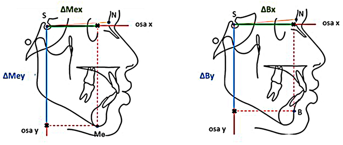
Obr. 1 / Fig. 1
Referenční osy x a y, parametry k nim vztažené / references x and y and the parameters related to them.
Tab. 2 / Tab. 2
Změny ve vybraných skeletálních parametrech v průběhu jednotlivých etap léčby / Significant changes in selected parameters across the entire dataset at consecutive time points.
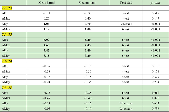
ΔBx – sagitální změna bodu B promítnutá na osu x, ΔMex – sagitální změna bodu Me promítnutá na osu x, ΔBy – vertikální změna bodu B promítnutá na osu y, ΔMey – vertikální změna bodu Me promítnutá na osu y; T1 – před léčbou, T2 – před operací, T3 – 6 týdnů po operaci, T4 – 6 měsíců po operaci, T5 – 2 roky po operaci. / ΔBx – sagittal change of point B relative to the x-axis, ΔMex – sagittal change of point Me relative to the x-axis, ΔBy – vertical change of point B relative to the y-axis, ΔMey – change of point Me relative to the y-axis; T1 – before treatment, T2 – before surgery, T3 – 6 weeks after surgery, T4 – 6 months after surgery, T5 – 2 years after surgery.
Tab. 3 / Tab. 3
Změny ve vybraných skeletálních parametrech ve skupinách A, N, P v prvním půlroce po ortognátní operaci (z času T3 do času T4) / changes between groups A, N, and P in the first six months after surgery (from time point T3 to time point T4).
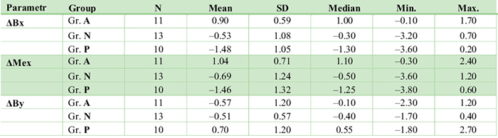
Typ růstové rotace čelisti dle hodnoty úhlu SN/ML: Gr. A Gr. A 27° (výrazná anteriorotace), Gr. N = 27°–39° (průměrná anteriorotace), Gr. P 39° (růst blízký posteriorotaci). ΔBx – sagitální změna bodu B promítnutá na osu x, ΔMex – sagitální změna bodu Me promítnutá na osu x, ΔBy – vertikální změna bodu B promítnutá na osu y, ΔMey – vertikální změna bodu Me promítnutá na osu y. / Patterns of growth rotation acc. to Nasion-Sella line to Mandibular line angle: ΔBx – sagittal change of point B relative to the x-axis, ΔMex – sagittal change of point Me relative to the x-axis, ΔBy – vertical change of point B relative to the y-axis, ΔMey – change of point Me relative to the y-axis.
Tab. 4 / Tab. 4
Test významnosti odlišností změn vybraných sagitálních skeletálních parametrů mezi skupinami A, N, P v prvním půlroce po ortognátní operaci (z času T3 do času T4). / Significance of some changes between groups A, N, P in the first six months following orthognathic surgery (from time T3 to time T4).

ΔBx – sagitální změna bodu B promítnutá na osu x, ΔMex – sagitální změna bodu Me promítnutá na osu x, ΔBy – vertikální změna bodu B promítnutá na osu y, ΔMey – vertikální změna bodu Me promítnutá na osu y. / ΔBx – sagittal change of point B projected onto the x-axis, ΔMex – sagittal change of point Me projected onto the x-axis, ΔBy – vertical change of point B projected onto the y-axis, ΔMey – vertical change of point Me projected onto the y-axis.

Literatura / References
1. Proffit WR. Contemporary Orthodontics. 6th ed. St. Louis: Mosby; 2018.
2. Naini FB, Gill DS, editors. Orthognathic Surgery: Principles, Planning and Practice. 1st ed. Chichester: Wiley-Blackwell; 2017. doi: 10.1002/9781119004370
3. Arnett WG, McLaughlin RP. Facial and Dental Planning for Orthodontists and Oral Surgeons. 1st ed. Edinburgh: Mosby; 2005.
4. Fonseca RJ. Oral and Maxillofacial Surgery: Volume 2. 2nd ed. St. Louis: Saunders; 2008.
5. Proffit W, White RP, Sarver D. Contemporary Treatment of Dentofacial Deformity. St. Louis: Mosby; 2002.
6. Kamínek M, et al. Ortodoncie. 1st ed. Praha: Galén; 2014.
7. Proffit WR, Turvey TA, Phillips C. The hierarchy of stability and predictability in orthognathic surgery with rigid fixation: an update and extension. Head Face Med. 2007;3(1):21. doi: 10.1186/1746-160X-3-21
8. Van Sickels JE, Dolce C, Keeling S, Tiner BD, Clark GM, Rugh JD. Technical factors accounting for stability of a bilateral sagittal split osteotomy advancement: wire osteosynthesis versus rigid fixation. Oral Surg Oral Med Oral Pathol Oral Radiol Endod. 2000;89(1):19–23. doi: 10.1016/S1079-2104(00)80008-6
9. Mobarak KA, Espeland L, Krogstad O, Lyberg T. Mandibular advancement surgery in high-angle and low-angle class II patients: different long-term skeletal responses. Am J Orthod Dentofac Orthop 2001;119(4):368–81. doi: 10.1067/mod.2001.110983
10. Eggensperger N, Smolka K, Luder J, Iizuka T. Short- and long-term skeletal relapse after mandibular advancement surgery. Int J Oral Maxillofac Surg. 2006;35(1):36–42. doi: 10.1016/j.ijom.2005.04.008
11. Eggensperger N, Smolka W, Rahal A, Iizuka T. Skeletal relapse after mandibular advancement and setback in single-jaw surgery. J Oral Maxillofac Surg. 2004;62(12):1486–96. doi: 10.1016/j.joms.2004.07.007
12. Ruslin M, Hajrah Yusuf AS, Forouzanfar T, Greebe RB, Tuinzing DB, Thamrin SA, et al. One-year stability of the mandibular advancement and counterclockwise rotation for correction of the skeletal class II malocclusion and high mandibular plane angle: Dental and skeletal aspect. Biomed J. 2021;45(1):206–14. doi: 10.1016/j.bj.2021.02.005
13. Frey DR, Hatch JP, Van Sickels JE, Dolce C, Rugh JD. Alteration of the mandibular plane during sagittal split advancement: short- and long-term stability. Oral Surg Oral Med Oral Pathol Oral Radiol Endod. 2007;104(2):160–9. doi: 10.1016/j.tripleo.2006.12.023
14. Weiland FJ, Bantleon HP, Droschl H. Evaluation of continuous arch and segmented arch leveling techniques in adult patients--a clinical study. Am J Orthod Dentofac Orthop. 1996;110(6):647–52. doi: 10.1016/S0889-5406(96)80042-4
15. de Coul FO, Oosterkamp BCM, Jansma J, Bierman MWJ, Pruim GJ, Sandham A. Maintenance of a deep bite prior to surgical mandibular advancement. Eur J Orthod. 2010;32(3):342–5. doi: 10.1093/ejo/cjp087
16. Mommaerts MY, López-Arcas JM, Schurmans AM. Correction of skeletal Class II— anterior deep bite and prominent chin by forced surgical rotation of the mandible. Br J Oral Maxillofac Surg. 2010;48(5):349–51. doi: 10.1016/j.bjoms.2009.08.041
17. Torgersbråten N, Stenvik A, Espeland L. Bimaxillary surgery to correct high-angle class II malocclusion: does a simultaneous genioplasty affect long-term stability? Eur J Orthod. 2020;42(4):426–33. doi: 10.1093/ejo/cjz055
18. Mihalik CA, Proffit WR, Phillips C. Long-term follow-up of Class II adults treated with orthodontic camouflage: a comparison with orthognathic surgery outcomes. Am J Orthod Dentofac Orthop. 2003;123(3):266–78. doi: 10.1067/mod.2003.43
19. Paunonen J, Helminen M, Peltomäki T. Long-term stability of mandibular advancement with bilateral sagittal split osteotomy. J Cranio-Maxillo-fac Surg. 2018;46(9):1421–6. doi: 10.1016/j.jcms.2018.05.023
MDDr. Tereza Šulcová
Stomatologická klinika FN Plzeň
Alej Svobody 80, 304 60 Plzeň